Rat Cortical Stem Cell Culture Expansion Protocol
This protocol must be read in its entirety before using Catalog # NSC001. Caution: The rat cortical stem cells used in this protocol contain trace amounts of human transferrin and DMSO, and the media used in this protocol contains trace amounts of transferrin. The transferrin was purified from donor plasma and tested at the donor level using an FDA licensed method and found to be non-reactive for anti HIV-1/2 and Hepatitis B surface antigen.
Contents
- Rat Cortical Stem Cell Culture Expansion Protocol (Monolayer System)
- Rat Cortical Stem Cell Culture Expansion Protocol (Neurosphere System)
Rat Cortical Stem Cell Culture Expansion Protocol (Monolayer System)
Materials Required
Reagents:
- Rat Cortical Stem Cells (R&D Systems, Catalog # NSC001)
- DMEM/F-12 (ThermoFisher Scientific®, Catalog # 12500-062) or a basal media (e.g., Neurobasal Media from ThermoFisher Scientific, Catalog # 21103-049)
- N2-MAX Media Supplement (Catalog # AR009)
- Glucose
- NaHCO3
- Glutamine
- Recombinant Human Fibroblast Growth Factor basic (FGF basic) (R&D Systems, Catalog # 3718-FB or 4114-TC)
- Purified Bovine Fibronectin (R&D Systems, Catalog # 1030-FN)
- PBS (ThermoFisher Scientific, Catalog # 10010-023 or equivalent)
- Penicillin-Streptomycin, 100X (ThermoFisher Scientific, Catalog # 15140-148 or equivalent)
- Poly-L-ornithine (Sigma, Catalog # P3655 or equivalent)
- Hank’s Balanced Salt Solution (HBSS) (Ca2+ /Mg2+ -free), 10X (ThermoFisher Scientific, Catalog # 14185 052 or equivalent)
- HEPES (Sigma, Catalog # H7006 or equivalent)
- BSA, very low endotoxin (Millipore, Catalog # 81-068-3 or equivalent)
- Trypan blue (ThermoFisher Scientific, Catalog # 15250-061)
- Deionized water
Materials:
- 10 cm tissue culture dishes (Falcon, Catalog # 353003 or equivalent)
- 15 mL tubes (Corning, Catalog # 430052 or equivalent)
- 50 mL Falcon tubes (Falcon, Catalog # 352070, Fisher, Catalog # 05-539-7, or equivalent)
- 0.2 µm, 1000 mL filter unit (Nalgene, VWR Catalog # 28198-496 or equivalent)
- Plastic cell scraper (Costar cell lifter, Catalog # 3008 or equivalent)
- Pipettes and pipette tips
Equipment:
- 37° C and 5% CO2 incubator
- Centrifuge
- Hemocytometer
- Microscope
- Water bath
| Completed N-2 MAX neural cell medium | Add N-2 MAX Media Supplement (5mL), NaHCO3 (0.854 g), Glutamine (0.0365 g), Glucose (0.775 g), Penicillin-Streptomycin (100X) |
|---|---|
| Buffered HBSS (1X) | Add 100 mL of Hanks Balanced Salt Solution (10X) and 3.9 g HEPES to 900 mL of deionized water to make 1000 mL of Buffered HBSS 1X. Adjust the pH to 7.2 ± 0.2. Sterile filter the solution using a 0.2 µm filter unit. Store at room temperature for up to 6 months. |
| FGF basic Stock (1000X) | Add sterile 0.1% BSA in PBS to the Human FGF basic vial to make a 20 µg/mL stock. Aliquot and store at < -20° C in a manual defrost freezer for up to 6 months. Avoid repeated freeze-thaw cycles. |
Poly-L-ornithine and Fibronectin Coated Plates
- Dissolve Poly-L-ornithine in sterile PBS to make a 15 mg/mL stock (1000X). Aliquot and store at < 20° C in a manual defrost freezer for up to 6 months. Avoid repeated freeze-thaw cycles.
- Dilute the 1000X Poly-L-ornithine stock 1000-fold in sterile PBS to make a 15 µg/mL (1X) solution. Prepare fresh as needed.
- Add 10 mL of the (1X) Poly-L-ornithine solution to each 10 cm tissue culture dish. Incubate overnight at 37° C and 5% CO2.
- Discard the Poly-L-ornithine solution. Wash each dish 3 times with 10 mL of PBS each time.
- Add 10 mL of PBS to each dish. Incubate overnight at 37° C and 5% CO2.
- Allow the vial of Bovine Fibronectin to warm to room temperature. Do not agitate. Make a 1 µg/mL solution by pipetting the Bovine Fibronectin into sterile PBS and gently inverting the tubes. Prepare fresh as needed.
- Discard the PBS from each dish. Wash each dish once with 10 mL of PBS.
- Add 10 mL of 1 µg/mL Bovine Fibronectin solution to each dish. Incubate at 37° C and 5% CO2 for 3 - 30 hours.
- Discard the Bovine Fibronectin solution. Wash each dish once with 10 mL of PBS before use.
Thawing of Cryopreserved Cells
Review the following protocol in detail before thawing the cells.
- Warm 20 mL of N-2 MAX-supplemented neural cell medium containing FGF basic in a 50 mL tube in a 37° C water bath.
- Remove the cryovial containing frozen rat cortical stem cells (R&D Systems, Catalog # NSC001) from the liquid nitrogen. Using a 2 mL pipette, immediately add 1 mL of fresh pre-warmed media to the vial by gently pipetting up and down. As cells begin to thaw, transfer the thawed portion into the pre-warmed media in the 50 mL tube. Repeat this process with the warmed media until all of the cells have thawed.
Note: Most of the frozen cells will be at the bottom of the cryovial. Rapid resuspension of frozen cells in warmed media during thawing is critical. Allowing cells to thaw slowly in the DMSO will dramatically reduce viability. Around 90% cell viability is expected from the freshly thawed cells when the appropriate thawing procedure is followed. - Seed cells at the appropriate density (i.e. 1.0 - 1.5 x 10⁶ per 10 cm plate for monolayer expansion).
- After the cells have attached to the plate (3 hours to overnight), replace the medium with fresh N-2 MAX-supplemented neural cell medium with FGF basic.
Procedure
Use serological pipettes to transfer and remove solutions.
Expansion
- Seed 1 - 1.5 x 10⁶ rat cortical stem cells in 10 mL of N-2 MAX-supplemented neural cell medium with 20 ng/mL of FGF basic on a Poly-L-ornithine and Fibronectin coated plate.
- Incubate the cells at 37° C and 5% CO2. Cells should become adherent within 24 hours.
- After 24 hours, add 10 µL of 1000X FGF basic stock to the culture.
- Every second day, replace the medium with fresh N-2 MAX-supplemented neural cell medium.
- Supplement the medium with 10 µL of 1000X stock (20 ng/mL) of FGF basic each day.
- Passage the cells when they reach 70 - 80% confluency (approximately 4 days after initial plating) according to the procedure described below.
Passage
- Warm the buffered HBSS (1X) and N-2 MAX-supplemented neural cell medium containing FGF basic to 37° C.
- Remove the Medium from the cells, and wash once in 10 mL of buffered HBSS (1X).
- Add 5 mL of buffered HBSS (1X). Incubate at room temperature for 15 - 45 minutes until cells round up (check frequently).
- Dislodge as many of the cells as possible from the plate by pipetting. Any remaining cells can be scraped with a hard plastic cell scraper. Transfer the cells to a 15 mL conical tube.
- Centrifuge for 5 minutes at 200 x g. Remove the supernatant.
- Resuspend the cells with 5 mL of N-2 MAX-supplemented neural cell medium containing FGF basic by slowly pipetting up and down approximately 5 times with a 5 mL pipette.
- Mix 10 µL of the cell suspension with 10 µL of Trypan blue and count the live cells on a hemocytometer.
- Seed 0.8 - 1.0 x 10⁶ viable cells in 10 mL of N-2 MAX-supplemented neural cell medium containing FGF basic on a Poly-L-ornithine, and Fibronectin coated plate.
- Incubate the cells at 37° C and 5% CO2. Repeat steps 4 and 5 in the Expansion section (see above). Passage the cells after 3 days or when cells reach 70 - 80% confluence.
Sample Data

Figure 1: Rat cortical stem cells cultured using the monolayer system.
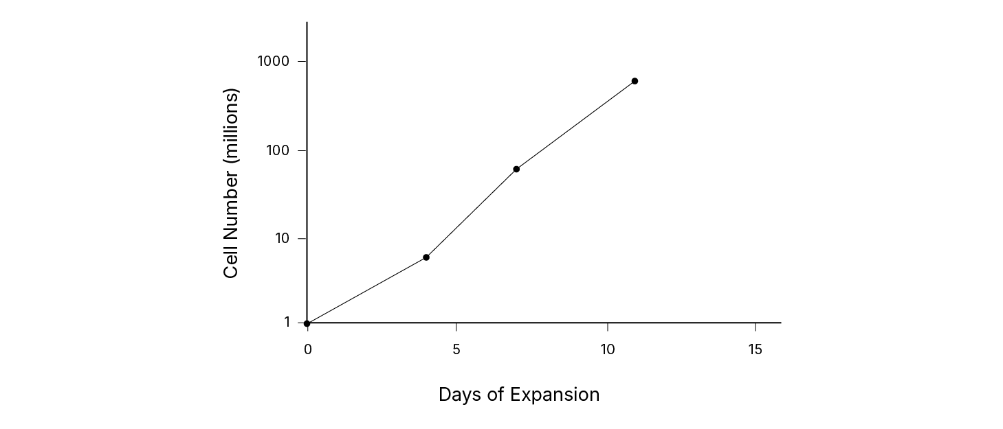
Figure 2: Expansion of rat cortical stem cells using the monolayer system.
Rat Cortical Stem Cell Culture Expansion Protocol (Neurosphere System)
Materials Required
Reagents:
- Rat Cortical Stem Cells (R&D Systems, Catalog # NSC001)
- DMEM/F-12 (ThermoFisher Scientific, Catalog # 12500-062) or a basal media (e.g., Neurobasal Media from ThermoFisher Scientific, Catalog # 21103-049)
- N2-MAX Media Supplement (Catalog # AR009)
- Glucose
- NaHCO3
- Glutamine
- Recombinant Human Fibroblast Growth Factor basic (FGF basic) (R&D Systems, Catalog # 233-FB or 4114-TC)
- Recombinant Human Epidermal Growth Factor (EGF) (R&D Systems, Catalog # 236-EG)
- PBS (ThermoFisher Scientific, Catalog # 10010-023 or equivalent)
- Penicillin-Streptomycin, 100X (ThermoFisher Scientific, Catalog # 15140-148 or equivalent)
- BSA, very low endotoxin (Millipore, Catalog # 81-068-3 or equivalent)
- Acetic acid (J.T. Baker, Catalog # 9508-03 or equivalent)
- Trypan blue (ThermoFisher Scientific, Catalog # 15250-061)
- Deionized water
Materials:
- 10 cm tissue culture dishes (Falcon, Catalog # 353003 or equivalent)
- 15 mL tubes (Corning, Catalog # 430052 or equivalent)
- 50 mL Falcon tubes (Falcon, Catalog # 352070, Fisher, Catalog # 05-539-7, or equivalent)
- 0.2 µm, 1000 mL filter unit (Nalgene, VWR Catalog # 28198-496 or equivalent)
- Plastic cell scraper (Costar cell lifter, Catalog # 3008 or equivalent)
- Pipettes and pipette tips
Equipment:
- 37° C and 5% CO2 incubator
- Centrifuge
- Hemocytometer
- Microscope
- Water bath
| Completed N-2 MAX neural cell medium | add N-2 MAX Media Supplement (5mL), NaHCO3 (0.854 g), Glutamine (0.0365 g), Glucose (0.775 g), Penicillin-Streptomycin (100X), and DMEM/F-12 (6 g) to 500 mL of deionized water. |
|---|---|
| FGF basic Stock (1000X) | Add sterile 0.1% BSA in PBS to the Human FGF basic vial to make a 20 µg/mL stock. Aliquot and store at < -20° C in a manual defrost freezer for up to 6 months. Avoid repeated freeze-thaw cycles. |
| EGF Stock (1000X) | Add sterile 0.1% BSA in 10 mM acetic acid to the Human EGF vial to make a 20 µg/mL stock. Aliquot and store at < -20° C in a manual defrost freezer for up to 6 months. Avoid repeated freeze-thaw cycles. |
Thawing of Cryopreserved Cells
Review the following protocol in detail before thawing the cells.
- Warm 30 mL of completed N-2 MAX-supplemented neural cell medium containing the mitogens in a 37° C water bath.
- Add 20 mL of pre-warmed completed N-2 MAX-supplemented neural cell medium with mitogens to a 50 mL tube. Reserve the remaining 10 mL pre-warmed completed N-2 MAX-supplemented neural cell medium for step # 5.
- Remove the cryovial containing frozen rat cortical stem cells (R&D Systems, Catalog # NSC001) from the liquid nitrogen. Using a 2 mL pipette, immediately add 1 mL of fresh pre-warmed media to the vial by gently pipetting up and down. As cells begin to thaw, transfer the thawed portion into the pre-warmed media in the 50 mL tube. Repeat this process with the warmed media until all of the cells have thawed.
Note: Most of the frozen cells will be at the bottom of the cryovial. - Centrifuge the cells at 200 x g for 5 minutes.
- Aspirate off 95% of the supernatant carefully and resuspend by gently pipetting the cell pellet up and down with 10 mL of completed N-2 MAX-supplemented neural cell medium with the mitogens.
Note: Rapid resuspension of frozen cells in warmed media during thawing is critical. Allowing cells to thaw slowly in the DMSO will dramatically reduce viability. Around 90% cell viability is expected from the freshly thawed cells when the appropriate thawing procedure is followed. - Seed cells at a density according to the appropriate expansion protocol.
Procedure
Use serological pipettes to transfer and remove solutions.
Expansion
- Seed approximately 1 x 10⁵ rat cortical stem cells in 5 mL of completed N-2 MAX-supplemented neural cell medium containing 20 ng/mL of EGF and 20 ng/mL of FGF basic per well in a 6-well plate.
- Incubate the cells at 37° C and 5% CO2.
- Add fresh EGF (20 ng/mL) and FGF basic (20 ng/mL) each day to the medium. Every fourth day, based on the number of neurospheres, replace the media according to the steps described below.
- 50 neurospheres or more - Transfer the medium containing the neurospheres to a 15 mL tube. Centrifuge for 5 minutes at 100 x g and remove the media (gently resuspend the pellet using a small quantity of fresh completed N-2 MAX-supplemented neural cell medium containing EGF (20 ng/mL) and FGF basic (20 ng/mL). Add the neurosphere suspension to 5 mL of fresh N-2 MAX-supplemented neural cell medium containing EGF (20 ng/mL) and FGF basic (20 ng/mL) in one well of a 6-well plate.
- Less than 50 neurospheres - Transfer the neurospheres, using a Pasteur pipette, directly into 2.5 mL of completed N-2 MAX-supplemented neural cell medium containing EGF (20 ng/mL) and FGF basic (20 ng/mL) in one well of a 6-well plate. Do not discard the conditioned medium. Add 2.5 mL of this conditioned medium to the well. When there are fewer neurospheres, conditioned medium is required. Only half of the medium is replaced with fresh completed N-2 MAX-supplemented neural cell medium containing EGF (20 ng/mL) and FGF basic (20 ng/mL).
- Pass the cells at 5 - 7 days, or when the neurospheres have a dark clump inside or ruffling on the outside of the neurosphere, according to the procedure described below.
Passage
- Transfer the media containing the floating neurospheres to a 15 mL tube. Do not dislodge attached neurospheres for passage.
- Centrifuge for 5 minutes at 100 x g.
- Partially dissociate the neurospheres by pipetting up and down 20 times, being careful not to create bubbles in the suspension.
Note: For optimal dissociation of the neurospheres, it is recommended that a P200 pipette be used. - At passages 1 and 2, the cells should be split 1:1. After passage 2, the cells can be split 1:2.
Sample Data

Figure 1: Rat cortical stem cells cultured using the neurosphere system.

Figure 2: Expansion of rat cortical stem cells using the neurosphere system.
References
- Johe, K.K. et al. (1996) Genes and Development 10:3129.
- Kim, J.H. et al. (2003) Methods Enzymol. 365:303.