CD8 Antibody (53-6.7) - BSA Free
Novus Biologicals | Catalog # NBP1-49045
Key Product Details
Species Reactivity
Validated:
Mouse
Cited:
Human, Mouse
Applications
Validated:
Immunohistochemistry, Immunohistochemistry-Paraffin, Immunohistochemistry-Frozen, Flow Cytometry, Immunocytochemistry/ Immunofluorescence, Immunoprecipitation, Cell depletion, Inhibition of T Cell Function, CyTOF-ready
Cited:
Immunohistochemistry, Immunohistochemistry-Paraffin, Immunohistochemistry-Frozen, Flow Cytometry, Immunocytochemistry/ Immunofluorescence, IF/IHC
Label
Unconjugated
Antibody Source
Monoclonal Rat IgG2a Kappa Clone # 53-6.7
Format
BSA Free
Loading...
Product Specifications
Immunogen
CD8 Antibody (53-6.7) was developed against mouse thymus or spleen.
Localization
Most thymocytes, T cell subset, some NK cells
Clonality
Monoclonal
Host
Rat
Isotype
IgG2a Kappa
Theoretical MW
27 kDa.
Disclaimer note: The observed molecular weight of the protein may vary from the listed predicted molecular weight due to post translational modifications, post translation cleavages, relative charges, and other experimental factors.
Disclaimer note: The observed molecular weight of the protein may vary from the listed predicted molecular weight due to post translational modifications, post translation cleavages, relative charges, and other experimental factors.
Description
This CD8 alpha antibody serves as an effective marker of cytotoxic T lymphocytes by binding to the CD8 co-receptor expressed on the cell surface of cytotoxic T cells, recognizing the topological domain of CD8 alpha. Because this CD8 antibody is made to the alpha chain it will recognize both the CD8 alpha - CD8 beta heterodimer, the most common form, as well as the CD8 alpha - CD8 alpha homodimer. The CD8A gene is also expressed in natural killer cells (NK cells), dendritic cells and cortical thymocytes making the CD8 alpha antibody a potential marker for these cells.
Scientific Data Images for CD8 Antibody (53-6.7) - BSA Free
![Flow Cytometry: CD8 Antibody (53-6.7) - BSA Free [NBP1-49045]](https://resources.rndsystems.com/images/products/CD8-Antibody-53-6-7-Flow-Cytometry-NBP1-49045-img0014.jpg "Flow Cytometry: CD8 Antibody (53-6.7) - BSA Free [NBP1-49045]")
Flow Cytometry: CD8 Antibody (53-6.7) - BSA Free [NBP1-49045]
Flow Cytometry: CD8 Antibody (53-6.7) [NBP1-49045] - CD8 alpha Antibody (53-6.7) [NBP1-49045] - Analysis of lymph nodes by multiple staining.![Immunohistochemistry-Paraffin: CD8 Antibody (53-6.7) - BSA Free [NBP1-49045]](https://resources.rndsystems.com/images/products/CD8-Antibody-53-6-7-Immunohistochemistry-Paraffin-NBP1-49045-img0004.jpg "Immunohistochemistry-Paraffin: CD8 Antibody (53-6.7) - BSA Free [NBP1-49045]")
Immunohistochemistry-Paraffin: CD8 Antibody (53-6.7) - BSA Free [NBP1-49045]
Immunohistochemistry-Paraffin: CD8 Antibody (53-6.7) [NBP1-49045] - CD8 alpha Antibody (53-6.7) [NBP1-49045] - CD8 alpha expression in mouse spleen tissue using anti-CD8 alpha antibody. Image from verified customer review.![Immunocytochemistry/ Immunofluorescence: CD8 Antibody (53-6.7) - BSA Free [NBP1-49045]](https://resources.rndsystems.com/images/products/CD8-Antibody-53-6-7-Immunocytochemistry-Immunofluorescence-NBP1-49045-img0006.jpg "Immunocytochemistry/ Immunofluorescence: CD8 Antibody (53-6.7) - BSA Free [NBP1-49045]")
Immunocytochemistry/ Immunofluorescence: CD8 Antibody (53-6.7) - BSA Free [NBP1-49045]
Immunocytochemistry/Immunofluorescence: CD8 Antibody (53-6.7) [NBP1-49045] - CD8 alpha Antibody (53-6.7) [NBP1-49045] - Analysis of bone marrow tissue by single and multiple staining.![Flow Cytometry: CD8 Antibody (53-6.7) - BSA Free [NBP1-49045]](https://resources.rndsystems.com/images/products/CD8-Antibody-53-6-7-Flow-Cytometry-NBP1-49045-img0013.jpg "Flow Cytometry: CD8 Antibody (53-6.7) - BSA Free [NBP1-49045]")
Flow Cytometry: CD8 Antibody (53-6.7) - BSA Free [NBP1-49045]
Flow Cytometry: CD8 Antibody (53-6.7) [NBP1-49045] - CD8 alpha Antibody (53-6.7) [NBP1-49045] - Staining of peripheral blood leukocytes.![Immunocytochemistry/ Immunofluorescence: CD8 Antibody (53-6.7) - BSA Free [NBP1-49045]](https://resources.rndsystems.com/images/products/CD8-Antibody-53-6-7-Immunocytochemistry-Immunofluorescence-NBP1-49045-img0011.jpg "Immunocytochemistry/ Immunofluorescence: CD8 Antibody (53-6.7) - BSA Free [NBP1-49045]")
Immunocytochemistry/ Immunofluorescence: CD8 Antibody (53-6.7) - BSA Free [NBP1-49045]
Immunocytochemistry/Immunofluorescence: CD8 Antibody (53-6.7) [NBP1-49045] - CD8 alpha Antibody (53-6.7) [NBP1-49045] - Analysis of immersion fixed splenocytes. Primary antibody was used at a dilution of 10 ug/mL and incubated for 3 hours at room temperature.![Immunohistochemistry: CD8 Antibody (53-6.7) - BSA Free [NBP1-49045]](https://resources.rndsystems.com/images/products/CD8-Antibody-53-6-7-Immunohistochemistry-NBP1-49045-img0016.jpg "Immunohistochemistry: CD8 Antibody (53-6.7) - BSA Free [NBP1-49045]")
Immunohistochemistry: CD8 Antibody (53-6.7) - BSA Free [NBP1-49045]
CD8-Antibody-53-6-7-Immunohistochemistry-NBP1-49045-img0016.jpg![Flow Cytometry: CD8 Antibody (53-6.7) - BSA Free [NBP1-49045]](https://resources.rndsystems.com/images/products/CD8-Antibody-53-6-7-BSA-Free-Flow-Cytometry-NBP1-49045-img0017.jpg "Flow Cytometry: CD8 Antibody (53-6.7) - BSA Free [NBP1-49045]")
Flow Cytometry: CD8 Antibody (53-6.7) - BSA Free [NBP1-49045]
Flow Cytometry: CD8 Antibody (53-6.7) - BSA Free [NBP1-49045] - Analysis of isolated mouse splenic CD8+ T cells using CD8 antibody (53-6.7) [Allophycocyanin] (NBP1-49045APC). Image from verified customer review.![Immunohistochemistry-Frozen: CD8 Antibody (53-6.7) - BSA Free [NBP1-49045]](https://resources.rndsystems.com/images/products/CD8-Antibody-53-6-7-Immunohistochemistry-Frozen-NBP1-49045-img0008.jpg "Immunohistochemistry-Frozen: CD8 Antibody (53-6.7) - BSA Free [NBP1-49045]")
Immunohistochemistry-Frozen: CD8 Antibody (53-6.7) - BSA Free [NBP1-49045]
Immunohistochemistry-Frozen: CD8 Antibody (53-6.7) [NBP1-49045] - CD8 alpha Antibody (53-6.7) [NBP1-49045] - Analysis of mosue spleen sections. CD8+ lymphocytes are marked by brown labeling of the cell surface.![Immunohistochemistry-Frozen: CD8 Antibody (53-6.7) - BSA Free [NBP1-49045]](https://resources.rndsystems.com/images/products/CD8-Antibody-53-6-7-Immunohistochemistry-Frozen-NBP1-49045-img0015.jpg "Immunohistochemistry-Frozen: CD8 Antibody (53-6.7) - BSA Free [NBP1-49045]")
Immunohistochemistry-Frozen: CD8 Antibody (53-6.7) - BSA Free [NBP1-49045]
Immunohistochemistry-Frozen: CD8 Antibody (53-6.7) [NBP1-49045] - Analysis of CD8+ T cells in allogeneic skin grafted onto a mouse.![Flow Cytometry: CD8 Antibody (53-6.7) - BSA Free [NBP1-49045]](https://resources.rndsystems.com/images/products/CD8-Antibody-53-6-7-Flow-Cytometry-NBP1-49045-img0009.jpg "Flow Cytometry: CD8 Antibody (53-6.7) - BSA Free [NBP1-49045]")
Flow Cytometry: CD8 Antibody (53-6.7) - BSA Free [NBP1-49045]
Flow Cytometry: CD8 Antibody (53-6.7) [NBP1-49045] - Analysis of fixed murine splenocytes by multiple staining.![CD8 Antibody (53-6.7) - BSA Free Immunohistochemistry-Paraffin: CD8 Antibody (53-6.7) - BSA Free [NBP1-49045]](https://resources.rndsystems.com/images/products/antibody/nbp1-49045_rat-monoclonal-cd8-antibody-53-6-7-immunohistochemistry-paraffin-2210202583119.jpg "Immunohistochemistry-Paraffin: CD8 Antibody (53-6.7) - BSA Free [NBP1-49045]")
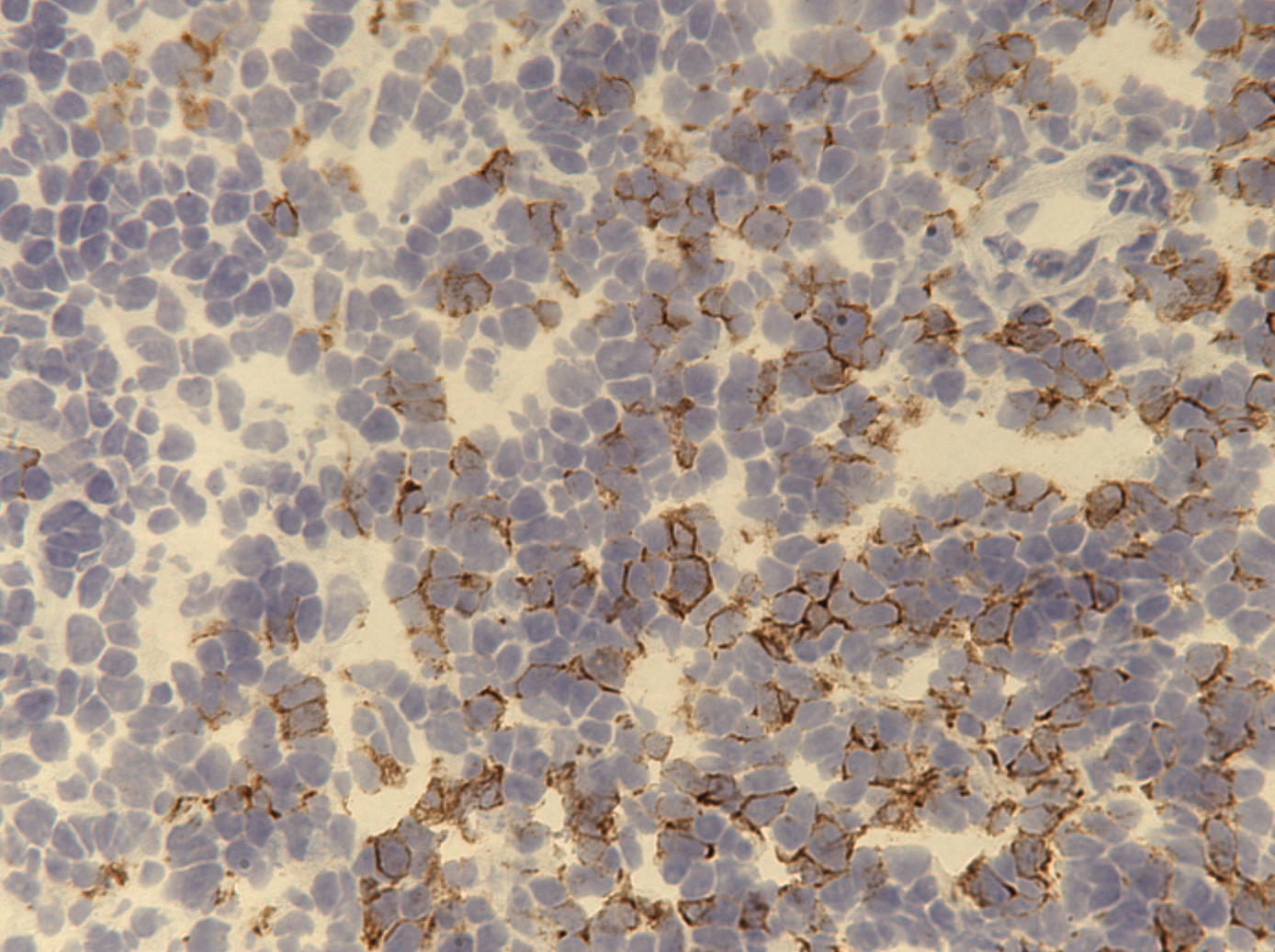
Immunohistochemistry-Paraffin: CD8 Antibody (53-6.7) - BSA Free [NBP1-49045]
Immunohistochemistry-Paraffin: CD8 Antibody (53-6.7) [NBP1-49045] - CD8 alpha Antibody (53-6.7) [NBP1-49045] - CD8 alpha expression in human spleen tissue using anti-CD8 alpha antibody.Applications for CD8 Antibody (53-6.7) - BSA Free
Application
Recommended Usage
Flow Cytometry
1:10 - 1:1000
Immunocytochemistry/ Immunofluorescence
1:10-1:500. Use reported in scientific literature
Immunohistochemistry
1:10-1:500
Immunohistochemistry-Frozen
1:10-1:500
Immunohistochemistry-Paraffin
1:10-1:500
Immunoprecipitation
1:10 - 1:500. Use reported in scientific literature (PMID 24565643)
Application Notes
Each lot of this CD8a antibody is quality control tested by immunofluorescent staining with flow cytometric analysis. For immunofluorescent staining, the suggested use of this reagent is <0.25 ug/10^6 cells in 100 uL volume. It is recommended that the reagent be titrated for optimal performance for each application. The 53-6.7 antibody has been reported to block antigen presentation via MHC class I and inhibit T cell responses to IL-2. This antibody has also been used for depletion of CD8a+ cells. Additional reported applications (for the relevant formats) include: immunoprecipitation, in vivo and in vitro cell depletion, inhibition of CD8 T cell proliferation, blocking of cytotoxicity, and immunohistochemical staining of both acetone-fixed frozen sections and zinc-fixed paraffin-embedded sections.
Reviewed Applications
Read 3 reviews rated 3.7 using NBP1-49045 in the following applications:
Flow Cytometry Panel Builder
Bio-Techne Knows Flow Cytometry
Save time and reduce costly mistakes by quickly finding compatible reagents using the Panel Builder Tool.
Advanced Features
- Spectra Viewer - Custom analysis of spectra from multiple fluorochromes
- Spillover Popups - Visualize the spectra of individual fluorochromes
- Antigen Density Selector - Match fluorochrome brightness with antigen density

Formulation, Preparation, and Storage
Purification
Protein A or G purified
Formulation
PBS
Format
BSA Free
Preservative
0.02% Sodium Azide
Concentration
1 mg/ml
Shipping
The product is shipped with polar packs. Upon receipt, store it immediately at the temperature recommended below.
Stability & Storage
Store at 4C short term. Aliquot and store at -20C long term. Avoid freeze-thaw cycles.
Background: CD8
Given its role in the immune system, CD8-deficiency in T-cells is a hallmark of many diseases and pathologies (8-10). Specifically, CD8+ T-cell deficiency is prevalent in chronic autoimmune diseases including multiple sclerosis, rheumatoid arthritis, ulcerative colitis, Crohn's disease, type 1 diabetes mellitus, and Graves' disease (8). Furthermore, cancers or chronic infection can lead to CD8 T-cell exhaustion as the continual antigen presentation and inflammatory signals eventually cause the CD8+ T-cells to lose functionality (9, 10). However, animal models and clinical studies have suggested that T-cells are capable of being reinvigorated using inhibitory receptor blockade resulting in better disease outcomes and these exhausted T-cells may be a potential therapeutic target (9, 10).
Alternative names for CD8 includes CD antigen: CD8a, CD8 antigen, alpha polypeptide (p32), CD8a molecule, CD8A, Leu2 T-lymphocyte antigen, LEU2, MAL, OKT8 T-cell antigen, p32, T cell co-receptor, T8 T-cell antigen, T-cell antigen Leu2, T-cell surface glycoprotein CD8 alpha chain, and T-lymphocyte differentiation antigen T8/Leu-2.
References
1. Littman D. R. (1987). The structure of the CD4 and CD8 genes. Annual review of immunology. https://doi.org/10.1146/annurev.iy.05.040187.003021
2. Naeim F. (2008). Chapter 2- Principles of Immunophenotyping. Hematopathology. https://doi.org/10.1016/B978-0-12-370607-2.00002-8.
3. Gao, G. F., & Jakobsen, B. K. (2000). Molecular interactions of coreceptor CD8 and MHC class I: the molecular basis for functional coordination with the T-cell receptor. Immunology today. https://doi.org/10.1016/s0167-5699(00)01750-3
4. UniProt (P01732)
5. UniProt (P01731)
6. Kappes D. J. (2007). CD4 and CD8: hogging all the Lck. Immunity. https://doi.org/10.1016/j.immuni.2007.11.002
7. Gangadharan, D., & Cheroutre, H. (2004). The CD8 isoform CD8alphaalpha is not a functional homologue of the TCR co-receptor CD8alphabeta. Current opinion in immunology. https://doi.org/10.1016/j.coi.2004.03.015
8. Pender M. P. (2012). CD8+ T-Cell Deficiency, Epstein-Barr Virus Infection, Vitamin D Deficiency, and Steps to Autoimmunity: A Unifying Hypothesis. Autoimmune diseases. https://doi.org/10.1155/2012/189096
9. Kurachi M. (2019). CD8+ T cell exhaustion. Seminars in immunopathology. https://doi.org/10.1007/s00281-019-00744-5
10. Hashimoto, M., Kamphorst, A. O., Im, S. J., Kissick, H. T., Pillai, R. N., Ramalingam, S. S., Araki, K., & Ahmed, R. (2018). CD8 T Cell Exhaustion in Chronic Infection and Cancer: Opportunities for Interventions. Annual review of medicine. https://doi.org/10.1146/annurev-med-012017-043208
Alternate Names
CD8, CD8A, 53-6.7, 53-6.7 CD8, 53-6.7 CD8 Alpha, 53-6.7 CD8 Aplha, 53-6.7 Clone, CD8 Antibody, Cytotoxic T cell marker, Cytotoxic T lymphocyte marker, T cell marker
Gene Symbol
CD8A
Additional CD8 Products
Product Documents for CD8 Antibody (53-6.7) - BSA Free
Certificate of Analysis
To download a Certificate of Analysis, please enter a lot or batch number in the search box below.
Product Specific Notices for CD8 Antibody (53-6.7) - BSA Free
This product is for research use only and is not approved for use in humans or in clinical diagnosis. Primary Antibodies are guaranteed for 1 year from date of receipt.
Citations for CD8 Antibody (53-6.7) - BSA Free
 Powered by Bioz
Customer Reviews for CD8 Antibody (53-6.7) - BSA Free (3)
3.7 out of 5
3 Customer Ratings
Have you used CD8 Antibody (53-6.7) - BSA Free?
Submit a review and receive an Amazon gift card!
$25/€18/£15/$25CAN/¥2500 Yen for a review with an image
$10/€7/£6/$10CAN/¥1110 Yen for a review without an image
Submit a review
Customer Images
Showing
1
-
3 of
3 reviews
Showing All
Filter By:
-
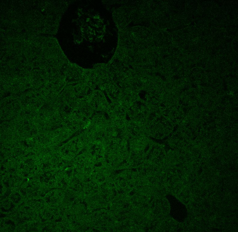
Application: Immunohistochemistry-ParaffinSample Tested: FFPE mouse liverSpecies: MouseVerified Customer | Posted 03/02/2021No visible staining with multiple secondaries. CD8 cells confirmed to be present with another antibodyAttempted IF with this primary antibody against CD8 at concentrations 1:500, 1:200, 1:100, 1:50, 1:20, and 1:10. At concentrations >1:100 we were able to see dim positivity for IHC with HRP, but we were never able to visualize CD8 cells with immunofluorescence using either Alexa 488 and Alexa 468 secondaries (both secondaries confirmed working against other rat primaries). Unfortunately we needed to costain against two other targets so IHC alone didn't work for our purposes.
 Bio-Techne ResponseThank you for reviewing our product. We are sorry to hear that this product did not perform as expected. We have been in touch with the customer to resolve this issue according to our Product Guarantee and to the customer’s satisfaction.
 -
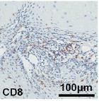
Application: Immunohistochemistry-ParaffinSample Tested: mouse Head and neck squamous cell carcinoma (HNSCC)Species: MouseVerified Customer | Posted 10/23/2017sections were incubated with antibody for CD8 alpha (Novus, 1:200, for mouse HNSCC samples) at 4 °C for 12 h.sections were incubated with antibody for CD8 alpha (Novus, 1:200, for mouse HNSCC samples) at 4 °C for 12 h.
-
Application: Immunohistochemistry-ParaffinSample Tested: Mouse SpleenSpecies: MouseVerified Customer | Posted 12/15/2014CD8 in mouse spleen OCT
There are no reviews that match your criteria.
Protocols
Find general support by application which include: protocols, troubleshooting, illustrated assays, videos and webinars.
- 7-Amino Actinomycin D (7-AAD) Cell Viability Flow Cytometry Protocol
- Antigen Retrieval Protocol (PIER)
- Antigen Retrieval for Frozen Sections Protocol
- Appropriate Fixation of IHC/ICC Samples
- Cellular Response to Hypoxia Protocols
- Chromogenic IHC Staining of Formalin-Fixed Paraffin-Embedded (FFPE) Tissue Protocol
- Chromogenic Immunohistochemistry Staining of Frozen Tissue
- ClariTSA™ Fluorophore Kits
- Detection & Visualization of Antibody Binding
- Extracellular Membrane Flow Cytometry Protocol
- Flow Cytometry Protocol for Cell Surface Markers
- Flow Cytometry Protocol for Staining Membrane Associated Proteins
- Flow Cytometry Staining Protocols
- Flow Cytometry Troubleshooting Guide
- Fluorescent IHC Staining of Frozen Tissue Protocol
- Graphic Protocol for Heat-induced Epitope Retrieval
- Graphic Protocol for the Preparation and Fluorescent IHC Staining of Frozen Tissue Sections
- Graphic Protocol for the Preparation and Fluorescent IHC Staining of Paraffin-embedded Tissue Sections
- Graphic Protocol for the Preparation of Gelatin-coated Slides for Histological Tissue Sections
- ICC Cell Smear Protocol for Suspension Cells
- ICC Immunocytochemistry Protocol Videos
- ICC for Adherent Cells
- IHC Sample Preparation (Frozen sections vs Paraffin)
- Immunocytochemistry (ICC) Protocol
- Immunocytochemistry Troubleshooting
- Immunofluorescence of Organoids Embedded in Cultrex Basement Membrane Extract
- Immunofluorescent IHC Staining of Formalin-Fixed Paraffin-Embedded (FFPE) Tissue Protocol
- Immunohistochemistry (IHC) and Immunocytochemistry (ICC) Protocols
- Immunohistochemistry Frozen Troubleshooting
- Immunohistochemistry Paraffin Troubleshooting
- Immunoprecipitation Protocol
- Intracellular Flow Cytometry Protocol Using Alcohol (Methanol)
- Intracellular Flow Cytometry Protocol Using Detergents
- Intracellular Nuclear Staining Flow Cytometry Protocol Using Detergents
- Intracellular Staining Flow Cytometry Protocol Using Alcohol Permeabilization
- Intracellular Staining Flow Cytometry Protocol Using Detergents to Permeabilize Cells
- Preparing Samples for IHC/ICC Experiments
- Preventing Non-Specific Staining (Non-Specific Binding)
- Primary Antibody Selection & Optimization
- Propidium Iodide Cell Viability Flow Cytometry Protocol
- Protocol for Heat-Induced Epitope Retrieval (HIER)
- Protocol for Liperfluo
- Protocol for Making a 4% Formaldehyde Solution in PBS
- Protocol for VisUCyte™ HRP Polymer Detection Reagent
- Protocol for the Characterization of Human Th22 Cells
- Protocol for the Characterization of Human Th9 Cells
- Protocol for the Fluorescent ICC Staining of Cell Smears - Graphic
- Protocol for the Fluorescent ICC Staining of Cultured Cells on Coverslips - Graphic
- Protocol for the Preparation & Fixation of Cells on Coverslips
- Protocol for the Preparation and Chromogenic IHC Staining of Frozen Tissue Sections
- Protocol for the Preparation and Chromogenic IHC Staining of Frozen Tissue Sections - Graphic
- Protocol for the Preparation and Chromogenic IHC Staining of Paraffin-embedded Tissue Sections
- Protocol for the Preparation and Chromogenic IHC Staining of Paraffin-embedded Tissue Sections - Graphic
- Protocol for the Preparation and Fluorescent ICC Staining of Cells on Coverslips
- Protocol for the Preparation and Fluorescent ICC Staining of Non-adherent Cells
- Protocol for the Preparation and Fluorescent ICC Staining of Stem Cells on Coverslips
- Protocol for the Preparation and Fluorescent IHC Staining of Frozen Tissue Sections
- Protocol for the Preparation and Fluorescent IHC Staining of Paraffin-embedded Tissue Sections
- Protocol for the Preparation of Gelatin-coated Slides for Histological Tissue Sections
- Protocol for the Preparation of a Cell Smear for Non-adherent Cell ICC - Graphic
- Protocol: Annexin V and PI Staining by Flow Cytometry
- Protocol: Annexin V and PI Staining for Apoptosis by Flow Cytometry
- TUNEL and Active Caspase-3 Detection by IHC/ICC Protocol
- The Importance of IHC/ICC Controls
- Troubleshooting Guide: Fluorokine Flow Cytometry Kits
- Troubleshooting Guide: Immunohistochemistry
- View all Protocols, Troubleshooting, Illustrated assays and Webinars
FAQs for CD8 Antibody (53-6.7) - BSA Free
Showing
1
-
1 of
1 FAQ
Showing All
-
Q: Could you provide me with any suggestions for CD8 antibodies that work well with a CD4 antibody?
A: NBP1-28254, NBP1-28336, NBP1-28238, NBP1-49045 and NBP2-80658 have images of mostly Flow Cytometry analyses for CD4 and CD8. NB200-578 induces transplantation tolerance when used in conjunction with CD4 antibodies. I would suggest taking a look at these products to determine if they would be suitable for your experiments.


