Ferritin Light Chain Antibody (FTL/1386) - Azide and BSA Free
Novus Biologicals | Catalog # NBP2-54574
![Western Blot: Ferritin Light Chain Antibody (FTL/1386)Azide and BSA Free [NBP2-54574]](https://resources.rndsystems.com/images/products/Ferritin-Light-Chain-Antibody-FTL-1386-Azide-and-BSA-Free-Western-Blot-NBP2-54574-img0005.jpg "Western Blot: Ferritin Light Chain Antibody (FTL/1386)Azide and BSA Free [NBP2-54574]")
Key Product Details
Species Reactivity
Human
Applications
Immunohistochemistry, Immunohistochemistry-Paraffin, Western Blot, ELISA, Protein Array, CyTOF-ready
Label
Unconjugated
Antibody Source
Monoclonal Mouse IgG2b Kappa Clone # FTL/1386
Format
Azide and BSA Free
Loading...
Product Specifications
Immunogen
Recombinant fragment (around aa 38-165) of human Ferritin Light Chain protein (exact sequence is proprietary) (Uniprot: P02792)
Localization
Cytoplasmic
Marker
Node-Negative Breast Tumor Prognostic Marker
Clonality
Monoclonal
Host
Mouse
Isotype
IgG2b Kappa
Description
1.0 mg/ml of antibody purified from Bioreactor Concentrate by Protein A/G. Prepared in 10mM PBS WITHOUT BSA & azide. Also available at 200 ug/ml WITH BSA & azide (NBP2-53295).
Antibody with azide - store at 2 to 8C. Antibody without azide - store at -20 to -80C.
Antibody with azide - store at 2 to 8C. Antibody without azide - store at -20 to -80C.
Scientific Data Images for Ferritin Light Chain Antibody (FTL/1386) - Azide and BSA Free
Western Blot: Ferritin Light Chain Antibody (FTL/1386)Azide and BSA Free [NBP2-54574]
Western Blot: Ferritin Light Chain Antibody (FTL/1386) - Azide and BSA Free [NBP2-54574] - Western Blot Analysis of human HeLa cell lysate using Ferritin Light Chain Antibody (FTL/1386).![Immunohistochemistry-Paraffin: Ferritin Light Chain Antibody (FTL/1386) - Azide and BSA Free [NBP2-54574]](https://resources.rndsystems.com/images/products/Ferritin-Light-Chain-Antibody-FTL-1386-Azide-and-BSA-Free-Immunohistochemistry-Paraffin-NBP2-54574-img0009.jpg "Immunohistochemistry-Paraffin: Ferritin Light Chain Antibody (FTL/1386) - Azide and BSA Free [NBP2-54574]")
Immunohistochemistry-Paraffin: Ferritin Light Chain Antibody (FTL/1386) - Azide and BSA Free [NBP2-54574]
Immunohistochemistry-Paraffin: Ferritin Light Chain Antibody (FTL/1386) - Azide and BSA Free [NBP2-54574] - Formalin-fixed, paraffin-embedded Human Testicular Carcinoma stained with Ferritin Light Chain Mouse Monoclonal Antibody (FTL/1386).![Western Blot: Ferritin Light Chain Antibody (FTL/1386)Azide and BSA Free [NBP2-54574]](https://resources.rndsystems.com/images/products/Ferritin-Light-Chain-Antibody-FTL-1386-Azide-and-BSA-Free-Western-Blot-NBP2-54574-img0003.jpg "Western Blot: Ferritin Light Chain Antibody (FTL/1386)Azide and BSA Free [NBP2-54574]")
Western Blot: Ferritin Light Chain Antibody (FTL/1386)Azide and BSA Free [NBP2-54574]
Western Blot: Ferritin Light Chain Antibody (FTL/1386) - Azide and BSA Free [NBP2-54574] - A431, HeLa, Liver and Testis Lysate using Ferritin, Light Chain Monoclonal Antibody (FTL/1386).![Immunohistochemistry-Paraffin: Ferritin Light Chain Antibody (FTL/1386) - Azide and BSA Free [NBP2-54574]](https://resources.rndsystems.com/images/products/Ferritin-Light-Chain-Antibody-FTL-1386-Azide-and-BSA-Free-Immunohistochemistry-Paraffin-NBP2-54574-img0001.jpg "Immunohistochemistry-Paraffin: Ferritin Light Chain Antibody (FTL/1386) - Azide and BSA Free [NBP2-54574]")
Immunohistochemistry-Paraffin: Ferritin Light Chain Antibody (FTL/1386) - Azide and BSA Free [NBP2-54574]
Immunohistochemistry-Paraffin: Ferritin Light Chain Antibody (FTL/1386) - Azide and BSA Free [NBP2-54574] - Formalin-fixed, paraffin-embedded Human Pancreas stained with Ferritin, Light Chain Monoclonal Antibody (FTL/1386).![Immunohistochemistry-Paraffin: Ferritin Light Chain Antibody (FTL/1386) - Azide and BSA Free [NBP2-54574]](https://resources.rndsystems.com/images/products/Ferritin-Light-Chain-Antibody-FTL-1386-Azide-and-BSA-Free-Immunohistochemistry-Paraffin-NBP2-54574-img0002.jpg "Immunohistochemistry-Paraffin: Ferritin Light Chain Antibody (FTL/1386) - Azide and BSA Free [NBP2-54574]")
Immunohistochemistry-Paraffin: Ferritin Light Chain Antibody (FTL/1386) - Azide and BSA Free [NBP2-54574]
Immunohistochemistry-Paraffin: Ferritin Light Chain Antibody (FTL/1386) - Azide and BSA Free [NBP2-54574] - Formalin-fixed, paraffin-embedded Human Pancreas stained with Ferritin, Light Chain Monoclonal Antibody (FTL/1386).![Immunohistochemistry-Paraffin: Ferritin Light Chain Antibody (FTL/1386) - Azide and BSA Free [NBP2-54574]](https://resources.rndsystems.com/images/products/Ferritin-Light-Chain-Antibody-FTL-1386-Azide-and-BSA-Free-Immunohistochemistry-Paraffin-NBP2-54574-img0008.jpg "Immunohistochemistry-Paraffin: Ferritin Light Chain Antibody (FTL/1386) - Azide and BSA Free [NBP2-54574]")
Immunohistochemistry-Paraffin: Ferritin Light Chain Antibody (FTL/1386) - Azide and BSA Free [NBP2-54574]
Immunohistochemistry-Paraffin: Ferritin Light Chain Antibody (FTL/1386) - Azide and BSA Free [NBP2-54574] - Formalin-fixed, paraffin-embedded Human Pancreas stained with Ferritin Light Chain Mouse Monoclonal Antibody (FTL/1386).![Protein Array: Ferritin Light Chain Antibody (FTL/1386) - Azide and BSA Free [NBP2-54574]](https://resources.rndsystems.com/images/products/Ferritin-Light-Chain-Antibody-FTL-1386-Azide-and-BSA-Free-Protein-Array-NBP2-54574-img0004.jpg "Protein Array: Ferritin Light Chain Antibody (FTL/1386) - Azide and BSA Free [NBP2-54574]")
Protein Array: Ferritin Light Chain Antibody (FTL/1386) - Azide and BSA Free [NBP2-54574]
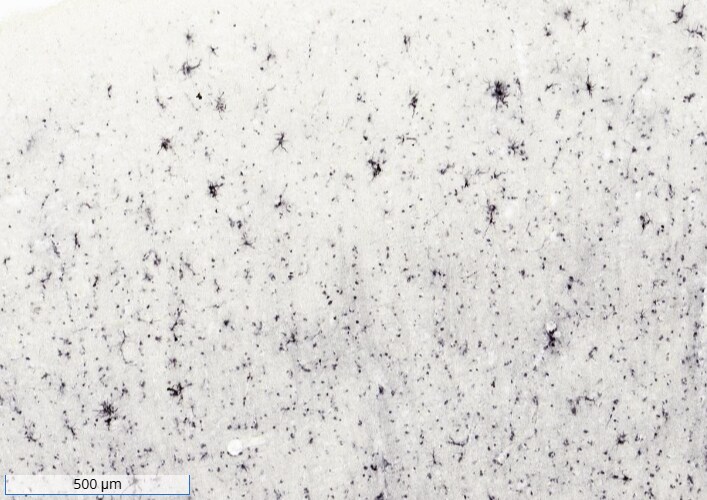
Protein Array: Ferritin Light Chain Antibody (FTL/1386) - Azide and BSA Free [NBP2-54574] - Analysis of Protein Array containing more than 19,000 full-length human proteins using Ferritin Light Chain Antibody (FTL/1386) Z- and S- Score: The Z-score represents the strength of a signal that a monoclonal antibody (MAb) (in combination with a fluorescently-tagged anti-IgG secondary antibody) produces when binding to a particular protein on the HuProt(TM) array. Z-scores are described in units of standard deviations (SDs) above the mean value of all signals generated on that array. If targets on HuProt(TM) are arranged in descending order of the Z-score, the S-score is the difference (also in units of SDs) between the Z-score. S-score therefore represents the relative target specificity of a MAb to its intended target. A MAb is considered to specific to its intended target, if the MAb has an S-score of at least 2.5.![Ferritin Light Chain Antibody (FTL/1386) - Azide and BSA Free Immunohistochemistry-Frozen: Mouse Monoclonal Ferritin Light Chain Antibody (FTL/1386) - Azide and BSA Free [NBP2-54574]](https://resources.rndsystems.com/images/products/antibody/nbp2-54574_mouse-monoclonal-ferritin-light-chain-antibody-ftl-1386-azide-and-bsa-free-immunohistochemistry-frozen-3112025151752.jpg "Immunohistochemistry-Frozen: Mouse Monoclonal Ferritin Light Chain Antibody (FTL/1386) - Azide and BSA Free [NBP2-54574]")
Immunohistochemistry-Frozen: Mouse Monoclonal Ferritin Light Chain Antibody (FTL/1386) - Azide and BSA Free [NBP2-54574]
Ferritin (Light chain) staining in human AD brain. Image from a verified customer review.Applications for Ferritin Light Chain Antibody (FTL/1386) - Azide and BSA Free
Application
Recommended Usage
Immunohistochemistry-Paraffin
0.1 - 0.2 ug/ml
Western Blot
0.1 - 0.2 ug/ml
Application Notes
ELISA: For coating, order Ab without BSA.
Immunohistology (Formalin-fixed): 0.25-0.5ug/ml for 30 min at RT. Staining of formalin-fixed tissues requires boiling tissue sections in 10mM Citrate Buffer, pH 6.0, for 10-20 min followed by cooling at RT for 20 minutes.
Optimal dilution for a specific application should be determined.
Immunohistology (Formalin-fixed): 0.25-0.5ug/ml for 30 min at RT. Staining of formalin-fixed tissues requires boiling tissue sections in 10mM Citrate Buffer, pH 6.0, for 10-20 min followed by cooling at RT for 20 minutes.
Optimal dilution for a specific application should be determined.
Reviewed Applications
Read 1 review rated 5 using NBP2-54574 in the following applications:
Formulation, Preparation, and Storage
Purification
Protein A or G purified
Formulation
10 mM PBS
Format
Azide and BSA Free
Preservative
No Preservative
Concentration
1 mg/ml
Shipping
The product is shipped with polar packs. Upon receipt, store it immediately at the temperature recommended below.
Stability & Storage
Store at -20 to -80C. Avoid freeze-thaw cycles.
Background: Ferritin Light Chain
Alternate Names
Ferritin L subunit, ferritin L-chain, ferritin light chain, ferritin light polypeptide-like 3, ferritin, light polypeptide, MGC71996, NBIA3
Gene Symbol
FTL
Additional Ferritin Light Chain Products
Product Documents for Ferritin Light Chain Antibody (FTL/1386) - Azide and BSA Free
Certificate of Analysis
To download a Certificate of Analysis, please enter a lot or batch number in the search box below.
Product Specific Notices for Ferritin Light Chain Antibody (FTL/1386) - Azide and BSA Free
This product is for research use only and is not approved for use in humans or in clinical diagnosis. Primary Antibodies are guaranteed for 1 year from date of receipt.
Customer Reviews for Ferritin Light Chain Antibody (FTL/1386) - Azide and BSA Free (1)
5 out of 5
1 Customer Rating
Have you used Ferritin Light Chain Antibody (FTL/1386) - Azide and BSA Free?
Submit a review and receive an Amazon gift card!
$25/€18/£15/$25CAN/¥2500 Yen for a review with an image
$10/€7/£6/$10CAN/¥1110 Yen for a review without an image
Submit a review
Customer Images
Showing
1
-
1 of
1 review
Showing All
Filter By:
-
Application: Immunohistochemistry-FrozenSample Tested: Human BrainSpecies: HumanVerified Customer | Posted 10/27/2025Ferritin (Light chain) staining in human AD brain.

There are no reviews that match your criteria.
Protocols
Find general support by application which include: protocols, troubleshooting, illustrated assays, videos and webinars.
- Antigen Retrieval Protocol (PIER)
- Antigen Retrieval for Frozen Sections Protocol
- Appropriate Fixation of IHC/ICC Samples
- Cellular Response to Hypoxia Protocols
- Chromogenic IHC Staining of Formalin-Fixed Paraffin-Embedded (FFPE) Tissue Protocol
- Chromogenic Immunohistochemistry Staining of Frozen Tissue
- ClariTSA™ Fluorophore Kits
- Detection & Visualization of Antibody Binding
- ELISA Sample Preparation & Collection Guide
- ELISA Troubleshooting Guide
- Fluorescent IHC Staining of Frozen Tissue Protocol
- Graphic Protocol for Heat-induced Epitope Retrieval
- Graphic Protocol for the Preparation and Fluorescent IHC Staining of Frozen Tissue Sections
- Graphic Protocol for the Preparation and Fluorescent IHC Staining of Paraffin-embedded Tissue Sections
- Graphic Protocol for the Preparation of Gelatin-coated Slides for Histological Tissue Sections
- How to Run an R&D Systems DuoSet ELISA
- How to Run an R&D Systems Quantikine ELISA
- How to Run an R&D Systems Quantikine™ QuicKit™ ELISA
- IHC Sample Preparation (Frozen sections vs Paraffin)
- Immunofluorescent IHC Staining of Formalin-Fixed Paraffin-Embedded (FFPE) Tissue Protocol
- Immunohistochemistry (IHC) and Immunocytochemistry (ICC) Protocols
- Immunohistochemistry Frozen Troubleshooting
- Immunohistochemistry Paraffin Troubleshooting
- Preparing Samples for IHC/ICC Experiments
- Preventing Non-Specific Staining (Non-Specific Binding)
- Primary Antibody Selection & Optimization
- Protocol for Heat-Induced Epitope Retrieval (HIER)
- Protocol for Making a 4% Formaldehyde Solution in PBS
- Protocol for VisUCyte™ HRP Polymer Detection Reagent
- Protocol for the Preparation & Fixation of Cells on Coverslips
- Protocol for the Preparation and Chromogenic IHC Staining of Frozen Tissue Sections
- Protocol for the Preparation and Chromogenic IHC Staining of Frozen Tissue Sections - Graphic
- Protocol for the Preparation and Chromogenic IHC Staining of Paraffin-embedded Tissue Sections
- Protocol for the Preparation and Chromogenic IHC Staining of Paraffin-embedded Tissue Sections - Graphic
- Protocol for the Preparation and Fluorescent IHC Staining of Frozen Tissue Sections
- Protocol for the Preparation and Fluorescent IHC Staining of Paraffin-embedded Tissue Sections
- Protocol for the Preparation of Gelatin-coated Slides for Histological Tissue Sections
- Quantikine HS ELISA Kit Assay Principle, Alkaline Phosphatase
- Quantikine HS ELISA Kit Principle, Streptavidin-HRP Polymer
- R&D Systems Quality Control Western Blot Protocol
- Sandwich ELISA (Colorimetric) – Biotin/Streptavidin Detection Protocol
- Sandwich ELISA (Colorimetric) – Direct Detection Protocol
- TUNEL and Active Caspase-3 Detection by IHC/ICC Protocol
- The Importance of IHC/ICC Controls
- Troubleshooting Guide: ELISA
- Troubleshooting Guide: Immunohistochemistry
- Troubleshooting Guide: Western Blot Figures
- Western Blot Conditions
- Western Blot Protocol
- Western Blot Protocol for Cell Lysates
- Western Blot Troubleshooting
- Western Blot Troubleshooting Guide
- View all Protocols, Troubleshooting, Illustrated assays and Webinars
Loading...