GFP Antibody - BSA Free
Novus Biologicals | Catalog # NB600-303
![Immunocytochemistry/ Immunofluorescence: GFP Antibody [NB600-303]](https://resources.rndsystems.com/images/products/GFP-Antibody-Immunocytochemistry-Immunofluorescence-NB600-303-img0023.jpg "Immunocytochemistry/ Immunofluorescence: GFP Antibody [NB600-303]")
Key Product Details
Species Reactivity
Validated:
Non-species specific
Cited:
Jellyfish
Applications
Validated:
Immunohistochemistry, Immunohistochemistry-Paraffin, Immunohistochemistry-Frozen, Western Blot, ELISA, Flow Cytometry, Immunocytochemistry/ Immunofluorescence, Simple Western, Immunoprecipitation, Chromatin Immunoprecipitation (ChIP), Electron Microscopy
Cited:
Immunohistochemistry-Paraffin, Immunohistochemistry-Frozen, Western Blot, ELISA, Flow Cytometry, Immunocytochemistry/ Immunofluorescence, Simple Western, Immunoprecipitation, Chemotaxis, IF/IHC, Electron Microscopy
Label
Unconjugated
Antibody Source
Polyclonal Rabbit IgG
Format
BSA Free
Loading...
Product Specifications
Immunogen
The immunogen is a Green Fluorescent Protein (GFP) fusion protein corresponding to the full length amino acid sequence (246aa) derived from the jellyfish Aequorea victoria.
Clonality
Polyclonal
Host
Rabbit
Isotype
IgG
Description
This product was prepared from monospecific antiserum by immunoaffinity chromatography using Green Fluorescent Protein (Aequorea victoria) coupled to agarose beads followed by solid phase adsorption(s) to remove any unwanted reactivities.
Store vial at -20C prior to opening. Aliquot contents and freeze at -20C or below for extended storage. Avoid cycles of freezing and thawing. Centrifuge product if not completely clear after standing at room temperature. This product is stable for several weeks at 4C as an undiluted liquid. Dilute only prior to immediate use.
Store vial at -20C prior to opening. Aliquot contents and freeze at -20C or below for extended storage. Avoid cycles of freezing and thawing. Centrifuge product if not completely clear after standing at room temperature. This product is stable for several weeks at 4C as an undiluted liquid. Dilute only prior to immediate use.
Scientific Data Images for GFP Antibody - BSA Free
Immunocytochemistry/ Immunofluorescence: GFP Antibody [NB600-303]
Immunocytochemistry/Immunofluorescence: GFP Antibody [NB600-303] - Analysis of FITC conjugate of NB600-303. Staining of mouse spleen cells Tissue: spleen cells infected with MHV68-H2bYFP. As seen in: Collins CM, Speck SH (2012) Tracking Murine Gammaherpesvirus 68 Infection of Germinal Center B Cells.![Western Blot: GFP Antibody [NB600-303]](https://resources.rndsystems.com/images/products/GFP-Antibody-Western-Blot-NB600-303-img0018.jpg "Western Blot: GFP Antibody [NB600-303]")
Western Blot: GFP Antibody [NB600-303]
Western Blot: GFP Antibody [NB600-303] - Analysis using the FITC conjugate of NB600-303. Detection of Lane 1: GFP. Load: 50 ug per lane. Primary antibody: none. Secondary antibody: Fluorescein conjugated Anti-GFP at 1:1000 for 60 min at RT. Block: 1% BSA-TTBS for 30 min at RT. Predicted/Observed size: 28 kDa, 28 kDa.![Western Blot: GFP Antibody [NB600-303]](https://resources.rndsystems.com/images/products/GFP-Antibody-Western-Blot-NB600-303-img0005.jpg "Western Blot: GFP Antibody [NB600-303]")
Western Blot: GFP Antibody [NB600-303]
Western Blot: GFP Antibody [NB600-303] - WB analysis of GFP in various lysates. Image courtesy of product review by Emilie Leclerc.![Immunoprecipitation: GFP Antibody [NB600-303]](https://resources.rndsystems.com/images/products/GFP-Antibody-Immunoprecipitation-NB600-303-img0007.jpg "Immunoprecipitation: GFP Antibody [NB600-303]")
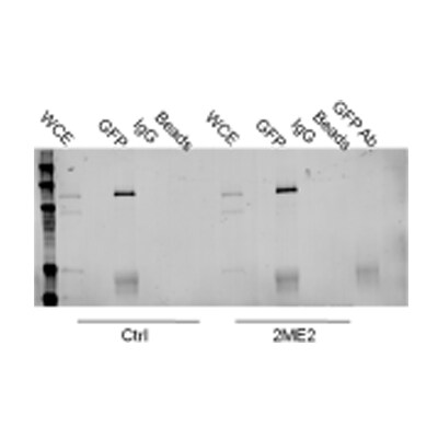
Immunoprecipitation: GFP Antibody [NB600-303]
Immunoprecipitation: GFP Antibody [NB600-303] - IP analysis of GFP in HeLa cell lysate. Image courtesy of anonymous customer review.![Western Blot: GFP Antibody [NB600-303]](https://resources.rndsystems.com/images/products/GFP-Antibody-Western-Blot-NB600-303-img0014.jpg "Western Blot: GFP Antibody [NB600-303]")
Western Blot: GFP Antibody [NB600-303]
Western Blot: GFP Antibody [NB600-303] - Analysis of GFP HRP expression in HeLa/GFP cell lysate.![Western Blot: GFP Antibody [NB600-303]](https://resources.rndsystems.com/images/products/GFP-Antibody-Western-Blot-NB600-303-img0019.jpg "Western Blot: GFP Antibody [NB600-303]")
Western Blot: GFP Antibody [NB600-303]
Western Blot: GFP Antibody [NB600-303] - Analysis using Biotin conjugate of NB600-303. Lane 1: 50ng of GFP. Lane 2: none. Primary antibody: none. Secondary antibody: Anti- Biotin Conjugated secondary antibody was used at 1:5000 in Blocking Buffer for Fluorescent Western Blotting for 45 min at RT. HRP Streptavidin was used at 1:40,000 in incubated with blocking buffer for 30 min at 20C. Block: 5% Blotto 30 min at 20C. Predicted/Observed size: 28 kDa for GFP. Other band(s): none.![Immunohistochemistry: GFP Antibody [NB600-303]](https://resources.rndsystems.com/images/products/GFP-Antibody-Immunohistochemistry-NB600-303-img0024.jpg "Immunohistochemistry: GFP Antibody [NB600-303]")
Immunohistochemistry: GFP Antibody [NB600-303]
GFP-Antibody-Immunohistochemistry-NB600-303-img0024.jpg![Western Blot: GFP Antibody [NB600-303]](https://resources.rndsystems.com/images/products/GFP-Antibody-Western-Blot-NB600-303-img0008.jpg "Western Blot: GFP Antibody [NB600-303]")
Western Blot: GFP Antibody [NB600-303]
Western Blot: GFP Antibody [NB600-303] - Lane 1: 293FT cells transfected with CDK4 dominant negative. Lane 2: 293FT cells poitive control. Load: 25 ug per lane. Primary antibody: GFP antibody at 1:400 for overnight at 4C. Secondary antibody: IRDye800 (TM) rabbit secondary antibody at 1:10,000 for 45 min at RT. Block: 5% BLOTTO overnight at 4C. Predicted/Observed size: 27 kDa for GFP.![Immunocytochemistry/ Immunofluorescence: GFP Antibody [NB600-303]](https://resources.rndsystems.com/images/products/GFP-Antibody-Immunocytochemistry-Immunofluorescence-NB600-303-img0022.jpg "Immunocytochemistry/ Immunofluorescence: GFP Antibody [NB600-303]")
Immunocytochemistry/ Immunofluorescence: GFP Antibody [NB600-303]
Immunocytochemistry/Immunofluorescence: GFP Antibody [NB600-303]
GFP Antibody
Western Blot of GFP Antibody. Lane 1: 293FT cells transfected with CDK4 dominant negative. Lane 2: 293FT cells poitive control. Load: 25 ug per lane. Primary antibody: GFP antibody at 1:400 for overnight at 4C. Secondary antibody: IRDye800(TM) rabbit secondary antibody at 1:10,000 for 45 min at RT. Block: 5% BLOTTO overnight at 4C. Predicted/Observed size: 27 kDa for GFP.
GFP Antibody
Western Blot of Rabbit anti-GFP antibody. Lane 1: Wild type GFP (0.1 ug) was used to spike HeLa whole cell lysate. Lane 2: none. Load: 30 ug per lane. Primary antibody: GFP antibody at 1:1000 for overnight at 4C. Secondary antibody: IRDye800(TM) Goat-a-Rabbit IgG [H&L] MX10 () at 1:10,000 for 45 min at RT. Block: 5% BLOTTO in PBS overnight at 4C. Predicted/Observed size: 27 kDa for epitope tag GFP. Other band(s): none.
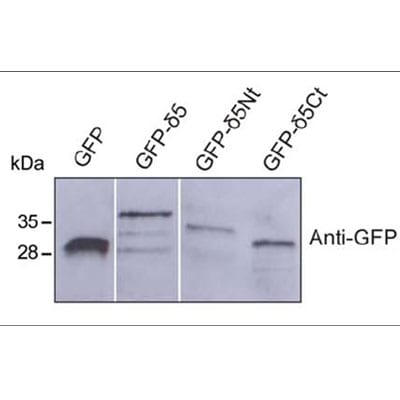
Western Blot: GFP Antibody [NB600-303] -
The polo-boxes of Plk4 are not sufficient to mediate STIL binding.(A) Scheme of Flag-Plk4 fragments. (B) After overexpression of Flag-Plk4-fragments (A) and GFP-STIL in HEK293T cells, cell lysates were subjected to immunoprecipitations using anti-Flag antibodies. Coprecipitation of GFP-STIL with Flag-Plk4 fragments was detected by western blotting using anti-GFP and anti-Flag antibodies.
Western Blot: GFP Antibody [NB600-303] -
Western Blot: GFP Antibody [NB600-303] - The polo-boxes of Plk4 are not sufficient to mediate STIL binding.(A) Scheme of Flag-Plk4 fragments. (B) After overexpression of Flag-Plk4-fragments (A) & GFP-STIL in HEK293T cells, cell lysates were subjected to immunoprecipitations using anti-Flag antibodies. Coprecipitation of GFP-STIL with Flag-Plk4 fragments was detected by western blotting using anti-GFP & anti-Flag antibodies. Image collected & cropped by CiteAb from the following publication (https://journals.biologists.com/bio/article/4/3/370/1390/Plk4-dependent…), licensed under a CC-BY license. Not internally tested by Novus Biologicals.
Immunocytochemistry/ Immunofluorescence: GFP Antibody [NB600-303] -
Immunocytochemistry/ Immunofluorescence: GFP Antibody [NB600-303] - Lgr5+ liver stem cells transplantation decreased liver fibrosis. a Schematic overview of the experimental setup. Eight-week-old wild-type C57 mice were i.p. injected with CCL4 (2 ml/kg, Sigma-Aldrich) dissolved in olive oil at a ratio of 1:4, or olive oil alone (2 ml/kg) twice a week for 6 weeks. Lgr5-GFP+ liver stem cells or primary hepatocyte (PH) derived from Lgr5-GFP mice were transplanted into the liver fibrosis mice by intrasplenical injection on day 0. b Lgr5+ cells were isolated from Lgr5-GFP mice treated with CCL4 by FACS assay for transplantation (left). The liver Lgr5 expression was stained using anti-GFP antibody in mice with Lgr5+ cells or PH transplantation (right) on day 40. n = 10 mice, scale bars, 200 μm. c, d Lgr5+ liver stem cells transplantation decreased CCL4-induced liver fibrosis & recovered liver functions. Lgr5-GFP+ liver stem cells or PH transplantation were described in a; the livers were harvested & stained using H&E & Sirius Red for fibrosis analysis & quantification of positive-staining areas measured by Image J software (c). The serum was harvested for ALT & AST analysis (d, e). For c, scale bars, 200 μm, the results are shown as mean ± s.d. of five independent sections taken randomly per mice & a total of 50 tissue specimens in each group (n = 10 mice) *p < 0.05, **p < 0.01. For d, e, triplicates for each condition were analyzed. The results are shown as mean ± s.d. of three independent experiments. *p < 0.05, **p < 0.01 Image collected & cropped by CiteAb from the following publication (https://pubmed.ncbi.nlm.nih.gov/29079780), licensed under a CC-BY license. Not internally tested by Novus Biologicals.Applications for GFP Antibody - BSA Free
Application
Recommended Usage
ELISA
1:10000-1:40000
Flow Cytometry
1:10 - 1:1000
Immunocytochemistry/ Immunofluorescence
1:10 - 1:500
Immunohistochemistry
1:10 - 1:500
Immunohistochemistry-Frozen
1:10 - 1:500
Immunohistochemistry-Paraffin
1:10 - 1:500
Immunoprecipitation
1:10 - 1:500
Simple Western
1:500
Western Blot
1:1000-1:3000
Application Notes
This product has been tested in ELISA and western blot. Expect ~27kDa band. Although not tested, this antibody is suitable for IP, IF, ChIP, and Flow. Specific conditions for reactivity should be optimized by the end user.
Immunoprecipitation reported successful by customer review.
Immunohistochemistry-Paraffin (PMID: 21311774) Immunocytochemistry/Immunofluorescence was reported in scientific literature.
Use in Flow Cytometry reported in scientific literature (PMID: 23903657).
Use in Electron Microscopy reported in scientific literature (PMID 25538186).
Use in Immunohistochemistry-Frozen reported in multiple pieces of scientific literature.
Use in chromatin immunoprecipitation reported in scientific literature (PMID 28174757).
See Simple Western Antibody Database for Simple Western validation: tested in human melanoma cells; antibody dilution of 1:500; separated by size
Reviewed Applications
Read 3 reviews rated 4 using NB600-303 in the following applications:
Flow Cytometry Panel Builder
Bio-Techne Knows Flow Cytometry
Save time and reduce costly mistakes by quickly finding compatible reagents using the Panel Builder Tool.
Advanced Features
- Spectra Viewer - Custom analysis of spectra from multiple fluorochromes
- Spillover Popups - Visualize the spectra of individual fluorochromes
- Antigen Density Selector - Match fluorochrome brightness with antigen density

Formulation, Preparation, and Storage
Purification
Immunogen affinity purified
Formulation
0.02 M Potassium Phosphate, 0.15 M Sodium Chloride, pH 7.2
Format
BSA Free
Preservative
0.01% Sodium Azide
Concentration
Please see the vial label for concentration. If unlisted please contact technical services.
Shipping
The product is shipped with polar packs. Upon receipt, store it immediately at the temperature recommended below.
Stability & Storage
Store at -20C short term. Aliquot and store at -80C long term. Avoid freeze-thaw cycles.
Background: GFP
References
1. Shi, C., Pan, F. C., Kim, J. N., Washington, M. K., Padmanabhan, C., Meyer, C. T.,... Means, A. L. (2019). Differential Cell Susceptibilities to Kras(G12D) in the Setting of Obstructive Chronic Pancreatitis. Cell Mol Gastroenterol Hepatol. doi:10.1016/j.jcmgh.2019.07.001
2. Zhao, S., Fortier, T. M., & Baehrecke, E. H. (2018). Autophagy Promotes Tumor-like Stem Cell Niche Occupancy. Curr Biol, 28(19), 3056-3064.e3053. doi:10.1016/j.cub.2018.07.075
3. Zusso, M., Lunardi, V., Franceschini, D., Pagetta, A., Lo, R., Stifani, S.,... Moro, S. (2019). Ciprofloxacin and levofloxacin attenuate microglia inflammatory response via TLR4/NF-kB pathway. J Neuroinflammation, 16(1), 148. doi:10.1186/s12974-019-1538-9
Long Name
Green Fluorescent Protein
Alternate Names
eGFP, GFPuv
Additional GFP Products
Product Documents for GFP Antibody - BSA Free
Certificate of Analysis
To download a Certificate of Analysis, please enter a lot or batch number in the search box below.
Product Specific Notices for GFP Antibody - BSA Free
This product is for research use only and is not approved for use in humans or in clinical diagnosis. Primary Antibodies are guaranteed for 1 year from date of receipt.
Citations for GFP Antibody - BSA Free
 Powered by Bioz
Customer Reviews for GFP Antibody - BSA Free (3)
4 out of 5
3 Customer Ratings
Have you used GFP Antibody - BSA Free?
Submit a review and receive an Amazon gift card!
$25/€18/£15/$25CAN/¥2500 Yen for a review with an image
$10/€7/£6/$10CAN/¥1110 Yen for a review without an image
Submit a review
Customer Images
Showing
1
-
3 of
3 reviews
Showing All
Filter By:
-
Application: Western BlotSample Tested: Hela whole cell lysateSpecies: HumanVerified Customer | Posted 11/18/2011

-
Application: ImmunoprecipitationSample Tested: HeLa cellsSpecies: HumanVerified Customer | Posted 09/27/2010
-
Application: Western BlotSample Tested: recombinant protein, Sample Amount: 0.5 - 1 ugSpecies: OtherVerified Customer | Posted 03/02/2009
There are no reviews that match your criteria.
Protocols
Find general support by application which include: protocols, troubleshooting, illustrated assays, videos and webinars.
- 7-Amino Actinomycin D (7-AAD) Cell Viability Flow Cytometry Protocol
- Antigen Retrieval Protocol (PIER)
- Antigen Retrieval for Frozen Sections Protocol
- Appropriate Fixation of IHC/ICC Samples
- Cellular Response to Hypoxia Protocols
- ChIP Protocol Video
- Chromatin Immunoprecipitation (ChIP) Protocol
- Chromatin Immunoprecipitation Protocol
- Chromogenic IHC Staining of Formalin-Fixed Paraffin-Embedded (FFPE) Tissue Protocol
- Chromogenic Immunohistochemistry Staining of Frozen Tissue
- ClariTSA™ Fluorophore Kits
- Detection & Visualization of Antibody Binding
- ELISA Sample Preparation & Collection Guide
- ELISA Troubleshooting Guide
- Extracellular Membrane Flow Cytometry Protocol
- Flow Cytometry Protocol for Cell Surface Markers
- Flow Cytometry Protocol for Staining Membrane Associated Proteins
- Flow Cytometry Staining Protocols
- Flow Cytometry Troubleshooting Guide
- Fluorescent IHC Staining of Frozen Tissue Protocol
- Graphic Protocol for Heat-induced Epitope Retrieval
- Graphic Protocol for the Preparation and Fluorescent IHC Staining of Frozen Tissue Sections
- Graphic Protocol for the Preparation and Fluorescent IHC Staining of Paraffin-embedded Tissue Sections
- Graphic Protocol for the Preparation of Gelatin-coated Slides for Histological Tissue Sections
- How to Run an R&D Systems DuoSet ELISA
- How to Run an R&D Systems Quantikine ELISA
- How to Run an R&D Systems Quantikine™ QuicKit™ ELISA
- ICC Cell Smear Protocol for Suspension Cells
- ICC Immunocytochemistry Protocol Videos
- ICC for Adherent Cells
- IHC Sample Preparation (Frozen sections vs Paraffin)
- Immunocytochemistry (ICC) Protocol
- Immunocytochemistry Troubleshooting
- Immunofluorescence of Organoids Embedded in Cultrex Basement Membrane Extract
- Immunofluorescent IHC Staining of Formalin-Fixed Paraffin-Embedded (FFPE) Tissue Protocol
- Immunohistochemistry (IHC) and Immunocytochemistry (ICC) Protocols
- Immunohistochemistry Frozen Troubleshooting
- Immunohistochemistry Paraffin Troubleshooting
- Immunoprecipitation Protocol
- Intracellular Flow Cytometry Protocol Using Alcohol (Methanol)
- Intracellular Flow Cytometry Protocol Using Detergents
- Intracellular Nuclear Staining Flow Cytometry Protocol Using Detergents
- Intracellular Staining Flow Cytometry Protocol Using Alcohol Permeabilization
- Intracellular Staining Flow Cytometry Protocol Using Detergents to Permeabilize Cells
- Preparing Samples for IHC/ICC Experiments
- Preventing Non-Specific Staining (Non-Specific Binding)
- Primary Antibody Selection & Optimization
- Propidium Iodide Cell Viability Flow Cytometry Protocol
- Protocol for Heat-Induced Epitope Retrieval (HIER)
- Protocol for Liperfluo
- Protocol for Making a 4% Formaldehyde Solution in PBS
- Protocol for VisUCyte™ HRP Polymer Detection Reagent
- Protocol for the Characterization of Human Th22 Cells
- Protocol for the Characterization of Human Th9 Cells
- Protocol for the Fluorescent ICC Staining of Cell Smears - Graphic
- Protocol for the Fluorescent ICC Staining of Cultured Cells on Coverslips - Graphic
- Protocol for the Preparation & Fixation of Cells on Coverslips
- Protocol for the Preparation and Chromogenic IHC Staining of Frozen Tissue Sections
- Protocol for the Preparation and Chromogenic IHC Staining of Frozen Tissue Sections - Graphic
- Protocol for the Preparation and Chromogenic IHC Staining of Paraffin-embedded Tissue Sections
- Protocol for the Preparation and Chromogenic IHC Staining of Paraffin-embedded Tissue Sections - Graphic
- Protocol for the Preparation and Fluorescent ICC Staining of Cells on Coverslips
- Protocol for the Preparation and Fluorescent ICC Staining of Non-adherent Cells
- Protocol for the Preparation and Fluorescent ICC Staining of Stem Cells on Coverslips
- Protocol for the Preparation and Fluorescent IHC Staining of Frozen Tissue Sections
- Protocol for the Preparation and Fluorescent IHC Staining of Paraffin-embedded Tissue Sections
- Protocol for the Preparation of Gelatin-coated Slides for Histological Tissue Sections
- Protocol for the Preparation of a Cell Smear for Non-adherent Cell ICC - Graphic
- Protocol: Annexin V and PI Staining by Flow Cytometry
- Protocol: Annexin V and PI Staining for Apoptosis by Flow Cytometry
- Quantikine HS ELISA Kit Assay Principle, Alkaline Phosphatase
- Quantikine HS ELISA Kit Principle, Streptavidin-HRP Polymer
- R&D Systems Quality Control Western Blot Protocol
- Sandwich ELISA (Colorimetric) – Biotin/Streptavidin Detection Protocol
- Sandwich ELISA (Colorimetric) – Direct Detection Protocol
- TUNEL and Active Caspase-3 Detection by IHC/ICC Protocol
- The Importance of IHC/ICC Controls
- Troubleshooting Guide: ELISA
- Troubleshooting Guide: Fluorokine Flow Cytometry Kits
- Troubleshooting Guide: Immunohistochemistry
- Troubleshooting Guide: Western Blot Figures
- Western Blot Conditions
- Western Blot Protocol
- Western Blot Protocol for Cell Lysates
- Western Blot Troubleshooting
- Western Blot Troubleshooting Guide
- View all Protocols, Troubleshooting, Illustrated assays and Webinars
FAQs for GFP Antibody - BSA Free
Showing
1
-
4 of
4 FAQs
Showing All
-
Q: Can you tell me the concentration for the latest lot of product NB600-303.
A: The current lot (31271) of NB600-303 is provided at a concentration of 6.05mg/ml.
-
Q: Does this antibody also detect ECFP?
A: NB600-303 has been validated for detection of S65T-GFP, RS-GFP, YFP and EGFP. Based on an alignment to a ECFP sequence that I found on NCBI (ncbi site), the GFP protein this product was raised against does show a good amount of homology. Therefore, there would be a good chance for cross reaction of this product against ECFP.
-
Q: I would like to know if ab290 from Abcam is identical to NB600-303?
A: Both antibodies (i.e. # ab290 from Abcam and # NB600-303 from Novus) use the same immunogen to develop this antibody. Both companies used highly purified recombinant full length protein made in E. coli as immunogen and this antibody is directed against the entire GFP molecule. We stand behind our products 100% and would be there to help in case you comes up with any concerns. Moreover our antibody is also well tested and has been cited in recent research papers in journals of high repute (Dis Model Mech. 2011 May;4(3):359-67; PLoS ONE. 2011; J Cell Sci. 2010 May 15;123(Pt 10):1732-41; Acta Anat (Basel). 1991;141(1):82-4; FASEB J. 2009 Nov 25. and many more...).
-
Q: We are looking for a rabbit anti-GFP antibody as a primary antibody for immunofluorescence and WB. I've seen that you carry several anti-GFP antibodies and I would like to ask which one you would recommend. The one with Cat.No NB600-308 has been used in a lot of publications and I reckon it is a solid antibody. Is this our best choice, or is there a better alternative?
A: NB600-308 is an excellent GFP antibody and I think it would work very well for you. It has been reviewed and published with which usually makes our customer feel much more confident, seeing as it worked in other people's hand.
-
Q: Can you tell me the concentration for the latest lot of product NB600-303.
A: The current lot (31271) of NB600-303 is provided at a concentration of 6.05mg/ml.
-
Q: Does this antibody also detect ECFP?
A: NB600-303 has been validated for detection of S65T-GFP, RS-GFP, YFP and EGFP. Based on an alignment to a ECFP sequence that I found on NCBI (ncbi site), the GFP protein this product was raised against does show a good amount of homology. Therefore, there would be a good chance for cross reaction of this product against ECFP.
-
Q: I would like to know if ab290 from Abcam is identical to NB600-303?
A: Both antibodies (i.e. # ab290 from Abcam and # NB600-303 from Novus) use the same immunogen to develop this antibody. Both companies used highly purified recombinant full length protein made in E. coli as immunogen and this antibody is directed against the entire GFP molecule. We stand behind our products 100% and would be there to help in case you comes up with any concerns. Moreover our antibody is also well tested and has been cited in recent research papers in journals of high repute (Dis Model Mech. 2011 May;4(3):359-67; PLoS ONE. 2011; J Cell Sci. 2010 May 15;123(Pt 10):1732-41; Acta Anat (Basel). 1991;141(1):82-4; FASEB J. 2009 Nov 25. and many more...).
-
Q: We are looking for a rabbit anti-GFP antibody as a primary antibody for immunofluorescence and WB. I've seen that you carry several anti-GFP antibodies and I would like to ask which one you would recommend. The one with Cat.No NB600-308 has been used in a lot of publications and I reckon it is a solid antibody. Is this our best choice, or is there a better alternative?
A: NB600-308 is an excellent GFP antibody and I think it would work very well for you. It has been reviewed and published with which usually makes our customer feel much more confident, seeing as it worked in other people's hand.
Loading...