GFP Antibody - Azide and BSA Free
Novus Biologicals | Catalog # NBP2-37821
![Western Blot: GFP Antibody [NBP2-37821]](https://resources.rndsystems.com/images/products/eGFP-Antibody-Western-Blot-NBP2-37821-img0004.jpg "Western Blot: GFP Antibody [NBP2-37821]")
Loading...
Key Product Details
Species Reactivity
Validated:
Non-species specific
Cited:
Jellyfish
Applications
Validated:
Western Blot, Immunocytochemistry/ Immunofluorescence
Cited:
Western Blot
Label
Unconjugated
Antibody Source
Polyclonal Rabbit IgG
Format
Azide and BSA Free
Loading...
Product Specifications
Immunogen
This GFP antibody was developed against purified recombinant eGFP
Specificity
GFP antibody detects eGFP in samples. This product has been successfully used in Western Blot and ICC/IF applications. The immunogen is purified recombinant eGFP.
Clonality
Polyclonal
Host
Rabbit
Isotype
IgG
Scientific Data Images for GFP Antibody - Azide and BSA Free
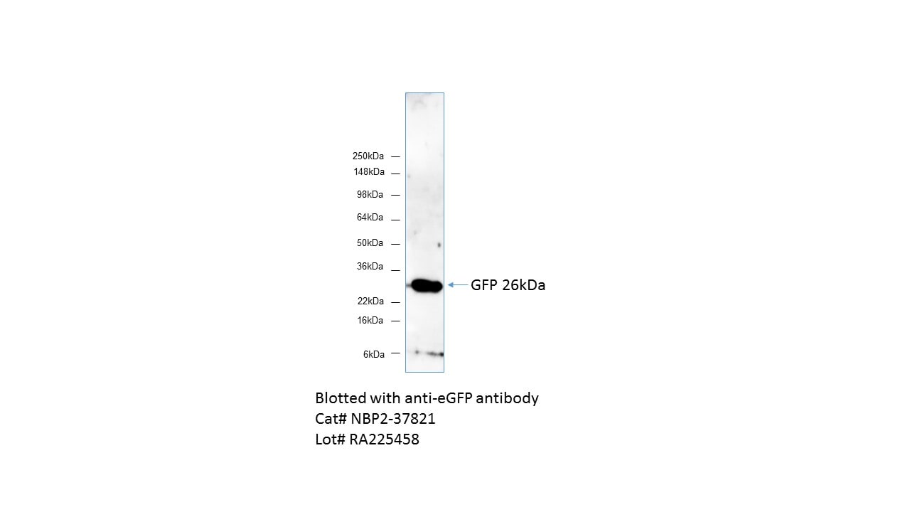
Western Blot: GFP Antibody [NBP2-37821]
Western Blot: eGFP Antibody [NBP2-37821] - WB analysis on HEK293 cells transfected with Green Fluorescent Protein (GFP) using eGFP antibody. 20ug whole cells lyate were loaded. Image from verified customer review.![Immunocytochemistry/ Immunofluorescence: GFP Antibody [NBP2-37821]](https://resources.rndsystems.com/images/products/eGFP-Antibody-Immunocytochemistry-Immunofluorescence-NBP2-37821-img0003.jpg "Immunocytochemistry/ Immunofluorescence: GFP Antibody [NBP2-37821]")
Immunocytochemistry/ Immunofluorescence: GFP Antibody [NBP2-37821]
Immunocytochemistry/Immunofluorescence: eGFP Antibody [NBP2-37821] - Analysis of eGFP using either natural fluorescence (green) or an eGFP antibody (red) in U2OS cells transfected with an EGFR-eGFP fusion protein.![Western Blot: GFP Antibody [NBP2-37821]](https://resources.rndsystems.com/images/products/eGFP-Antibody-Western-Blot-NBP2-37821-img0001.jpg "Western Blot: GFP Antibody [NBP2-37821]")
Western Blot: GFP Antibody [NBP2-37821]
Western Blot: eGFP Antibody [NBP2-37821] - 12% SDS-PAGE of HEK-293 cells transfected with pCMV-Myc-eGFP (0.25ug DNA lanes 2 and 4; 0.50ug DNA lanes 3 and 5) Coomassie stain (lanes 2 and 3) and western blot (lanes 4 and 5) with anti-eGFP antibody and goat anti-rabbit HRP secondaryApplications for GFP Antibody - Azide and BSA Free
Application
Recommended Usage
Immunocytochemistry/ Immunofluorescence
1:20 - 1:200
Western Blot
1 ug/ml
Reviewed Applications
Read 1 review rated 5 using NBP2-37821 in the following applications:
Formulation, Preparation, and Storage
Purification
Immunogen affinity purified
Formulation
PBS (pH 7.2) with 0.1% gelatin
Format
Azide and BSA Free
Preservative
No Preservative
Concentration
1 mg/ml
Shipping
The product is shipped with polar packs. Upon receipt, store it immediately at the temperature recommended below.
Stability & Storage
Store at 4C in the dark.
Background: GFP
References
1. Shi, C., Pan, F. C., Kim, J. N., Washington, M. K., Padmanabhan, C., Meyer, C. T.,... Means, A. L. (2019). Differential Cell Susceptibilities to Kras(G12D) in the Setting of Obstructive Chronic Pancreatitis. Cell Mol Gastroenterol Hepatol. doi:10.1016/j.jcmgh.2019.07.001
2. Zhao, S., Fortier, T. M., & Baehrecke, E. H. (2018). Autophagy Promotes Tumor-like Stem Cell Niche Occupancy. Curr Biol, 28(19), 3056-3064.e3053. doi:10.1016/j.cub.2018.07.075
3. Zusso, M., Lunardi, V., Franceschini, D., Pagetta, A., Lo, R., Stifani, S.,... Moro, S. (2019). Ciprofloxacin and levofloxacin attenuate microglia inflammatory response via TLR4/NF-kB pathway. J Neuroinflammation, 16(1), 148. doi:10.1186/s12974-019-1538-9
Long Name
Green Fluorescent Protein
Alternate Names
eGFP, GFPuv, anti-gfp (green fluorescent protein) pab, clone 9F9.F9, GFP immunofluorescence, GFP immunoprecipitation, GFP monoclonal, GFP mouse, GFP staining, GFP western blot
Additional GFP Products
Product Documents for GFP Antibody - Azide and BSA Free
Certificate of Analysis
To download a Certificate of Analysis, please enter a lot or batch number in the search box below.
Product Specific Notices for GFP Antibody - Azide and BSA Free
This product is for research use only and is not approved for use in humans or in clinical diagnosis. Primary Antibodies are guaranteed for 1 year from date of receipt.
Citations for GFP Antibody - Azide and BSA Free
 Powered by Bioz
Customer Reviews for GFP Antibody - Azide and BSA Free (1)
5 out of 5
1 Customer Rating
Have you used GFP Antibody - Azide and BSA Free?
Submit a review and receive an Amazon gift card!
$25/€18/£15/$25CAN/¥2500 Yen for a review with an image
$10/€7/£6/$10CAN/¥1110 Yen for a review without an image
Submit a review
Customer Images
Showing
1
-
1 of
1 review
Showing All
Filter By:
-
Application: Western BlotSample Tested: Transfected HEK 293 cellsSpecies: Non-species relatedVerified Customer | Posted 02/02/2017HEK293 cells were transfected with Green Fluorescent Protein (GFP), 20ug whole cells lyate were loaded. This is a clean antibody with almost no background at all.

There are no reviews that match your criteria.
Protocols
Find general support by application which include: protocols, troubleshooting, illustrated assays, videos and webinars.
- Appropriate Fixation of IHC/ICC Samples
- Cellular Response to Hypoxia Protocols
- ClariTSA™ Fluorophore Kits
- Detection & Visualization of Antibody Binding
- ICC Cell Smear Protocol for Suspension Cells
- ICC Immunocytochemistry Protocol Videos
- ICC for Adherent Cells
- Immunocytochemistry (ICC) Protocol
- Immunocytochemistry Troubleshooting
- Immunofluorescence of Organoids Embedded in Cultrex Basement Membrane Extract
- Immunohistochemistry (IHC) and Immunocytochemistry (ICC) Protocols
- Preparing Samples for IHC/ICC Experiments
- Preventing Non-Specific Staining (Non-Specific Binding)
- Primary Antibody Selection & Optimization
- Protocol for VisUCyte™ HRP Polymer Detection Reagent
- Protocol for the Fluorescent ICC Staining of Cell Smears - Graphic
- Protocol for the Fluorescent ICC Staining of Cultured Cells on Coverslips - Graphic
- Protocol for the Preparation and Fluorescent ICC Staining of Cells on Coverslips
- Protocol for the Preparation and Fluorescent ICC Staining of Non-adherent Cells
- Protocol for the Preparation and Fluorescent ICC Staining of Stem Cells on Coverslips
- Protocol for the Preparation of a Cell Smear for Non-adherent Cell ICC - Graphic
- R&D Systems Quality Control Western Blot Protocol
- TUNEL and Active Caspase-3 Detection by IHC/ICC Protocol
- The Importance of IHC/ICC Controls
- Troubleshooting Guide: Western Blot Figures
- Western Blot Conditions
- Western Blot Protocol
- Western Blot Protocol for Cell Lysates
- Western Blot Troubleshooting
- Western Blot Troubleshooting Guide
- View all Protocols, Troubleshooting, Illustrated assays and Webinars
FAQs for GFP Antibody - Azide and BSA Free
Showing
1
-
1 of
1 FAQ
Showing All
-
Q: We are looking for a rabbit anti-GFP antibody as a primary antibody for immunofluorescence and WB. I've seen that you carry several anti-GFP antibodies and I would like to ask which one you would recommend. The one with Cat.No NB600-308 has been used in a lot of publications and I reckon it is a solid antibody. Is this our best choice, or is there a better alternative?
A: NB600-308 is an excellent GFP antibody and I think it would work very well for you. It has been reviewed and published with which usually makes our customer feel much more confident, seeing as it worked in other people's hand.
Loading...