HtrA2/Omi is the mammalian homologue of bacterial high temperature requirement protein (HtrA). HtrA2/Omi localizes to the mitochondria and is processed to expose an amino-terminal Reaper-like motif similar to SMAC/Diablo. HtrA2/Omi is released from the mitochondria in response to apoptotic insult and can interact with the BIR2 or BIR3 domains of XIAP to relieve caspase-IAP inhibition. This effect can be measured by reversing XIAP-BIR2 (R&D Systems, Catalog # 786-XB) inhibition of Caspase-7 (R&D Systems, Catalog # 823-C7) cleavage of a fluorogenic peptide (DEVD-AFC, MP Bio, Catalog # AFC-138). IC50 values for this effect are typically between 0.2 and 1.5 μM. HtrA2/Omi is trimeric and functions as a serine protease. The serine protease activity may play a more central role in apoptosis than its IAP antagonizing function. A PDZ domain regulates the serine protease activity by blocking access to the active site. The specificity of the protease is yet to be defined and no endogenous substrates are known to date.

Key Product Details
Validated by
Biological Validation
Species Reactivity
Validated:
Human, Mouse, Rat
Cited:
Human, Mouse, Rat, Hamster
Applications
Validated:
Western Blot, Immunocytochemistry, Simple Western
Cited:
Immunohistochemistry, Immunohistochemistry-Frozen, Western Blot, Immunocytochemistry, Co-Immunoprecipitation, ELISA Capture
Label
Unconjugated
Antibody Source
Polyclonal Rabbit IgG
Loading...
Product Specifications
Immunogen
E. coli-derived recombinant human HTRA2/Omi
Ala134-Glu458
Accession # O43464
Ala134-Glu458
Accession # O43464
Specificity
Detects human, mouse, and rat full length and mitochondria-processed HTRA2/Omi.
Clonality
Polyclonal
Host
Rabbit
Isotype
IgG
Scientific Data Images for HTRA2/Omi Antibody
Detection of Human/Mouse/Rat HTRA2/Omi by Western Blot.
Western blot shows lysates of PC-12 rat adrenal pheochromocytoma cell line, Jurkat human acute T cell leukemia cell line, HeLa human cervical epithelial carcinoma cell line, L-929 mouse fibroblast cell line, and C2C12 mouse myoblast cell line. PVDF membrane was probed with 0.25 µg/mL of Rabbit Anti-Human/Mouse/Rat HTRA2/Omi Antigen Affinity-purified Polyclonal Antibody (Catalog # AF1458) followed by HRP-conjugated Anti-Rabbit IgG Secondary Antibody (Catalog # HAF008). Specific bands were detected for HTRA2/Omi at approximately 36 and 49 kDa (as indicated). This experiment was conducted under reducing conditions and using Immunoblot Buffer Group 2.
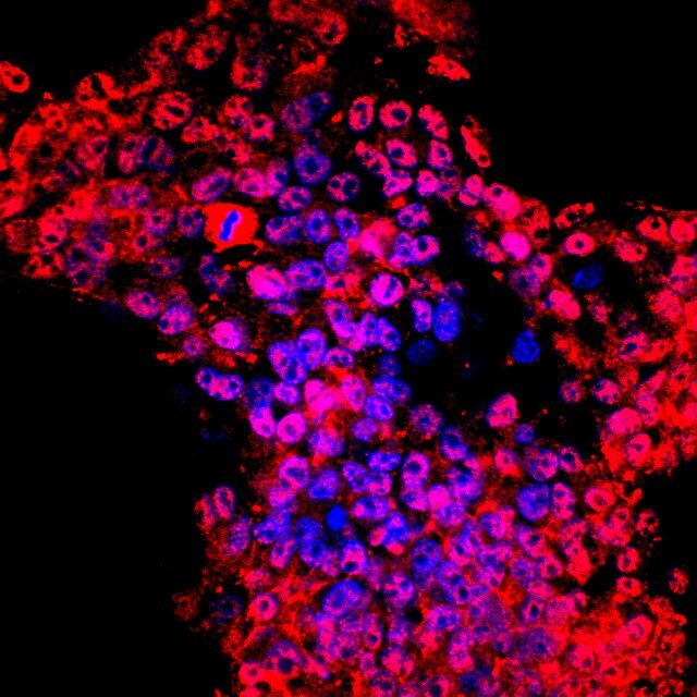
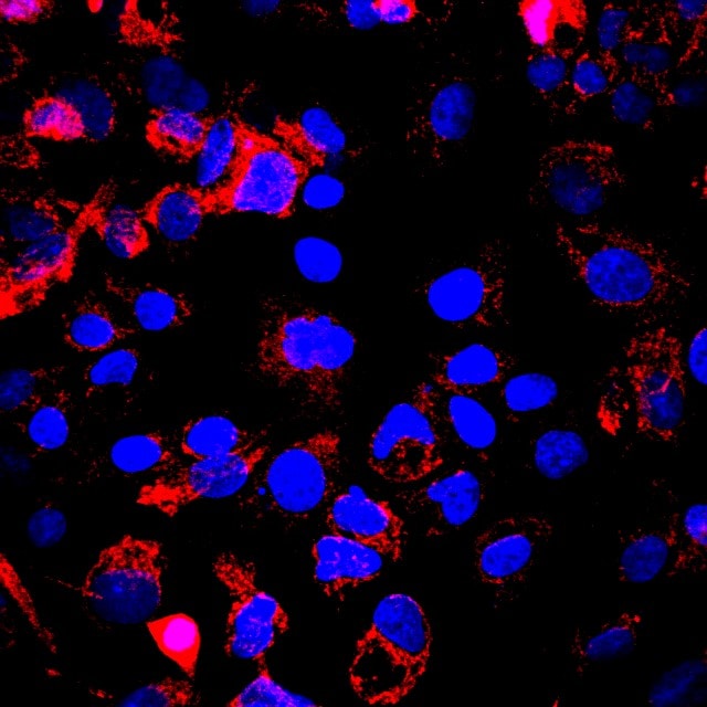
HTRA2/Omi in Jurkat Human Cell Line.
HTRA2/Omi was detected in immersion fixed Jurkat human acute T cell leukemia cell line stimulated with staurosporin using Rabbit Anti-Human/Mouse/Rat HTRA2/Omi Antigen Affinity-purified Polyclonal Antibody (Catalog # AF1458) at 10 µg/mL for 3 hours at room temperature. Cells were stained using the NorthernLights™ 557-conjugated Anti-Rabbit IgG Secondary Antibody (yellow; Catalog # NL004) and counterstained with DAPI (blue). View our protocol for Fluorescent ICC Staining of Cells on Coverslips.
Detection of Human and Mouse HTRA2/Omi by Simple WesternTM.
Simple Western lane view shows lysates of C2C12 mouse myoblast cell line and HeLa human cervical epithelial carcinoma cell line, loaded at 0.2 mg/mL. Specific bands were detected for HTRA2/Omi at approximately 51 kDa (precursor) and 41 kDa (processed) (as indicated) using 2.5 µg/mL of Rabbit Anti-Human/Mouse/Rat HTRA2/Omi Antigen Affinity-purified Polyclonal Antibody (Catalog # AF1458). This experiment was conducted under reducing conditions and using the 12-230 kDa separation system.
Detection of Human HTRA2/Omi by Western Blot
Caspases are not activated by FGFR inhibition in FGFR2‐mutant EC cells. (A) Western blots showing total caspase‐3 and caspase‐7 in response to treatment with 300 nm BGJ398 for up to 72 h. Tubulin serves as a loading control. *Denotes nonspecific band. (B) AN3CA and JHUEM2 cells were pretreated with 100 μm Z‐VAD‐FMK for 1 h prior to the addition of DMSO, 1 μm PD173074 (PD), 300 nm BGJ398 (BGJ) or 300 nm AZD4547 (AZD) for 72 h. Cell death was detected by staining cells with Annexin V. The mean percentage of Annexin V‐positive cells from three independent experiments (each performed in triplicate) is shown along with SD. (C) Western blot showing cleavage of caspase‐3 in AN3CA and JHUEM2 cells treated with 1 μm actinomycin D (Act D) or staurosporine (STS), respectively, for 24 h. (D) Mean percentage of AN3CA and JHUEM2 cells showing Annexin V‐positive staining following treatment with 100 μm Z‐VAD‐FMK alone or 1 h prior to treatment with 1 μm Act D and STS for 24 h. The mean from three independent experiments (each performed in triplicate) is shown along with SD. (E) Western blot showing staining of Bim, Bid, HTRA2/OMI and Smac/diablo in cytosolic and mitochondrial fractions of JHUEM2 cells treated with 300 nm BGJ398 for 24 and 48 h. Tom20 serves as a marker of the mitochondrial fraction, GRP78 as a marker of the ER, Lamin B1 as a marker of the nuclear fraction and GAPDH as a marker of the cytosolic fraction. (F) Western blot showing staining of AIF in mitochondrial and nuclear fractions of AN3CA cells treated with 1 μm PD173074 for 48 h. Cox IV and PARP serve as mitochondrial and nuclear markers, respectively. Image collected and cropped by CiteAb from the following open publication (https://pubmed.ncbi.nlm.nih.gov/30537101), licensed under a CC-BY license. Not internally tested by R&D Systems.
Detection of Mouse HTRA2/Omi by Western Blot
Neural deletion of Htra2 is sufficient to generate neurological phenotypes.(A) Exons 2 to 4 of Htra2 were flanked with loxP sites, with a FRT flanked neo cassette 3′ to exon 4. Expression of FlpE causes deletion of the selection cassette. Cre-mediated deletion causes excision of exons 2 to 4. Small arrows beneath the allele constructs denote the position of genotyping primers. (B) PCR from genomic DNA can distinguish WT (+, arrow, 279 bp), KO (–, filled arrowhead, 358 bp) and floxed (f, empty arrowhead, 313 bp) alleles of Htra2. (C) Western blot analysis confirmed loss of HTRA2 protein (arrow) in all tissues of HTRA2 KO mice and reduction in brain of NesKO mice (arrowheads denote non-specific bands). The levels of HTRA2 protein in NesKO spleen and thymus were comparable with NesWT. Cx: cortex, Mb: midbrain, Hb: hindbrain. PHB2 was used as a loading control. (D) HTRA2 KO mice and NesKO mice were smaller than WT littermates by comparison. The size of the thymus and spleen was reduced although brain was relatively normal in size (representative animals shown at P30, scale bar: 1 cm.). (E) Body weight of HTRA2 KO and NesKO mice did not increase beyond P18 (n = 56 (HTRA2 WT), 62 (HTRA2 KO), 35 (NesWT), 25 (NesKO), error bars indicate SEM). Image collected and cropped by CiteAb from the following open publication (https://pubmed.ncbi.nlm.nih.gov/25531304), licensed under a CC-BY license. Not internally tested by R&D Systems.
Detection of Mouse HTRA2/Omi by Western Blot
Neural deletion of Htra2 is sufficient to generate neurological phenotypes.(A) Exons 2 to 4 of Htra2 were flanked with loxP sites, with a FRT flanked neo cassette 3′ to exon 4. Expression of FlpE causes deletion of the selection cassette. Cre-mediated deletion causes excision of exons 2 to 4. Small arrows beneath the allele constructs denote the position of genotyping primers. (B) PCR from genomic DNA can distinguish WT (+, arrow, 279 bp), KO (–, filled arrowhead, 358 bp) and floxed (f, empty arrowhead, 313 bp) alleles of Htra2. (C) Western blot analysis confirmed loss of HTRA2 protein (arrow) in all tissues of HTRA2 KO mice and reduction in brain of NesKO mice (arrowheads denote non-specific bands). The levels of HTRA2 protein in NesKO spleen and thymus were comparable with NesWT. Cx: cortex, Mb: midbrain, Hb: hindbrain. PHB2 was used as a loading control. (D) HTRA2 KO mice and NesKO mice were smaller than WT littermates by comparison. The size of the thymus and spleen was reduced although brain was relatively normal in size (representative animals shown at P30, scale bar: 1 cm.). (E) Body weight of HTRA2 KO and NesKO mice did not increase beyond P18 (n = 56 (HTRA2 WT), 62 (HTRA2 KO), 35 (NesWT), 25 (NesKO), error bars indicate SEM). Image collected and cropped by CiteAb from the following open publication (https://pubmed.ncbi.nlm.nih.gov/25531304), licensed under a CC-BY license. Not internally tested by R&D Systems.Applications for HTRA2/Omi Antibody
Application
Recommended Usage
Immunocytochemistry
5-15 µg/mL
Sample: Immersion fixed non-apoptotic Jurkat human acute T cell leukemia cell line and Staurosporine treated apoptotic Jurkat human acute T cell leukemia cell line
Sample: Immersion fixed non-apoptotic Jurkat human acute T cell leukemia cell line and Staurosporine treated apoptotic Jurkat human acute T cell leukemia cell line
Simple Western
2.5 µg/mL
Sample: C2C12 mouse myoblast cell line and HeLa human cervical epithelial carcinoma cell line
Sample: C2C12 mouse myoblast cell line and HeLa human cervical epithelial carcinoma cell line
Western Blot
0.25 µg/mL
Sample: PC-12 rat adrenal pheochromocytoma cell line, Jurkat human acute T cell leukemia cell line, HeLa human cervical epithelial carcinoma cell line, L-929 mouse fibroblast cell line, and C2C12 mouse myoblast cell line
Sample: PC-12 rat adrenal pheochromocytoma cell line, Jurkat human acute T cell leukemia cell line, HeLa human cervical epithelial carcinoma cell line, L-929 mouse fibroblast cell line, and C2C12 mouse myoblast cell line
Reviewed Applications
Read 3 reviews rated 4.3 using AF1458 in the following applications:
Formulation, Preparation, and Storage
Purification
Antigen Affinity-purified
Reconstitution
Reconstitute at 0.2 mg/mL in sterile PBS. For liquid material, refer to CoA for concentration.
Loading...
Formulation
Lyophilized from a 0.2 μm filtered solution in PBS with Trehalose. *Small pack size (SP) is supplied either lyophilized or as a 0.2 µm filtered solution in PBS.
Shipping
Lyophilized product is shipped at ambient temperature. Liquid small pack size (-SP) is shipped with polar packs. Upon receipt, store immediately at the temperature recommended below.
Stability & Storage
Use a manual defrost freezer and avoid repeated freeze-thaw cycles.
- 12 months from date of receipt, -20 to -70 °C as supplied.
- 1 month, 2 to 8 °C under sterile conditions after reconstitution.
- 6 months, -20 to -70 °C under sterile conditions after reconstitution.
Calculators
Background: HTRA2/Omi
References
- Suzuki, Y. et al. (2001) Mol. Cell. 8:613.
- van Loo, G. et al. (2002) Cell Death & Diff. 9:20.
- Hedge, R. et al. (2001) J. Biol. Chem. 277:432.
- Verhagen, A. et al. (2001) J. Biol. Chem. 277:445.
- Martins, L. et al. (2002) J. Biol. Chem. 277:439.
- Silke, J., and A. Verhagen (2002) Cell Death & Diff. 9:362.
- Savopoulos, J. et al. (2000) Protein Expression & Purification 19:227.
Long Name
High Temperature Requirement Protein-2
Alternate Names
Omi, PRSS25
Gene Symbol
HTRA2
UniProt
Additional HTRA2/Omi Products
Product Documents for HTRA2/Omi Antibody
Certificate of Analysis
To download a Certificate of Analysis, please enter a lot or batch number in the search box below.
Note: Certificate of Analysis not available for kit components.
Product Specific Notices for HTRA2/Omi Antibody
For research use only
Related Research Areas
Citations for HTRA2/Omi Antibody
 Powered by Bioz
Customer Reviews for HTRA2/Omi Antibody (3)
4.3 out of 5
3 Customer Ratings
Have you used HTRA2/Omi Antibody?
Submit a review and receive an Amazon gift card!
$25/€18/£15/$25CAN/¥2500 Yen for a review with an image
$10/€7/£6/$10CAN/¥1110 Yen for a review without an image
Submit a review
Customer Images
Showing
1
-
3 of
3 reviews
Showing All
Filter By:
-
Application: ImmunocytochemistrySample Tested: pig SC cellsSpecies: PigVerified Customer | Posted 06/07/2017AF1458 HTRA2 ab labeled pig SC cells with Alexa594

-
Application: ImmunocytochemistrySample Tested: Hep2G cellsSpecies: PigVerified Customer | Posted 06/07/2017AF1458, HTRA2 antibody labeling Hep2G cells with Alexa594
-
Application: ImmunofluorescenceSample Tested: See PMID 23183826Species: HumanVerified Customer | Posted 01/05/2015
There are no reviews that match your criteria.
Protocols
Find general support by application which include: protocols, troubleshooting, illustrated assays, videos and webinars.
- Appropriate Fixation of IHC/ICC Samples
- Cellular Response to Hypoxia Protocols
- ClariTSA™ Fluorophore Kits
- Detection & Visualization of Antibody Binding
- ICC Cell Smear Protocol for Suspension Cells
- ICC Immunocytochemistry Protocol Videos
- ICC for Adherent Cells
- Immunocytochemistry (ICC) Protocol
- Immunocytochemistry Troubleshooting
- Immunofluorescence of Organoids Embedded in Cultrex Basement Membrane Extract
- Immunohistochemistry (IHC) and Immunocytochemistry (ICC) Protocols
- Preparing Samples for IHC/ICC Experiments
- Preventing Non-Specific Staining (Non-Specific Binding)
- Primary Antibody Selection & Optimization
- Protocol for VisUCyte™ HRP Polymer Detection Reagent
- Protocol for the Fluorescent ICC Staining of Cell Smears - Graphic
- Protocol for the Fluorescent ICC Staining of Cultured Cells on Coverslips - Graphic
- Protocol for the Preparation and Fluorescent ICC Staining of Cells on Coverslips
- Protocol for the Preparation and Fluorescent ICC Staining of Non-adherent Cells
- Protocol for the Preparation and Fluorescent ICC Staining of Stem Cells on Coverslips
- Protocol for the Preparation of a Cell Smear for Non-adherent Cell ICC - Graphic
- R&D Systems Quality Control Western Blot Protocol
- TUNEL and Active Caspase-3 Detection by IHC/ICC Protocol
- The Importance of IHC/ICC Controls
- Troubleshooting Guide: Western Blot Figures
- Western Blot Conditions
- Western Blot Protocol
- Western Blot Protocol for Cell Lysates
- Western Blot Troubleshooting
- Western Blot Troubleshooting Guide
- View all Protocols, Troubleshooting, Illustrated assays and Webinars
Loading...
Associated Pathways
