KC, a member of the alpha (CXC) chemokine subfamily, was initially identified as an immediate early gene induced in mouse fibroblasts by platelet‑derived growth factor. KC cDNA encodes a 96 amino acid (aa) residue precursor protein with a predicted secretory signal peptide that is removed to yield the mature protein. The protein sequence of mouse KC shows approximately 63% identity to that of mouse MIP-2. KC is also approximately 60% identical to the human GROs. It has been suggested that mouse KC and MIP-2 are the orthologs of the human GROs and rat CINCs. In addition to mouse fibroblasts, KC is expressed in macrophages and endothelial cells. Mouse KC is a potent neutrophil attractant and activator. The functional receptor for KC has been identified as CXCR2. Based on the pattern of KC expression in a number of inflammatory disease models, KC appears to have an important role in inflammation. KC was found to be involved in monocyte arrest on atherosclerotic endothelium and may also play a pathophysiological role in Alzheimer’s disease. Many chemokines are substrates for selective proteolysis at the amino-terminus by various proteases including dipeptidyl peptidase IV or matrix metalloproteases, resulting in truncated chemokine isoforms with different (both enhanced or reduced) bioactivities. The naturally occurring 68 aa N-terminal truncated isoform of mouse KC is reported to be a more potent synergistic growth stimulant for CFU-GM.
Mouse CXCL1/GRO alpha/KC/CINC-1 Antibody (1174A)
R&D Systems | Catalog # MAB4532
Recombinant Monoclonal Antibody.

Key Product Details
Validated by
Biological Validation
Species Reactivity
Validated:
Mouse
Cited:
Transgenic Mouse
Applications
Validated:
Intracellular Staining by Flow Cytometry
Cited:
Immunohistochemistry
Label
Unconjugated
Antibody Source
Recombinant Monoclonal Rabbit IgG Clone # 1174A
Loading...
Product Specifications
Immunogen
E. coli-derived recombinant mouse CXCL1/GRO alpha /KC/CINC‑1
Asn29-Lys96
Accession # P12850
Asn29-Lys96
Accession # P12850
Specificity
Detects mouse CXCL1/GRO alpha /KC/CINC‑1 in direct ELISAs.
Clonality
Monoclonal
Host
Rabbit
Isotype
IgG
Scientific Data Images for Mouse CXCL1/GRO alpha/KC/CINC-1 Antibody (1174A)
Detection of CXCL1/GRO alpha /KC/CINC‑1 in Mouse Splenocytes by Flow Cytometry.
Mouse splenocytes either (A) treated with 1 µg/mL LPS and Brefeldin A for 4 hours or (B) untreated were stained with Rabbit Anti-Mouse CXCL1/GROa/KC/CINC-1 Monoclonal Antibody (Catalog # MAB4532) followed by Phycoerythrin-conjugated Anti-Rabbit IgG Secondary Antibody (Catalog # F0110) and Rat Anti-Mouse TNF-a Fluorescein-conjugated Monoclonal Antibody (Catalog # IC410F). To facilitate intracellular staining, cells were fixed and permeabilized with FlowX FoxP3 Fixation & Permeabilization Buffer Kit (Catalog # FC012).Applications for Mouse CXCL1/GRO alpha/KC/CINC-1 Antibody (1174A)
Application
Recommended Usage
Intracellular Staining by Flow Cytometry
0.25-1 µg/106 cells
Sample: Mouse splenocytes treated with LPS and Brefeldin A were fixed and permeabilized with FlowX FoxP3 Fixation & Permeabilization Buffer Kit (Catalog # FC012).
Sample: Mouse splenocytes treated with LPS and Brefeldin A were fixed and permeabilized with FlowX FoxP3 Fixation & Permeabilization Buffer Kit (Catalog # FC012).
Reviewed Applications
Read 1 review rated 3 using MAB4532 in the following applications:
Flow Cytometry Panel Builder
Bio-Techne Knows Flow Cytometry
Save time and reduce costly mistakes by quickly finding compatible reagents using the Panel Builder Tool.
Advanced Features
- Spectra Viewer - Custom analysis of spectra from multiple fluorochromes
- Spillover Popups - Visualize the spectra of individual fluorochromes
- Antigen Density Selector - Match fluorochrome brightness with antigen density

Formulation, Preparation, and Storage
Purification
Protein A or G purified from cell culture supernatant
Reconstitution
Reconstitute at 0.5 mg/mL in sterile PBS. For liquid material, refer to CoA for concentration.
Loading...
Formulation
Lyophilized from a 0.2 μm filtered solution in PBS with Trehalose. *Small pack size (SP) is supplied either lyophilized or as a 0.2 µm filtered solution in PBS.
Shipping
Lyophilized product is shipped at ambient temperature. Liquid small pack size (-SP) is shipped with polar packs. Upon receipt, store immediately at the temperature recommended below.
Stability & Storage
Use a manual defrost freezer and avoid repeated freeze-thaw cycles.
- 12 months from date of receipt, -20 to -70 °C as supplied.
- 1 month, 2 to 8 °C under sterile conditions after reconstitution.
- 6 months, -20 to -70 °C under sterile conditions after reconstitution.
Calculators
Background: CXCL1/GRO alpha/KC/CINC-1
Alternate Names
CINC-1, CINC1, GRO alpha, KC, MGSA-alpha
Gene Symbol
CXCL1
UniProt
Additional CXCL1/GRO alpha/KC/CINC-1 Products
Product Documents for Mouse CXCL1/GRO alpha/KC/CINC-1 Antibody (1174A)
Certificate of Analysis
To download a Certificate of Analysis, please enter a lot or batch number in the search box below.
Note: Certificate of Analysis not available for kit components.
Product Specific Notices for Mouse CXCL1/GRO alpha/KC/CINC-1 Antibody (1174A)
For research use only
Related Research Areas
Citations for Mouse CXCL1/GRO alpha/KC/CINC-1 Antibody (1174A)
 Powered by Bioz
Customer Reviews for Mouse CXCL1/GRO alpha/KC/CINC-1 Antibody (1174A) (1)
3 out of 5
1 Customer Rating
Have you used Mouse CXCL1/GRO alpha/KC/CINC-1 Antibody (1174A)?
Submit a review and receive an Amazon gift card!
$25/€18/£15/$25CAN/¥2500 Yen for a review with an image
$10/€7/£6/$10CAN/¥1110 Yen for a review without an image
Submit a review
Customer Images
Showing
1
-
1 of
1 review
Showing All
Filter By:
-
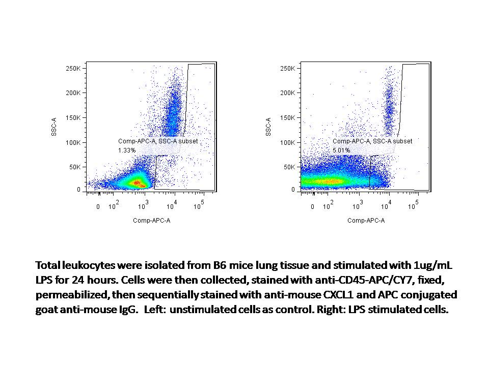
Application: Flow CytometrySample Tested: mouse lung total leukocytesSpecies: MouseVerified Customer | Posted 02/26/2016

There are no reviews that match your criteria.
Protocols
Find general support by application which include: protocols, troubleshooting, illustrated assays, videos and webinars.
- 7-Amino Actinomycin D (7-AAD) Cell Viability Flow Cytometry Protocol
- Extracellular Membrane Flow Cytometry Protocol
- Flow Cytometry Protocol for Cell Surface Markers
- Flow Cytometry Protocol for Staining Membrane Associated Proteins
- Flow Cytometry Staining Protocols
- Flow Cytometry Troubleshooting Guide
- Intracellular Flow Cytometry Protocol Using Alcohol (Methanol)
- Intracellular Flow Cytometry Protocol Using Detergents
- Intracellular Nuclear Staining Flow Cytometry Protocol Using Detergents
- Intracellular Staining Flow Cytometry Protocol Using Alcohol Permeabilization
- Intracellular Staining Flow Cytometry Protocol Using Detergents to Permeabilize Cells
- Propidium Iodide Cell Viability Flow Cytometry Protocol
- Protocol for Liperfluo
- Protocol for the Characterization of Human Th22 Cells
- Protocol for the Characterization of Human Th9 Cells
- Protocol: Annexin V and PI Staining by Flow Cytometry
- Protocol: Annexin V and PI Staining for Apoptosis by Flow Cytometry
- Troubleshooting Guide: Fluorokine Flow Cytometry Kits
- View all Protocols, Troubleshooting, Illustrated assays and Webinars
Loading...
Associated Pathways
