![Immunocytochemistry/ Immunofluorescence: Plasminogen Antibody [NB300-544]](https://resources.rndsystems.com/images/products/Plasminogen-Antibody-Immunocytochemistry-Immunofluorescence-NB300-544-img0001.jpg "Immunocytochemistry/ Immunofluorescence: Plasminogen Antibody [NB300-544]")
Loading...
Key Product Details
Species Reactivity
Validated:
Human, Mouse
Cited:
Mouse
Predicted:
Rat (95%). Backed by our 100% Guarantee.
Applications
Validated:
Immunohistochemistry, Immunohistochemistry-Paraffin, Western Blot, Immunocytochemistry/ Immunofluorescence
Cited:
Immunohistochemistry-Paraffin
Label
Unconjugated
Antibody Source
Polyclonal Rabbit IgG
Loading...
Product Specifications
Immunogen
Synthetic Peptide: V(98) Y L S E C K T G I G N G Y R G T M S(116)
Reactivity Notes
Human reactivity reported in scientific literature (PMID: 24646176).
Clonality
Polyclonal
Host
Rabbit
Isotype
IgG
Scientific Data Images for Plasminogen Antibody
Immunocytochemistry/ Immunofluorescence: Plasminogen Antibody [NB300-544]
Immunocytochemistry/Immunofluorescence: Plasminogen Antibody [NB300-544] - Analysis of Angiostatin (green) showing staining in the cytoplasm of NIH-3T3 cells (right) compared to a negative control without primary antibody (left).![Immunohistochemistry: Plasminogen Antibody [NB300-544]](https://resources.rndsystems.com/images/products/Plasminogen-Antibody-Immunohistochemistry-NB300-544-img0003.jpg "Immunohistochemistry: Plasminogen Antibody [NB300-544]")
Immunohistochemistry: Plasminogen Antibody [NB300-544]
Plasminogen-Antibody-Immunohistochemistry-NB300-544-img0003.jpg![Immunocytochemistry/ Immunofluorescence: Plasminogen Antibody [NB300-544]](https://resources.rndsystems.com/images/products/Plasminogen-Antibody-Immunocytochemistry-Immunofluorescence-NB300-544-img0002.jpg "Immunocytochemistry/ Immunofluorescence: Plasminogen Antibody [NB300-544]")
Immunocytochemistry/ Immunofluorescence: Plasminogen Antibody [NB300-544]
Immunocytochemistry/Immunofluorescence: Plasminogen Antibody [NB300-544] - Analysis of Angiostatin (green) showing staining in the cytoplasm of K562 cells (right) compared to a negative control without primary antibody (left).Applications for Plasminogen Antibody
Application
Recommended Usage
Immunocytochemistry/ Immunofluorescence
1:10 - 1:100
Western Blot
2 ug/ml
Application Notes
Use in Immunohistochemistry-paraffin reported in scientific literature (PMID: 30878040).
Reviewed Applications
Read 1 review rated 3 using NB300-544 in the following applications:
Formulation, Preparation, and Storage
Purification
Immunogen affinity purified
Formulation
PBS with 1 mg/ml BSA
Preservative
0.05% Sodium Azide
Concentration
1 mg/ml
Shipping
The product is shipped with polar packs. Upon receipt, store it immediately at the temperature recommended below.
Stability & Storage
Store at -20C. Avoid freeze-thaw cycles.
Background: Plasminogen
Alternate Names
Plg
Gene Symbol
PLG
UniProt
Additional Plasminogen Products
Product Documents for Plasminogen Antibody
Certificate of Analysis
To download a Certificate of Analysis, please enter a lot or batch number in the search box below.
Product Specific Notices for Plasminogen Antibody
This product is for research use only and is not approved for use in humans or in clinical diagnosis. Primary Antibodies are guaranteed for 1 year from date of receipt.
Citations for Plasminogen Antibody
 Powered by Bioz
Customer Reviews for Plasminogen Antibody (1)
3 out of 5
1 Customer Rating
Have you used Plasminogen Antibody?
Submit a review and receive an Amazon gift card!
$25/€18/£15/$25CAN/¥2500 Yen for a review with an image
$10/€7/£6/$10CAN/¥1110 Yen for a review without an image
Submit a review
Customer Images
Showing
1
-
1 of
1 review
Showing All
Filter By:
-
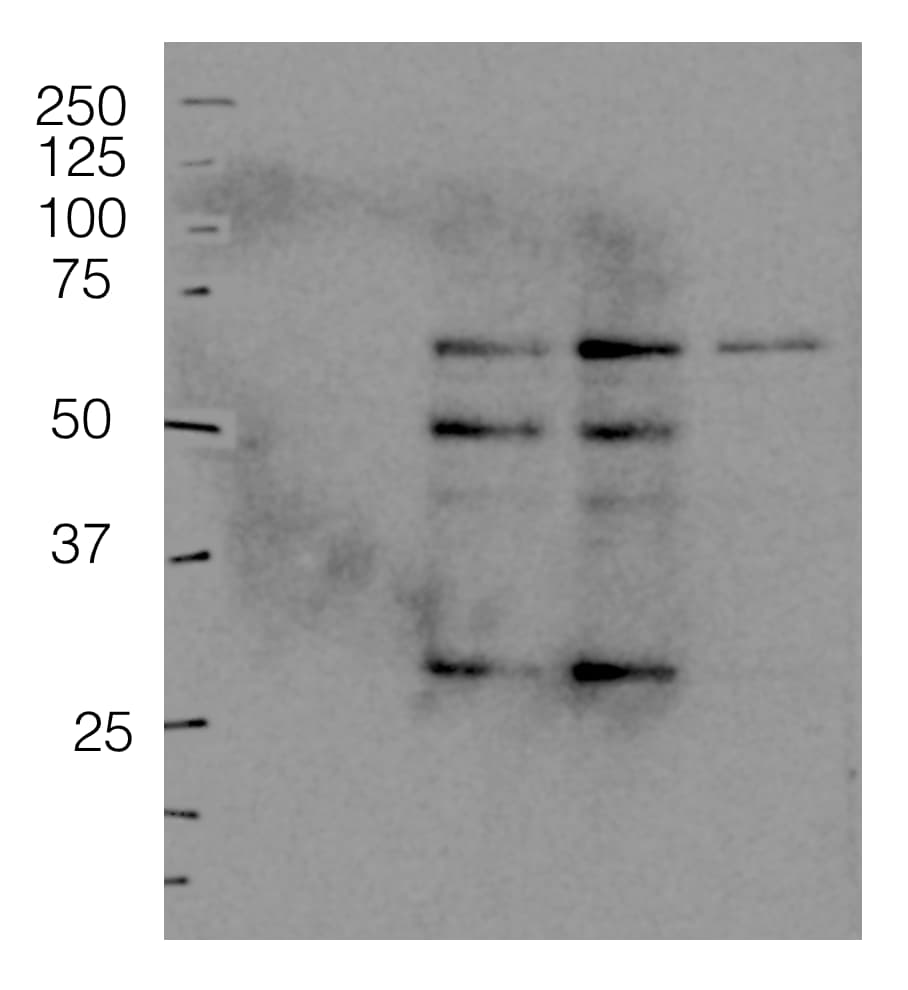
Application: Western BlotSample Tested: kidneySpecies: MouseVerified Customer | Posted 12/08/20171:500 dilution, 30ug of tissue, bands seen are likely angiostatin and its fragment. plasminogen appears weak or non-existent at times.

There are no reviews that match your criteria.
Protocols
Find general support by application which include: protocols, troubleshooting, illustrated assays, videos and webinars.
- Antigen Retrieval Protocol (PIER)
- Antigen Retrieval for Frozen Sections Protocol
- Appropriate Fixation of IHC/ICC Samples
- Cellular Response to Hypoxia Protocols
- Chromogenic IHC Staining of Formalin-Fixed Paraffin-Embedded (FFPE) Tissue Protocol
- Chromogenic Immunohistochemistry Staining of Frozen Tissue
- ClariTSA™ Fluorophore Kits
- Detection & Visualization of Antibody Binding
- Fluorescent IHC Staining of Frozen Tissue Protocol
- Graphic Protocol for Heat-induced Epitope Retrieval
- Graphic Protocol for the Preparation and Fluorescent IHC Staining of Frozen Tissue Sections
- Graphic Protocol for the Preparation and Fluorescent IHC Staining of Paraffin-embedded Tissue Sections
- Graphic Protocol for the Preparation of Gelatin-coated Slides for Histological Tissue Sections
- ICC Cell Smear Protocol for Suspension Cells
- ICC Immunocytochemistry Protocol Videos
- ICC for Adherent Cells
- IHC Sample Preparation (Frozen sections vs Paraffin)
- Immunocytochemistry (ICC) Protocol
- Immunocytochemistry Troubleshooting
- Immunofluorescence of Organoids Embedded in Cultrex Basement Membrane Extract
- Immunofluorescent IHC Staining of Formalin-Fixed Paraffin-Embedded (FFPE) Tissue Protocol
- Immunohistochemistry (IHC) and Immunocytochemistry (ICC) Protocols
- Immunohistochemistry Frozen Troubleshooting
- Immunohistochemistry Paraffin Troubleshooting
- Preparing Samples for IHC/ICC Experiments
- Preventing Non-Specific Staining (Non-Specific Binding)
- Primary Antibody Selection & Optimization
- Protocol for Heat-Induced Epitope Retrieval (HIER)
- Protocol for Making a 4% Formaldehyde Solution in PBS
- Protocol for VisUCyte™ HRP Polymer Detection Reagent
- Protocol for the Fluorescent ICC Staining of Cell Smears - Graphic
- Protocol for the Fluorescent ICC Staining of Cultured Cells on Coverslips - Graphic
- Protocol for the Preparation & Fixation of Cells on Coverslips
- Protocol for the Preparation and Chromogenic IHC Staining of Frozen Tissue Sections
- Protocol for the Preparation and Chromogenic IHC Staining of Frozen Tissue Sections - Graphic
- Protocol for the Preparation and Chromogenic IHC Staining of Paraffin-embedded Tissue Sections
- Protocol for the Preparation and Chromogenic IHC Staining of Paraffin-embedded Tissue Sections - Graphic
- Protocol for the Preparation and Fluorescent ICC Staining of Cells on Coverslips
- Protocol for the Preparation and Fluorescent ICC Staining of Non-adherent Cells
- Protocol for the Preparation and Fluorescent ICC Staining of Stem Cells on Coverslips
- Protocol for the Preparation and Fluorescent IHC Staining of Frozen Tissue Sections
- Protocol for the Preparation and Fluorescent IHC Staining of Paraffin-embedded Tissue Sections
- Protocol for the Preparation of Gelatin-coated Slides for Histological Tissue Sections
- Protocol for the Preparation of a Cell Smear for Non-adherent Cell ICC - Graphic
- R&D Systems Quality Control Western Blot Protocol
- TUNEL and Active Caspase-3 Detection by IHC/ICC Protocol
- The Importance of IHC/ICC Controls
- Troubleshooting Guide: Immunohistochemistry
- Troubleshooting Guide: Western Blot Figures
- Western Blot Conditions
- Western Blot Protocol
- Western Blot Protocol for Cell Lysates
- Western Blot Troubleshooting
- Western Blot Troubleshooting Guide
- View all Protocols, Troubleshooting, Illustrated assays and Webinars
Loading...
Associated Pathways

