
c-Fos Antibody (2H8M9)
Novus Biologicals | Catalog # NBP3-57387
Recombinant Monoclonal Antibody

Scientific images may be resized, cropped, or adjusted for brightness or contrast for presentation purposes. These updates are limited to presentation and do not affect the underlying scientific interpretation of the data. Where available, additional source data may be provided upon request.
Loading...
Key Product Details
Species Reactivity
Human, Mouse, Rat
Applications
Immunohistochemistry-Paraffin, Western Blot, ELISA
Label
Unconjugated
Antibody Source
Recombinant Monoclonal Rabbit IgG Clone # 2H8M9
Loading...
Product Specifications
Immunogen
Recombinant protein (or fragment).This information is considered to be commercially sensitive. (Swissprot# P01100)
Clonality
Monoclonal
Host
Rabbit
Isotype
IgG
Description
This antibody is highly specific and optimized for use in Immunohistochemistry.
Normal Tissues Tested
Fallopian tube, Pancreas, Lung, Kidney, Placenta, Stomach, Liver, Thyroid, Appendix, Testis, Prostate, Tonsil, Cervix, esophagus, Breast, Colon, Brain
Cancer Tissues Tested
Lung adenocarcinoma, Lung squamous carcinoma, Hepatocellular Carcinoma, Hepatocholangio carcinoma, Thyroid Cancer, Prostate cancer, cervical squamous cell carcinoma, Breast Cancer, Colon Cancer
Positive Controls
Low tissue specificity.
Negative Controls
Fallopian tube
Scientific Data Images for c-Fos Antibody (2H8M9)
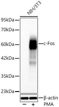
Western Blot: c-Fos Antibody (2H8M9) [NBP3-57387]
Western blot analysis of lysates from NIH/3T3 cells using NBP3-57387 at 1:1000 dilution incubated overnight at 4C. NIH/3T3 cells were treated with PMA(200 nM) at 37C for 30 minutes after serum-starvation overnight. Secondary antibody: HRP-conjugated Goat anti-Rabbit IgG (H+L) at 1:10000 dilution. Lysates/proteins: 30 ug per lane. Blocking buffer: 3 % nonfat dry milk in TBST. Exposure time: 45s.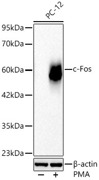
Western Blot: c-Fos Antibody (2H8M9) [NBP3-57387]
Western blot analysis of lysates from PC-12 cells using NBP3-57387 at 1:1000 dilution incubated overnight at 4C. PC-12 cells were treated with PMA(200 nM) at 37C for 30 minutes after serum-starvation overnight. Secondary antibody: HRP-conjugated Goat anti-Rabbit IgG (H+L) at 1:10000 dilution. Lysates/proteins: 30 ug per lane. Blocking buffer: 3 % nonfat dry milk in TBST. Exposure time: 60s.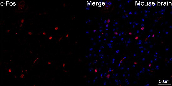
Immunohistochemistry-Paraffin: c-Fos Antibody (2H8M9) [NBP3-57387]
Confocal imaging of paraffin-embedded Mouse brain tissue using NBP3-57387 (dilution 1:200) followed by a further incubation with Cy3 Goat Anti-Rabbit IgG (H+L) (dilution 1:500) (Red). DAPI was used for nuclear staining (Blue). High pressure antigen retrieval performed with 0.01M Citrate Buffer (pH 6.0) prior to IF staining. Objective: 40x.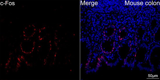
Immunohistochemistry-Paraffin: c-Fos Antibody (2H8M9) [NBP3-57387]
Confocal imaging of paraffin-embedded Mouse colon tissue using NBP3-57387 (dilution 1:200) followed by a further incubation with Cy3 Goat Anti-Rabbit IgG (H+L) (dilution 1:500) (Red). DAPI was used for nuclear staining (Blue). High pressure antigen retrieval performed with 0.01M Citrate Buffer (pH 6.0) prior to IF staining. Objective: 40x.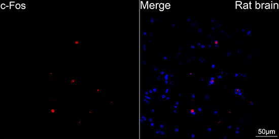
Immunohistochemistry-Paraffin: c-Fos Antibody (2H8M9) [NBP3-57387]
Confocal imaging of paraffin-embedded Rat brain tissue using NBP3-57387 (dilution 1:200) followed by a further incubation with Cy3 Goat Anti-Rabbit IgG (H+L) (dilution 1:500) (Red). DAPI was used for nuclear staining (Blue). High pressure antigen retrieval performed with 0.01M Citrate Buffer (pH 6.0) prior to IF staining. Objective: 40x.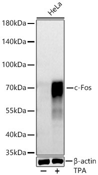
Western Blot: c-Fos Antibody (2H8M9) [NBP3-57387]
Western blot analysis of lysates from HeLa cells using NBP3-57387 at 1:1000 dilution incubated overnight at 4C. HeLa cells were treated with PMA(200 nM) at 37C for 30 minutes after serum-starvation overnight. Secondary antibody: HRP-conjugated Goat anti-Rabbit IgG (H+L) at 1:10000 dilution. Lysates/proteins: 25 ug per lane. Blocking buffer: 3 % nonfat dry milk in TBST. Exposure time: 45s.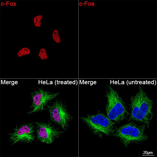
Immunocytochemistry/ Immunofluorescence: c-Fos Antibody (2H8M9) [NBP3-57387]
Confocal imaging of HeLa cells (treated with PMA) and HeLa cells (untreated) using NBP3-57387 (dilution 1:200) followed by a further incubation with Cy3-conjugated Goat anti-Rabbit IgG (H+L) (dilution 1:500) (Red). The cells were counterstained with alpha-Tubulin Mouse mAb (dilution 1:400) followed by incubation with conjugated Goat Anti-Mouse IgG (H+L) (dilution 1:500) (Green). DAPI was used for nuclear staining (Blue). Objective: 100x.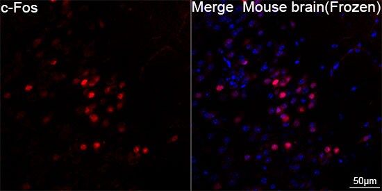
Immunohistochemistry-Frozen: c-Fos Antibody (2H8M9) [NBP3-57387]
Confocal imaging of frozen sections of Mouse brain tissue using NBP3-57387 (dilution 1:2000) followed by a further incubation with Cy3-conjugated Goat anti-Rabbit IgG (H+L) (dilution 1:500) (Red). DAPI was used for nuclear staining (Blue). Microwave antigen retrieval performed with 0.01M Citrate Buffer (pH 6.0) prior to IF staining. Objective: 40x.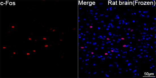
Immunohistochemistry-Frozen: c-Fos Antibody (2H8M9) [NBP3-57387]
Confocal imaging of frozen sections of Rat brain tissue using NBP3-57387 (dilution 1:2000) followed by a further incubation with Cy3-conjugated Goat anti-Rabbit IgG (H+L) (dilution 1:500) (Red). DAPI was used for nuclear staining (Blue). Microwave antigen retrieval performed with 0.01M Citrate Buffer (pH 6.0) prior to IF staining. Objective: 40x.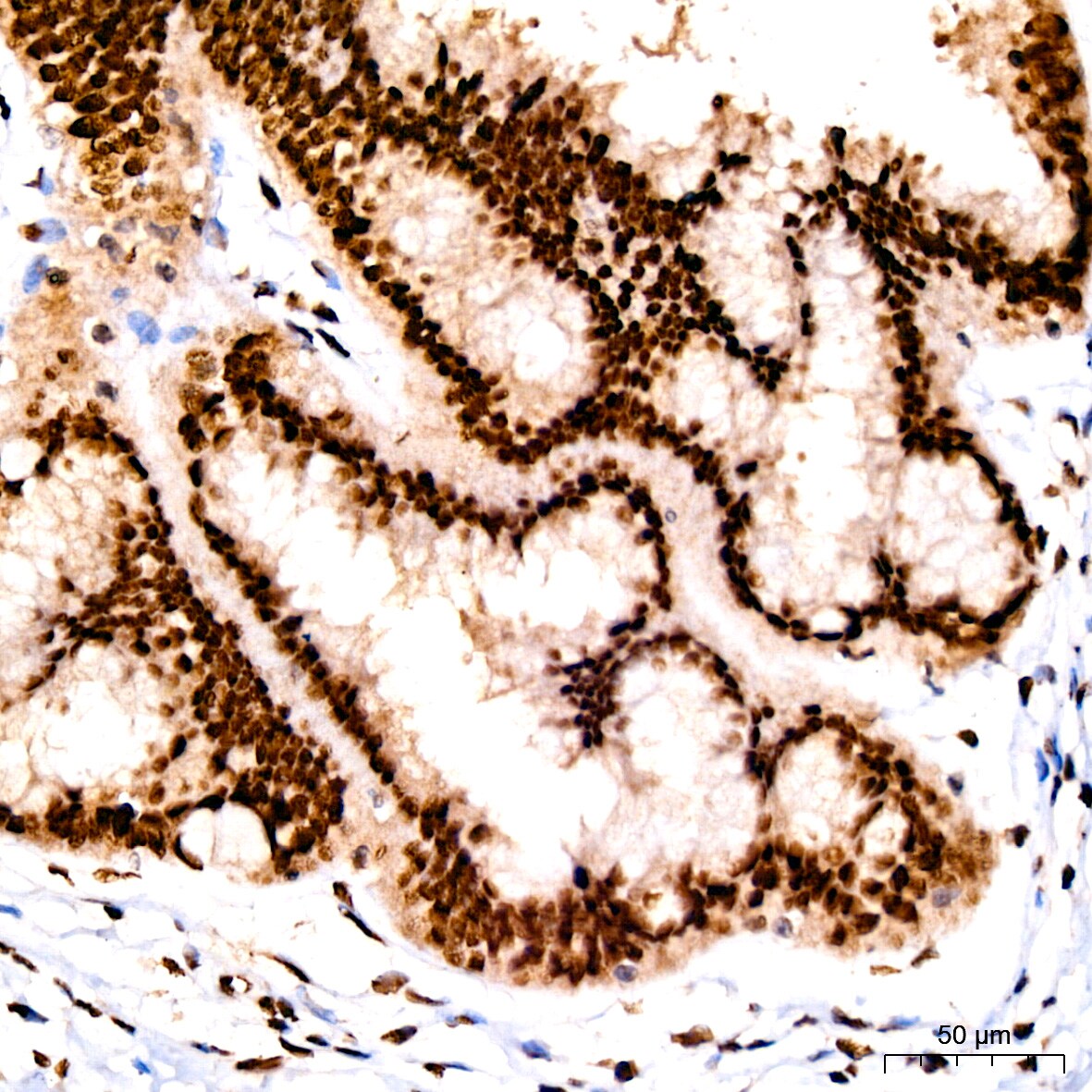
Immunohistochemistry-Paraffin: c-Fos Antibody (2H8M9) [NBP3-57387]
Immunohistochemistry analysis of paraffin-embedded Human cervix using NBP3-57387 at a dilution of 1:400 (40x lens). High pressure antigen retrieval performed with 0.01M Tris/EDTA Buffer (pH 9.0) prior to IHC staining.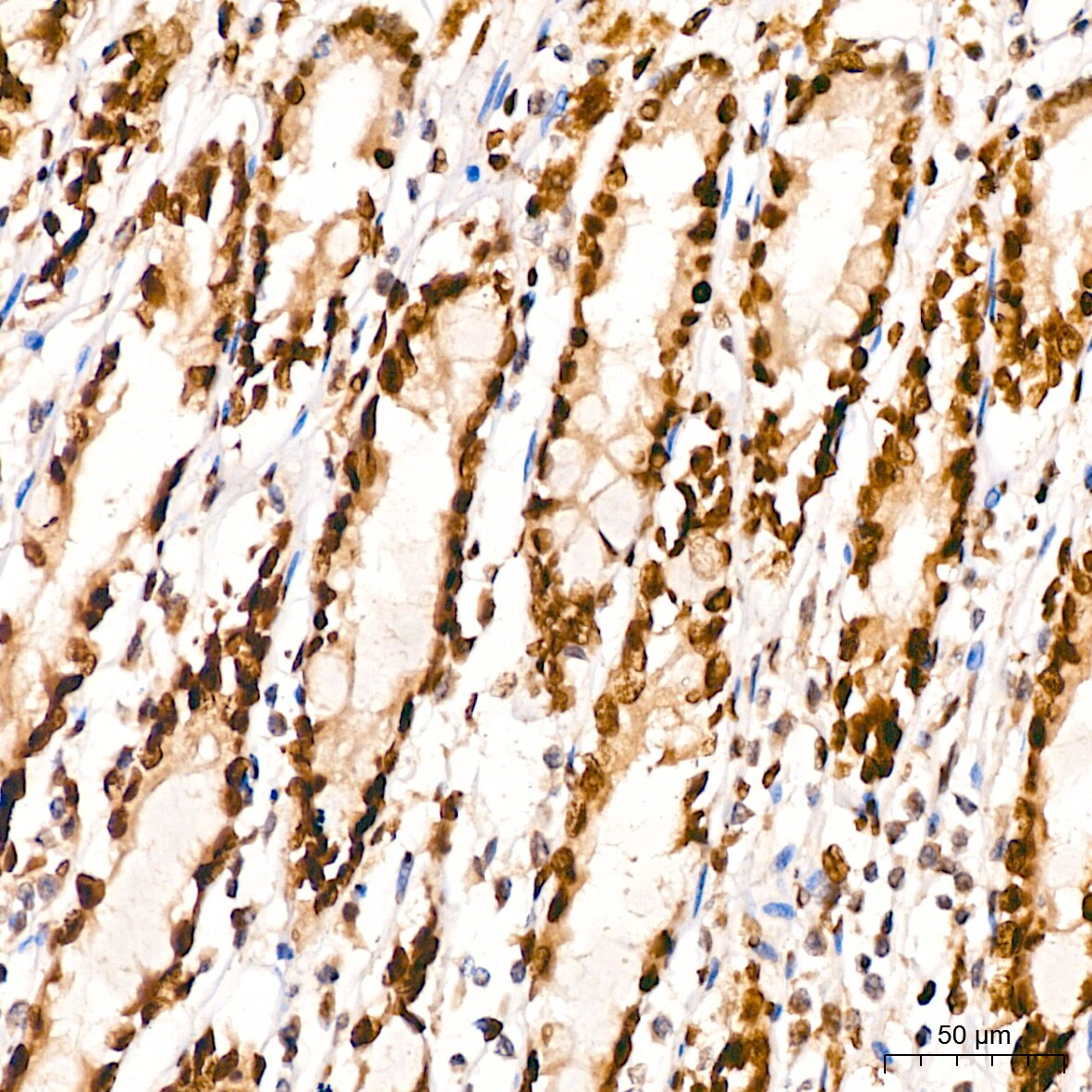
Immunohistochemistry-Paraffin: c-Fos Antibody (2H8M9) [NBP3-57387]
Immunohistochemistry analysis of paraffin-embedded Human colon using NBP3-57387 at a dilution of 1:400 (40x lens). High pressure antigen retrieval performed with 0.01M Tris/EDTA Buffer (pH 9.0) prior to IHC staining.Applications for c-Fos Antibody (2H8M9)
Application
Recommended Usage
ELISA
1 ug/mL
Immunohistochemistry-Paraffin
1:200 - 1:800
Western Blot
1:1000 - 1:4000
Application Notes
For immunofluorescence on paraffin-embedded tissues, the recommended concentration is 1:200 - 1:400
Formulation, Preparation, and Storage
Purification
Affinity purified
Formulation
PBS, 50% glycerol, 0.05% BSA, (pH 7.3)
Preservative
0.09% Sodium Azide
Concentration
Please see the vial label for concentration. If unlisted please contact technical services.
Shipping
The product is shipped with polar packs. Upon receipt, store it immediately at the temperature recommended below.
Stability & Storage
Store at -20C. Avoid freeze-thaw cycles.
Background: c-Fos
In response to stimuli, c-Fos, which is encoded by protooncogenes, has a role in cell proliferation, differentiation, and transformation (3,6). A variety of stimuli can increase c-Fos expression such as growth factors, proinflammatory cytokines, UV radiation, neurotransmitters, hormones, injury, and stress (1,6). c-Fos has long been used as a marker for neuronal activity and is associated with neural and behavioral responses following stimuli (1-3, 6-7). Mouse studies have revealed that c-Fos is important for efficient neurogenesis and cortical development (3). Additionally, c-Fos signal can be used as a molecular marker for learning and memory, such as recognition and fear (2,7). Studies have found that repeated positive stimuli result in increased Fos expression while, conversely, repeated negative value stimuli are indicated by decreased signal (7). Intermediate early genes have also been implicated in neuropsychiatric disorders including showing altered c-Fos expression in a schizophrenia animal model (2). Furthermore, antipsychotics and antidepressants are both capable of impacting c-Fos expression (2).
References
1. Kovacs K. J. (1998). c-Fos as a transcription factor: a stressful (re)view from a functional map. Neurochemistry International. https://doi.org/10.1016/s0197-0186(98)00023-0
2. Gallo, F. T., Katche, C., Morici, J. F., Medina, J. H., & Weisstaub, N. V. (2018). Immediate Early Genes, Memory and Psychiatric Disorders: Focus on c-Fos, Egr1 and Arc. Frontiers in Behavioral Neuroscience. https://doi.org/10.3389/fnbeh.2018.00079
3. Velazquez, F. N., Caputto, B. L., & Boussin, F. D. (2015). c-Fos importance for brain development. Aging. https://doi.org/10.18632/aging.100862
4. Uniprot (P01100)
5. Wu, Z., Nicoll, M., & Ingham, R. J. (2021). AP-1 family transcription factors: a diverse family of proteins that regulate varied cellular activities in classical hodgkin lymphoma and ALK+ ALCL. Experimental Hematology & Oncology. https://doi.org/10.1186/s40164-020-00197-9
6. Shaulian, E., & Karin, M. (2001). AP-1 in cell proliferation and survival. Oncogene. https://doi.org/10.1038/sj.onc.1204383
7. Chung L. (2015). A Brief Introduction to the Transduction of Neural Activity into Fos Signal. Development & Reproduction. https://doi.org/10.12717/DR.2015.19.2.061
Long Name
FBJ/Finkel–Biskis–Jinkins Murine Osteosarcoma Viral Oncogene Homolog
Alternate Names
cFos, FOS, G0S7
Gene Symbol
FOS
Additional c-Fos Products
Product Documents for c-Fos Antibody (2H8M9)
Certificate of Analysis
To download a Certificate of Analysis, please enter a lot or batch number in the search box below.
Product Specific Notices for c-Fos Antibody (2H8M9)
This product is for research use only and is not approved for use in humans or in clinical diagnosis. Primary Antibodies are guaranteed for 1 year from date of receipt.
Customer Reviews for c-Fos Antibody (2H8M9)
There are currently no reviews for this product. Be the first to review c-Fos Antibody (2H8M9) and earn rewards!
Have you used c-Fos Antibody (2H8M9)?
Submit a review and receive an Amazon gift card!
$25/€18/£15/$25CAN/¥2500 for a review with an image
$10/€7/£6/$10CAN/¥1110 for a review without an image
Submit a review
Protocols
Find general support by application which include: protocols, troubleshooting, illustrated assays, videos and webinars.
- Antigen Retrieval Protocol (PIER)
- Antigen Retrieval for Frozen Sections Protocol
- Appropriate Fixation of IHC/ICC Samples
- Cellular Response to Hypoxia Protocols
- Chromogenic IHC Staining of Formalin-Fixed Paraffin-Embedded (FFPE) Tissue Protocol
- Chromogenic Immunohistochemistry Staining of Frozen Tissue
- ClariTSA™ Fluorophore Kits
- Detection & Visualization of Antibody Binding
- ELISA Sample Preparation & Collection Guide
- ELISA Troubleshooting Guide
- Fluorescent IHC Staining of Frozen Tissue Protocol
- Graphic Protocol for Heat-induced Epitope Retrieval
- Graphic Protocol for the Preparation and Fluorescent IHC Staining of Frozen Tissue Sections
- Graphic Protocol for the Preparation and Fluorescent IHC Staining of Paraffin-embedded Tissue Sections
- Graphic Protocol for the Preparation of Gelatin-coated Slides for Histological Tissue Sections
- How to Run an R&D Systems DuoSet ELISA
- How to Run an R&D Systems Quantikine ELISA
- How to Run an R&D Systems Quantikine™ QuicKit™ ELISA
- IHC Sample Preparation (Frozen sections vs Paraffin)
- Immunofluorescent IHC Staining of Formalin-Fixed Paraffin-Embedded (FFPE) Tissue Protocol
- Immunohistochemistry (IHC) and Immunocytochemistry (ICC) Protocols
- Immunohistochemistry Frozen Troubleshooting
- Immunohistochemistry Paraffin Troubleshooting
- Preparing Samples for IHC/ICC Experiments
- Preventing Non-Specific Staining (Non-Specific Binding)
- Primary Antibody Selection & Optimization
- Protocol for Heat-Induced Epitope Retrieval (HIER)
- Protocol for Making a 4% Formaldehyde Solution in PBS
- Protocol for VisUCyte™ HRP Polymer Detection Reagent
- Protocol for the Preparation & Fixation of Cells on Coverslips
- Protocol for the Preparation and Chromogenic IHC Staining of Frozen Tissue Sections
- Protocol for the Preparation and Chromogenic IHC Staining of Frozen Tissue Sections - Graphic
- Protocol for the Preparation and Chromogenic IHC Staining of Paraffin-embedded Tissue Sections
- Protocol for the Preparation and Chromogenic IHC Staining of Paraffin-embedded Tissue Sections - Graphic
- Protocol for the Preparation and Fluorescent IHC Staining of Frozen Tissue Sections
- Protocol for the Preparation and Fluorescent IHC Staining of Paraffin-embedded Tissue Sections
- Protocol for the Preparation of Gelatin-coated Slides for Histological Tissue Sections
- Quantikine HS ELISA Kit Assay Principle, Alkaline Phosphatase
- Quantikine HS ELISA Kit Principle, Streptavidin-HRP Polymer
- R&D Systems Quality Control Western Blot Protocol
- Sandwich ELISA (Colorimetric) – Biotin/Streptavidin Detection Protocol
- Sandwich ELISA (Colorimetric) – Direct Detection Protocol
- TUNEL and Active Caspase-3 Detection by IHC/ICC Protocol
- The Importance of IHC/ICC Controls
- Troubleshooting Guide: ELISA
- Troubleshooting Guide: Immunohistochemistry
- Troubleshooting Guide: Western Blot Figures
- Western Blot Conditions
- Western Blot Protocol
- Western Blot Protocol for Cell Lysates
- Western Blot Troubleshooting
- Western Blot Troubleshooting Guide
- View all Protocols, Troubleshooting, Illustrated assays and Webinars
FAQs for c-Fos Antibody (2H8M9)
Showing
1
-
1 of
1 FAQ
Showing All
-
Q: I am looking for some c-fos antibodies that could work in IHC.
A:
Here are a list of products which might be of interest to you: Product List. We have additional filters on the left which may help you narrow down based on host species, target species, clonality, etc. We will guarantee these to work for all stated species and applications listed on the datasheet.
Loading...
Associated Pathways



NOD-like Receptor Signaling Pathways
 Pathogen or Damage-activated C-Type Lectin Receptor Signaling Pathways
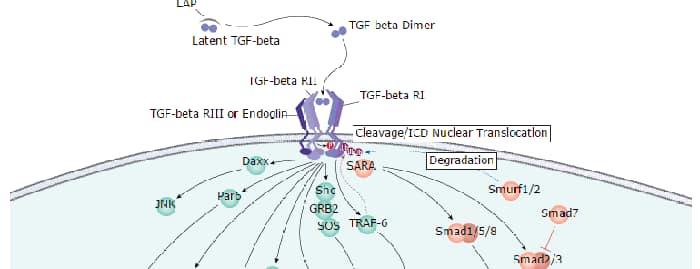
 TGF-beta Signaling Pathways
 Th1 Differentiation Pathway
 Toll-Like Receptor Signaling Pathways
 VEGF - VEGF R2 Signaling Pathways
