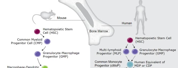
GM-CSF was initially characterized as a factor that can support the in vitro colony formation of granulocyte-macrophage progenitors. It is also a growth factor for erythroid, megakaryocyte, and eosinophil progenitors. GM-CSF is produced by a number of different cell types (including T cells, B cells, macrophages, mast cells, endothelial cells, fibroblasts, and adipocytes) in response to cytokine or inflammatory stimuli. On mature hematopoietic cells, GM-CSF is a survival factor for and activates the effector functions of granulocytes, monocytes/macrophages, and eosinophils (1, 2). GM-CSF promotes a Th1 biased immune response, angiogenesis, allergic inflammation, and the development of autoimmunity (3-5). It shows clinical effectiveness in ameliorating chemotherapy-induced neutropenia, and GM-CSF transfected tumor cells are utilized as cancer vaccines (6, 7). The 22 kDa glycosylated GM-CSF, similar to IL-3 and IL-5, is a cytokine with a core of four bundled
alpha ‑helices (8-10). Mature canine GM-CSF shares 49%‑57% amino acid sequence identity with mouse and rat GM-CSF and 69%‑72% with feline, human, and porcine
GM-CSF. GM-CSF exerts its biological effects through a heterodimeric receptor complex composed of GM-CSF R alpha /CD116 and the signal transducing common beta chain (CD131) which is also a component of the high-affinity receptors for IL-3 and IL-5 (11, 12). In addition, GM-CSF binds a naturally occurring soluble form of GM‑CSF R alpha (13). The activity of GM-CSF is species specific between human and mouse, although human GM-CSF is active on canine cells (14, 15).

Scientific images may be resized, cropped, or adjusted for brightness or contrast for presentation purposes. These updates are limited to presentation and do not affect the underlying scientific interpretation of the data. Where available, additional source data may be provided upon request.
Key Product Details
Species Reactivity
Applications
Label
Antibody Source
Product Specifications
Immunogen
Ala18-Lys144
Accession # P48749.1
Specificity
Clonality
Host
Isotype
Scientific Data Images for Canine GM-CSF Antibody [Biotin]
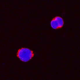
GM‑CSF in Canine PBMCs.
GM-CSF was detected in immersion fixed canine peripheral blood mononuclear cells (PBMCs) stimulated with calcium ionomycin and PMA using Goat Anti-Canine GM-CSF Biotinylated Antigen Affinity-purified Polyclonal Antibody (Catalog # BAF1546) at 15 µg/mL for 3 hours at room temperature. Cells were stained using the NorthernLights™ 557-conjugated Streptavidin (red; Catalog # NL999) and counterstained with DAPI (blue). Specific staining was localized to cytoplasm. View our protocol for Fluorescent ICC Staining of Non-adherent Cells.
Canine GM-CSF ELISA Standard Curve
Recombinant Canine GM‑CSF (Catalog # 1546-GM) was serially diluted and captured by Mouse Anti-Canine GM‑CSF Monoclonal Antibody (Catalog # MAB1546) coated on a Clear Polystyrene Microplate (Catalog # DY990). Goat Anti-Canine GM‑CSF Antigen Affinity-purified Polyclonal Antibody (Catalog # AF1546) was biotinylated and incubated with the protein captured on the plate. Detection of the standard curve was achieved by incubating Streptavidin-HRP (Catalog # DY998)Applications for Canine GM-CSF Antibody [Biotin]
Immunocytochemistry
Sample: Immersion fixed canine peripheral blood mononuclear cells (PBMCs) stimulated with calcium ionomycin and PMA
Western Blot
Sample: Recombinant Canine GM-CSF (Catalog # 1546-GM)
Canine GM-CSF Sandwich Immunoassay
Formulation, Preparation, and Storage
Purification
Reconstitution
Reconstitute at 0.2 mg/mL in sterile PBS.
Formulation
Shipping
Stability & Storage
- 12 months from date of receipt, -20 to -70 °C as supplied.
- 1 month, 2 to 8 °C under sterile conditions after reconstitution.
- 6 months, -20 to -70 °C under sterile conditions after reconstitution.
Calculators
Background: GM-CSF
References
- Martinez-Moczygemba, M. and D.P. Huston (2003) J. Allergy Clin. Immunol. 112:653.
- Barreda, D.R. et al. (2004) Dev. Comp. Immunol. 28:509.
- Eksioglu, E.A. et al. (2007) Exp. Hematol. 35:1163.
- Cao, Y. (2007) J. Clin. Invest. 117:2362.
- Fleetwood, A.J. et al. (2005) Crit. Rev. Immunol. 25:405.
- Heuser, M. et al. (2007) Semin. Hematol. 44:148.
- Hege, K.M. et al. (2006) Int. Rev. Immunol. 25:321.
- Kaushansky, K. et al. (1992) Biochemistry 31:1881.
- Diederichs, K. et al. (1991) Science 254:1779.
- Nash, R.A. et al. (1991) Blood 78:930.
- Onetto-Pothier, N. et al. (1990) Blood 75:59.
- Hayashida, K. et al. (1990) Proc. Natl. Acad. Sci. USA 87:9655.
- Pelley, J.L. et al. (2007) Exp. Hematol. 35:1483.
- Shanafelt, A.B. et al. (1991) J. Biol. Chem. 266:13804.
- Hogge, G.S. et al. (1990) Cancer Gene Ther. 6:26.
Long Name
Alternate Names
Entrez Gene IDs
Gene Symbol
UniProt
Additional GM-CSF Products
Product Documents for Canine GM-CSF Antibody [Biotin]
Certificate of Analysis
To download a Certificate of Analysis, please enter a lot or batch number in the search box below.
Note: Certificate of Analysis not available for kit components.
Product Specific Notices for Canine GM-CSF Antibody [Biotin]
For research use only
Related Research Areas
Customer Reviews for Canine GM-CSF Antibody [Biotin]
There are currently no reviews for this product. Be the first to review Canine GM-CSF Antibody [Biotin] and earn rewards!
Have you used Canine GM-CSF Antibody [Biotin]?
Submit a review and receive an Amazon gift card!
$25/€18/£15/$25CAN/¥2500 for a review with an image
$10/€7/£6/$10CAN/¥1110 for a review without an image
Submit a review
Protocols
Find general support by application which include: protocols, troubleshooting, illustrated assays, videos and webinars.
- Appropriate Fixation of IHC/ICC Samples
- Cellular Response to Hypoxia Protocols
- ClariTSA™ Fluorophore Kits
- Detection & Visualization of Antibody Binding
- ICC Cell Smear Protocol for Suspension Cells
- ICC Immunocytochemistry Protocol Videos
- ICC for Adherent Cells
- Immunocytochemistry (ICC) Protocol
- Immunocytochemistry Troubleshooting
- Immunofluorescence of Organoids Embedded in Cultrex Basement Membrane Extract
- Immunohistochemistry (IHC) and Immunocytochemistry (ICC) Protocols
- Preparing Samples for IHC/ICC Experiments
- Preventing Non-Specific Staining (Non-Specific Binding)
- Primary Antibody Selection & Optimization
- Protocol for VisUCyte™ HRP Polymer Detection Reagent
- Protocol for the Fluorescent ICC Staining of Cell Smears - Graphic
- Protocol for the Fluorescent ICC Staining of Cultured Cells on Coverslips - Graphic
- Protocol for the Preparation and Fluorescent ICC Staining of Cells on Coverslips
- Protocol for the Preparation and Fluorescent ICC Staining of Non-adherent Cells
- Protocol for the Preparation and Fluorescent ICC Staining of Stem Cells on Coverslips
- Protocol for the Preparation of a Cell Smear for Non-adherent Cell ICC - Graphic
- R&D Systems Quality Control Western Blot Protocol
- TUNEL and Active Caspase-3 Detection by IHC/ICC Protocol
- The Importance of IHC/ICC Controls
- Troubleshooting Guide: Western Blot Figures
- Western Blot Conditions
- Western Blot Protocol
- Western Blot Protocol for Cell Lysates
- Western Blot Troubleshooting
- Western Blot Troubleshooting Guide
- View all Protocols, Troubleshooting, Illustrated assays and Webinars