
CCBE1 Antibody - BSA Free
Novus Biologicals | Catalog # NBP1-79501

![Western Blot: CCBE1 Antibody [NBP1-79501]](https://resources.rndsystems.com/images/products/CCBE1-Antibody-Western-Blot-NBP1-79501-img0002.jpg "Western Blot: CCBE1 Antibody [NBP1-79501]")
Scientific images may be resized, cropped, or adjusted for brightness or contrast for presentation purposes. These updates are limited to presentation and do not affect the underlying scientific interpretation of the data. Where available, additional source data may be provided upon request.
Key Product Details
Species Reactivity
Validated:
Cited:
Applications
Validated:
Cited:
Label
Antibody Source
Format
Product Specifications
Immunogen
Clonality
Host
Isotype
Theoretical MW
Disclaimer note: The observed molecular weight of the protein may vary from the listed predicted molecular weight due to post translational modifications, post translation cleavages, relative charges, and other experimental factors.
Description
Scientific Data Images for CCBE1 Antibody - BSA Free
Western Blot: CCBE1 Antibody [NBP1-79501]
Western Blot: CCBE1 Antibody [NBP1-79501] - Titration: 1.0 ug/ml Positive Control: Mouse Kidney.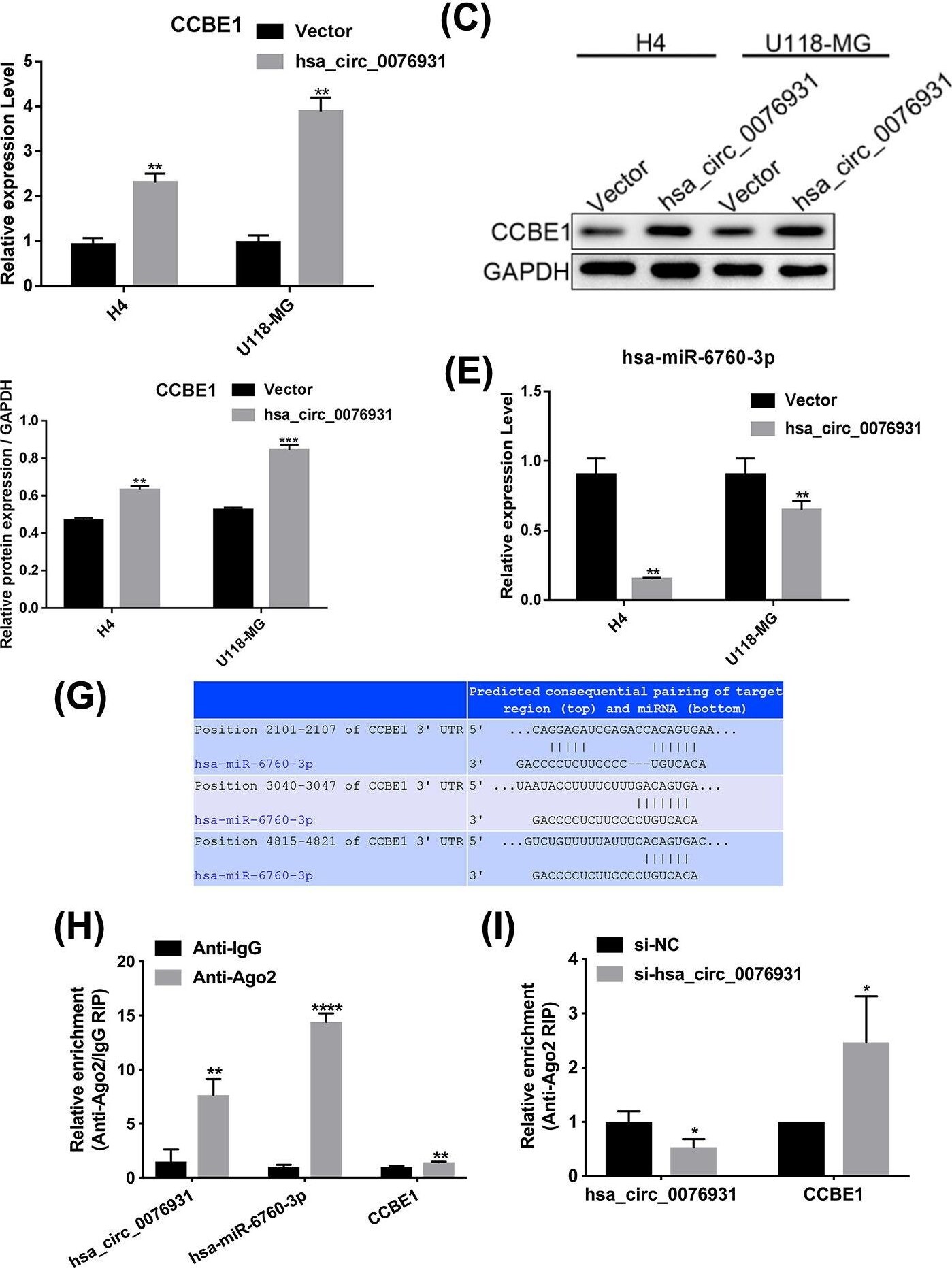
Western Blot: CCBE1 Antibody [NBP1-79501] -
Western Blot: CCBE1 Antibody [NBP1-79501] - Hsa_circ_0076931/miR-6760-3p/CCBE1 axis is predicted to play important roles in the progression of glioma(A) circRNA/miRNA/mRNA ceRNA network analysis. (B) The level of CCBE1 in H4 & U118-MG cells was detected by qRT-PCR after transfection with hsa_circ_0076931. (C) The level of CCBE1 in H4 & U118-MG cells was detected by Western blot after transfection with hsa_circ_0076931. (D) Quantification of CCBE1 protein is shown. (E) The level of miR-6067-3p in H4 & U118-MG cells was detected by qRT-PCR after transfection with hsa_circ_0076931. (F) Schematic of the binding site between hsa_circ_0076931 & miR-6067-3p, & the luciferase activity of WT hsa_circ_0076931 3′ UTR or mutant hsa_circ_0076931 3′ UTR after transfection with miR-6067-3p mimics in 293T cells. (G) Schematic of the binding site between miR-6067-3p & CCBE1. (H) AGO2-RIP assay was used to analyze the expression of hsa_circ_0076931, miR-6760-3p, & CCBE1 in U118-MG cells. (I) After interference with hsa_circ_0076931, the expression of CCBE1 was tested by AGO2-RIP assay in U118-MG cells; *P<0.05, **P<0.01, ***P<0.001. Image collected & cropped by CiteAb from the following publication (https://pubmed.ncbi.nlm.nih.gov/34931668), licensed under a CC-BY license. Not internally tested by Novus Biologicals.Applications for CCBE1 Antibody - BSA Free
Western Blot
Formulation, Preparation, and Storage
Purification
Formulation
Format
Preservative
Concentration
Shipping
Stability & Storage
Background: CCBE1
Long Name
Alternate Names
Gene Symbol
UniProt
Additional CCBE1 Products
Product Documents for CCBE1 Antibody - BSA Free
Certificate of Analysis
To download a Certificate of Analysis, please enter a lot or batch number in the search box below.
Product Specific Notices for CCBE1 Antibody - BSA Free
This product is for research use only and is not approved for use in humans or in clinical diagnosis. Primary Antibodies are guaranteed for 1 year from date of receipt.
Related Research Areas
Citations for CCBE1 Antibody - BSA Free
 Powered by Bioz
Customer Reviews for CCBE1 Antibody - BSA Free
There are currently no reviews for this product. Be the first to review CCBE1 Antibody - BSA Free and earn rewards!
Have you used CCBE1 Antibody - BSA Free?
Submit a review and receive an Amazon gift card!
$25/€18/£15/$25CAN/¥2500 for a review with an image
$10/€7/£6/$10CAN/¥1110 for a review without an image
Submit a review
Protocols
Find general support by application which include: protocols, troubleshooting, illustrated assays, videos and webinars.
- Cellular Response to Hypoxia Protocols
- R&D Systems Quality Control Western Blot Protocol
- Troubleshooting Guide: Western Blot Figures
- Western Blot Conditions
- Western Blot Protocol
- Western Blot Protocol for Cell Lysates
- Western Blot Troubleshooting
- Western Blot Troubleshooting Guide
- View all Protocols, Troubleshooting, Illustrated assays and Webinars