

![Western Blot: DNA-PKcs Antibody [NB100-658]](https://resources.rndsystems.com/images/products/DNA-PKcs-Antibody-Western-Blot-NB100-658-img0025.jpg "Western Blot: DNA-PKcs Antibody [NB100-658]")
Scientific images may be resized, cropped, or adjusted for brightness or contrast for presentation purposes. These updates are limited to presentation and do not affect the underlying scientific interpretation of the data. Where available, additional source data may be provided upon request.
Key Product Details
Validated by
Species Reactivity
Validated:
Cited:
Applications
Validated:
Cited:
Label
Antibody Source
Product Specifications
Immunogen
Clonality
Host
Isotype
Scientific Data Images for DNA-PKcs Antibody
Western Blot: DNA-PKcs Antibody [NB100-658]
Western Blot: DNA-PKcs Antibody [NB100-658] - MDA-MB-231, MCF-7, SUM149, SUM159, T47D, HCC1954 whole cell lysates (50 ug/lane). 10% SDS-PAGE. DNA-PKcs (NB100-658) primary antibody at 1:1000, 4C, overnight. Western blot image submitted by a verified customer review.![Western Blot: DNA-PKcs Antibody [NB100-658]](https://resources.rndsystems.com/images/products/DNA-PKcs-Antibody-Western-Blot-NB100-658-img0023.jpg "Western Blot: DNA-PKcs Antibody [NB100-658]")
![Western Blot: DNA-PKcs Antibody [NB100-658]](https://resources.rndsystems.com/images/products/DNA-PKcs-Antibody-Western-Blot-NB100-658-img0024.jpg "Western Blot: DNA-PKcs Antibody [NB100-658]")
Western Blot: DNA-PKcs Antibody [NB100-658]
Western Blot: DNA-PKcs Antibody [NB100-658] - Hep3B human hepatocellular carcinoma cell line. 50 ug cell lysate per lane. 10% SDS-PAGE. DNA-PKcs antibody at 1:1000 dilution O/N. Secondary antibody: HRP-conjugated Donkey anti-rabbit IgG polyclonal antibody at 1:2000 dilution. WB image submitted by a verified customer review.![Knockdown Validated: DNA-PKcs Antibody [NB100-658]](https://resources.rndsystems.com/images/products/DNA-PKcs-Antibody-Knockdown-Validated-NB100-658-img0026.jpg "Western Blot: DNA-PKcs Antibody [NB100-658]")
Western Blot: DNA-PKcs Antibody [NB100-658] -
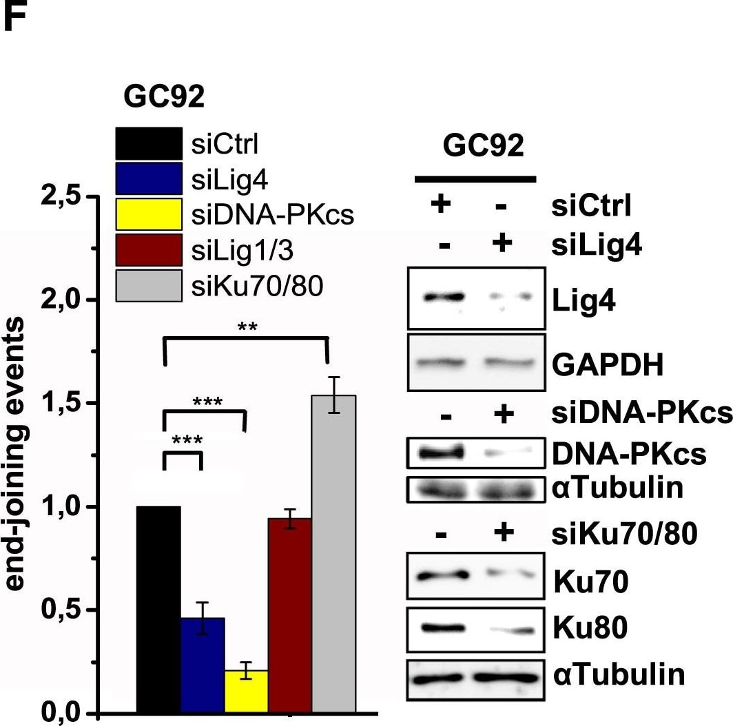
Western Blot: DNA-PKcs Antibody [NB100-658] - Molecular Characterization of G1 Resection(A) Schematic of the NHEJ reporter assay. The repair of two I-SceI-induced DSBs can result in loss of the intervening fragment, which is detected by a CD4+ signal (Rass et al., 2009). CD4+ clones were amplified by PCR (green arrows) across the repair site & sequenced. Repair of the two DSBs can also occur without loss of the intervening fragment, which escapes detection.(B) gamma H2AX foci in GC92 WT & Artemis KO cells treated with siDNA-PKcs or siCtIP. Cells were transfected with I-SceI, & foci were scored in I-SceI+ & I-SceI− cells (identified by immunofluorescence [IF] against I-SceI). Data are mean ± SEM.(C) End joining events in GC92 WT & Artemis KO cells containing the NHEJ reporter substrate. Cells were transfected with RFP or cMyc-Artemis constructs. Events were quantified by the fraction of CD4+ & RFP/cMyc+cells relative to all RFP/cMyc+cells, & results were normalized to WT cells. Data are mean ± SEM.(D) End joining events in GC92 WT & Artemis KO cells treated with siCtIP. Data are mean ± SEM.(E) Distribution of deletion sizes obtained from the sequence analysis of GC92 WT & siLig1/3-treated cells. nt, nucleotide.(F) End joining events in GC92 cells treated with siKu70/80, siLig4, siLig1/3, or siDNA-PKcs. Data are mean ± SEM.See also Table S1. Image collected & cropped by CiteAb from the following publication (https://pubmed.ncbi.nlm.nih.gov/28132842), licensed under a CC-BY license. Not internally tested by Novus Biologicals.Applications for DNA-PKcs Antibody
Immunoprecipitation
Western Blot
Reviewed Applications
Read 1 review rated 5 using NB100-658 in the following applications:
Formulation, Preparation, and Storage
Purification
Formulation
Preservative
Concentration
Shipping
Stability & Storage
Background: DNA-PKcs
Long Name
Alternate Names
Entrez Gene IDs
Gene Symbol
UniProt
Additional DNA-PKcs Products
Product Documents for DNA-PKcs Antibody
Certificate of Analysis
To download a Certificate of Analysis, please enter a lot or batch number in the search box below.
Product Specific Notices for DNA-PKcs Antibody
This product is for research use only and is not approved for use in humans or in clinical diagnosis. Primary Antibodies are guaranteed for 1 year from date of receipt.
Related Research Areas
Citations for DNA-PKcs Antibody
 Powered by Bioz
Customer Reviews for DNA-PKcs Antibody (1)
Have you used DNA-PKcs Antibody?
Submit a review and receive an Amazon gift card!
$25/€18/£15/$25CAN/¥2500 for a review with an image
$10/€7/£6/$10CAN/¥1110 for a review without an image
Submit a review
Customer Images
-
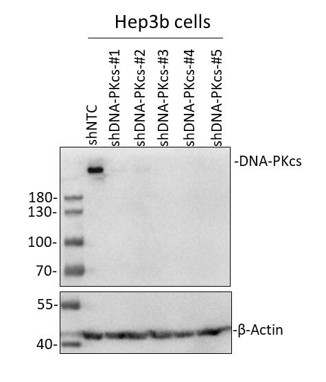
Application: Western BlotSample Tested: Hep3B human hepatocellular carcinoma cell lineSpecies: HumanVerified Customer | Posted 08/16/201950ug Hep3b cells lysate/lane, 10% SDS-PAGE; Primary antibody (DNA-PKcs Antibody (NB100-658), Lot: A1 ): 1:1000 diluted in 5% milk incubate at 4℃ for overnight; Secondary antibody: NA934V, Donkey Anti-Rabbit IgG Polyclonal Antibody (HRP (Horseradish Peroxidase)- GE Healthcare (Lot: 16908227) (1:2000 diluted in 5% milk). Incubate at room temperature for 1 hour.

There are no reviews that match your criteria.
Protocols
Find general support by application which include: protocols, troubleshooting, illustrated assays, videos and webinars.
- Cellular Response to Hypoxia Protocols
- Immunoprecipitation Protocol
- R&D Systems Quality Control Western Blot Protocol
- Troubleshooting Guide: Western Blot Figures
- Western Blot Conditions
- Western Blot Protocol
- Western Blot Protocol for Cell Lysates
- Western Blot Troubleshooting
- Western Blot Troubleshooting Guide
- View all Protocols, Troubleshooting, Illustrated assays and Webinars