
E6AP/UBE3A Antibody - BSA Free
Novus Biologicals | Catalog # NB500-239

Scientific images may be resized, cropped, or adjusted for brightness or contrast for presentation purposes. These updates are limited to presentation and do not affect the underlying scientific interpretation of the data. Where available, additional source data may be provided upon request.
Key Product Details
Validated by
Species Reactivity
Validated:
Predicted:
Applications
Label
Antibody Source
Format
Product Specifications
Immunogen
Reactivity Notes
Clonality
Host
Isotype
Scientific Data Images for E6AP/UBE3A Antibody - BSA Free
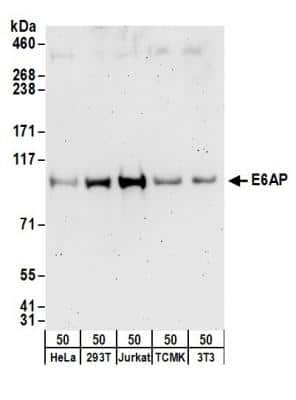
Western Blot: E6AP/UBE3A Antibody [NB500-239]
Western Blot: E6AP/UBE3A Antibody [NB500-239] - Detection of Human and Mouse E6AP by Western Blot. Samples: Whole cell lysate (50 ug) from HeLa, 293T, Jurkat, mouse TCMK-1, and mouse NIH3T3 cells. Antibodies: Affinity purified rabbit anti-E6AP antibody NB500-239 used for WB at 0.4 ug/ml. Detection: Chemiluminescence with an exposure time of 3 minutes.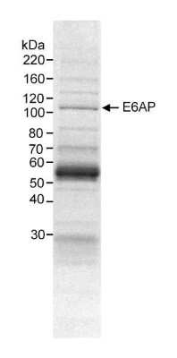
Western Blot: E6AP/UBE3A Antibody [NB500-239]
Western Blot: E6AP/UBE3A Antibody [NB500-239] - Cytoplasmic (S100) fraction (10 mg) from HeLa cells. Antibody used at 20 ug/10 mg extract.Applications for E6AP/UBE3A Antibody - BSA Free
Immunoprecipitation
Western Blot
Formulation, Preparation, and Storage
Purification
Formulation
Format
Preservative
Concentration
Shipping
Stability & Storage
Background: E6AP/UBE3A
Long Name
Alternate Names
Entrez Gene IDs
Gene Symbol
Additional E6AP/UBE3A Products
Product Documents for E6AP/UBE3A Antibody - BSA Free
Certificate of Analysis
To download a Certificate of Analysis, please enter a lot or batch number in the search box below.
Product Specific Notices for E6AP/UBE3A Antibody - BSA Free
This product is for research use only and is not approved for use in humans or in clinical diagnosis. Primary Antibodies are guaranteed for 1 year from date of receipt.
Related Research Areas
Citations for E6AP/UBE3A Antibody - BSA Free
 Powered by Bioz
Customer Reviews for E6AP/UBE3A Antibody - BSA Free
There are currently no reviews for this product. Be the first to review E6AP/UBE3A Antibody - BSA Free and earn rewards!
Have you used E6AP/UBE3A Antibody - BSA Free?
Submit a review and receive an Amazon gift card!
$25/€18/£15/$25CAN/¥2500 for a review with an image
$10/€7/£6/$10CAN/¥1110 for a review without an image
Submit a review
Protocols
View specific protocols for E6AP/UBE3A Antibody - BSA Free (NB500-239):
Nuclear Extract and Cytoplasmic Fraction Preparation
1. Nuclear extracts (NE) and cytoplasmic fractions (S100) were prepared by Dignam's method (Dignam, Lebovitz, and Roeder, Nucleic Acids Res. 11: 1475-1489. 1983).
2. 100 liters of HeLa cell culture were harvested and washed 3 times with cold PBS.
3. The packed-cell volume (PCV) was measured, and the cell pellet was gently resuspended with 5 PCVs of hypotonic buffer (10 mM HEPES-KOH [pH 8], 10 mM KCl, 1.5 mM MgCl2, 1 mM DTT, 0.2 mM PMSF).
4. Cells were incubated on ice for 10 minutes and then pelleted by centrifugation at 1,800xg for 10 minutes.
5. Hypotonic buffer was added to 2 PCVs, and cells were resuspended and then homogenized with 15 strokes using a pestle B in a Dounce glass homogenizer until the cells were more than 90% lysed, as determined by a light microscope.
6. The lysate was centrifuged at 20,000xg for 30 minutes at 4 degrees Celcius.
7. The supernatant was saved for S100 fraction, and the pellet was saved to measure the packed nuclear volume (PNV).
8. 0.4 ml of extraction buffer (20 mM HEPES-KOH [pH 8], 0.6 M KCl, 1.5 mM MgCl2, 0.2 mM EDTA, 25% [vol/vol] glycerol, 1 mM DTT, 0.2 mM PMSF) per ml of PNV was added.
9. Cell nuclei were homogenized with 10 strokes of pestle A in the homogenizer.
10. Suspension was stirred at 4 degrees Celcius for 30 minutes and centrifuged for 30 minutes at 20,000xg.
11. The supernatant (nuclear extract) was aliquotted for use.
12. The S100 fraction (resulting supernatant) was mixed with 0.11 volume of high-salt buffer (20 mM HEPES-KOH [pH 8], 1.2 M KCl, 1.5 mM MgCl2, 0.2 mM EDTA, 20% [vol/vol] glycerol, 1 mM DTT, 0.2 mM PMSF) and centrifuged at 100,000xg for 60 minutes at 4 degrees Celcius.
13. This supernatant was dialyzed for 2 hours at 4 degrees Celcius.
14. The sample was centrifuged for 30 minutes at 20,000xg and the supernatant (S100) was aliquotted for use.
Immunoprecipitation
Antibody characterization:
1. HeLa NE and S100 were diluted with 1 volume of RIPA buffer [150 mM NaCl, 1% NP-40, 0.5% DOC, 0.1% SDS, 50 mM Tris [pH 8]).
2. Cleared by spinning at 100,000 g for 20 minutes at 4 degrees Celcius.
3. 1 ml of supernatant (~10 mg total protein) was mixed with 20 ug of primary antibody (NB 500-239) and rotated overnight at 4 degrees Celcius.
4. Supernatant was mixed with 0.05 ml of protein A-sepharose beads (50% slurry) and rotated for 2 hours at 4 degrees Celcius.
5. Immunoprecipitates were washed 3 times with the 10% RIPA in PBS.
6. The washed beads were boiled with 0.04 ml of Laemmli buffer and subjected to SDS-PAGE (4-20% Tris-glycine gel).
Complex purification:
1. NE and S100 were cleared by spinning at 20,000 g for 30 minutes at 4 degrees Celcius.
2. 1.5 ml of supernatant (~15 mg total protein) was mixed with 20 ug of primary antibody (NB 500-239) and rotated for 4 hours at 4 degrees Celcius.
3. Sample and antibody mixture were centrifuged at 15,000 g for 20 minutes at 4 degrees Celcius.
4. Supernatant was mixed with 0.05 ml of protein A-sepharose beads (50% slurry) and rotated for 1 hour at 4 degrees Celcius.
5. Immunoprecipitates were washed 3 times with the NETN buffer (20 mM Tris-HCl [pH 8], 100 mM NaCl, 1 mM EDTA, 0.5% NP-40).
6. The washed beads were boiled with 0.04 ml of Laemmli buffer and subjected to SDS-PAGE (4-20% Tris-glycine gel).
*If an insufficient amount of protein is purified for identification from 15 mg of extract, carry out the same procedure using 50-100 mg of extract to increase the amount of purified protein yield.
Find general support by application which include: protocols, troubleshooting, illustrated assays, videos and webinars.
- Cellular Response to Hypoxia Protocols
- Immunoprecipitation Protocol
- R&D Systems Quality Control Western Blot Protocol
- Troubleshooting Guide: Western Blot Figures
- Western Blot Conditions
- Western Blot Protocol
- Western Blot Protocol for Cell Lysates
- Western Blot Troubleshooting
- Western Blot Troubleshooting Guide
- View all Protocols, Troubleshooting, Illustrated assays and Webinars
Associated Pathways
