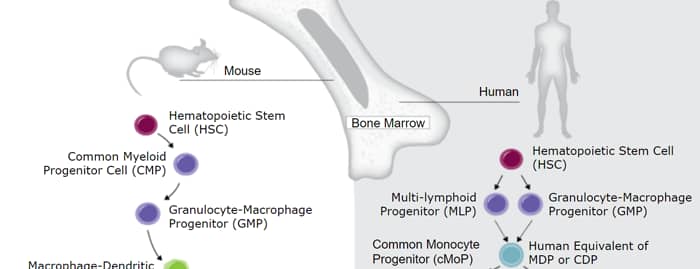
Flt-3 Ligand, also known as FL, is an alpha -helical cytokine that promotes the differentiation of multiple hematopoietic cell lineages (1‑3). Mature feline Flt-3 Ligand consists of a 156 amino acid (aa) extracellular region with a cytokine-like domain and a juxtamembrane tether region, a 21 aa transmembrane segment, and an 88 aa cytoplasmic tail (4, 5). Within the ECD, feline Flt-3 Ligand shares 92%, 81%, 70%, and 63% aa sequence identity with canine, human, mouse, and rat Flt-3 Ligand, respectively. Flt-3 Ligand is expressed as a noncovalently-linked dimer by T cells and bone marrow and thymic fibroblasts (1, 6). Each 36 kDa chain carries approximately 12 kDa of N- and O-linked carbohydrates (6). Alternate splicing and proteolytic cleavage of the transmembrane form can generate a soluble 30 kDa fragment that includes the cytokine domain (6, 7). Multiple splice forms of canine, human, and mouse Flt-3 Ligand have been described and may also exist in feline (4, 6‑10). Both transmembrane and soluble Flt-3 Ligand signal through the tyrosine kinase receptor Flt-3/Flk-2 (3, 5, 7, 10). Flt-3 Ligand induces the expansion of monocytes and immature dendritic cells as well as early B cell lineage differentiation (2, 11). It synergizes with IL-3, GM-CSF, and SCF to promote the mobilization and myeloid differentiation of hematopoietic stem cells (7, 8, 10). It cooperates with IL-2, -6, -7, and -15 to induce NK cell development and with IL-3, -7, and -11 to induce terminal B cell maturation (1, 12). Animal studies also show Flt-3 Ligand to reduce the severity of experimentally induced allergic inflammation (13).


Scientific images may be resized, cropped, or adjusted for brightness or contrast for presentation purposes. These updates are limited to presentation and do not affect the underlying scientific interpretation of the data. Where available, additional source data may be provided upon request.
Key Product Details
Species Reactivity
Applications
Label
Antibody Source
Product Specifications
Immunogen
Ser27-Pro185
Accession # NP_001009842
Specificity
Clonality
Host
Isotype
Endotoxin Level
Scientific Data Images for Feline Flt-3 Ligand/FLT3L Antibody
Cell Proliferation Induced by Flt-3 Ligand/FLT3L and Neutralization by Feline Flt-3 Ligand/FLT3L Antibody.
Recombinant Feline Flt-3 Ligand/FLT3L (Catalog # 2467-FL) stimulates proliferation in BaFlt3 mouse pro-B cell line transfected with mouse Flt-3 in a dose-dependent manner (orange line). Proliferation elicited by Recombinant Feline Flt-3 Ligand/FLT3L (2 ng/mL) is neutralized (green line) by increasing concentrations of Goat Anti-Feline Flt-3 Ligand/FLT3L Antigen Affinity-purified Polyclonal Antibody (Catalog # AF2467). The ND50 is typically 0.1-0.4 µg/mL.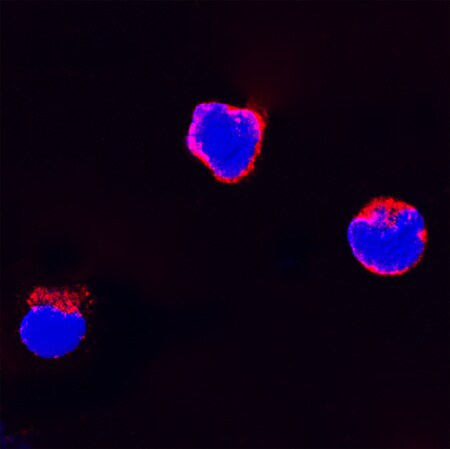
Flt-3 Ligand/FLT3L in Feline PBMCs.
Flt-3 Ligand/FLT3L was detected in immersion fixed feline peripheral blood mononuclear cells (PBMCs) using Goat Anti-Feline Flt-3 Ligand/FLT3L Antigen Affinity-purified Polyclonal Antibody (Catalog # AF2467) at 15 µg/mL for 3 hours at room temperature. Cells were stained using the NorthernLights™ 557-conjugated Anti-Goat IgG Secondary Antibody (red; Catalog # NL001) and counterstained with DAPI (blue). Specific staining was localized to cytoplasm. View our protocol for Fluorescent ICC Staining of Non-adherent Cells.Applications for Feline Flt-3 Ligand/FLT3L Antibody
Immunocytochemistry
Sample: Immersion fixed feline peripheral blood mononuclear cells (PBMCs)
Western Blot
Sample: Recombinant Feline Flt‑3 Ligand/FLT3L (Catalog # 2467-FL)
Neutralization
Formulation, Preparation, and Storage
Purification
Reconstitution
Reconstitute at 0.2 mg/mL in sterile PBS. For liquid material, refer to CoA for concentration.
Formulation
Shipping
Stability & Storage
- 12 months from date of receipt, -20 to -70 °C as supplied.
- 1 month, 2 to 8 °C under sterile conditions after reconstitution.
- 6 months, -20 to -70 °C under sterile conditions after reconstitution.
Calculators
Background: Flt-3 Ligand/FLT3L
References
- Wodnar-Filipowicz, A. (2003) News Physiol. Sci. 18:247.
- Dong, J. et al. (2002) Cancer Biol. Ther. 1:486.
- Gilliland, D.G. and J.D. Griffin (2002) Blood 100:1532.
- Yang, S. and G.-K. Sim (2000) DNA Seq. 11:163.
- Savvides, S.N. et al. (2000) Nat. Struct. Biol. 7:486.
- McClanahan, T. et al. (1996) Blood 88:3371.
- Hannum, C. et al. (1994) Nature 368:643.
- Lyman, S.D. et al. (1994) Blood 83:2795.
- Lyman, S.D. et al. (1995) Oncogene 10:149.
- Lyman, S.D. et al. (1993) Cell 75:1157.
- Diener, K.R. et al. (2008) Exp. Hematol. 36:51.
- Farag, S.S. and M.A. Caligiuri (2006) Blood Rev. 20:123.
- Edwan, J.H. et al. (2004) J. Immunol. 172:5016.
Long Name
Alternate Names
Gene Symbol
UniProt
Additional Flt-3 Ligand/FLT3L Products
Product Documents for Feline Flt-3 Ligand/FLT3L Antibody
Certificate of Analysis
To download a Certificate of Analysis, please enter a lot or batch number in the search box below.
Note: Certificate of Analysis not available for kit components.
Product Specific Notices for Feline Flt-3 Ligand/FLT3L Antibody
For research use only
Related Research Areas
Customer Reviews for Feline Flt-3 Ligand/FLT3L Antibody
There are currently no reviews for this product. Be the first to review Feline Flt-3 Ligand/FLT3L Antibody and earn rewards!
Have you used Feline Flt-3 Ligand/FLT3L Antibody?
Submit a review and receive an Amazon gift card!
$25/€18/£15/$25CAN/¥2500 for a review with an image
$10/€7/£6/$10CAN/¥1110 for a review without an image
Submit a review
Protocols
Find general support by application which include: protocols, troubleshooting, illustrated assays, videos and webinars.
- Appropriate Fixation of IHC/ICC Samples
- Cellular Response to Hypoxia Protocols
- ClariTSA™ Fluorophore Kits
- Detection & Visualization of Antibody Binding
- ICC Cell Smear Protocol for Suspension Cells
- ICC Immunocytochemistry Protocol Videos
- ICC for Adherent Cells
- Immunocytochemistry (ICC) Protocol
- Immunocytochemistry Troubleshooting
- Immunofluorescence of Organoids Embedded in Cultrex Basement Membrane Extract
- Immunohistochemistry (IHC) and Immunocytochemistry (ICC) Protocols
- Preparing Samples for IHC/ICC Experiments
- Preventing Non-Specific Staining (Non-Specific Binding)
- Primary Antibody Selection & Optimization
- Protocol for VisUCyte™ HRP Polymer Detection Reagent
- Protocol for the Fluorescent ICC Staining of Cell Smears - Graphic
- Protocol for the Fluorescent ICC Staining of Cultured Cells on Coverslips - Graphic
- Protocol for the Preparation and Fluorescent ICC Staining of Cells on Coverslips
- Protocol for the Preparation and Fluorescent ICC Staining of Non-adherent Cells
- Protocol for the Preparation and Fluorescent ICC Staining of Stem Cells on Coverslips
- Protocol for the Preparation of a Cell Smear for Non-adherent Cell ICC - Graphic
- R&D Systems Quality Control Western Blot Protocol
- TUNEL and Active Caspase-3 Detection by IHC/ICC Protocol
- The Importance of IHC/ICC Controls
- Troubleshooting Guide: Western Blot Figures
- Western Blot Conditions
- Western Blot Protocol
- Western Blot Protocol for Cell Lysates
- Western Blot Troubleshooting
- Western Blot Troubleshooting Guide
- View all Protocols, Troubleshooting, Illustrated assays and Webinars