
Heparan sulfate is a highly sulfated polysaccharide that can be found on the cell surface and extracellular matrix. It is usually covalently attached to a protein core as the glycan component of a proteoglycan. Heparan sulfate interacts with a variety of proteins, such as growth factors, protease inhibitors, cytokines, lipoprotein lipase and viral envelope proteins, therefore playing roles in cell growth, cell differentiation, cell motility, blood coagulation, lipid metabolism and viral infection. Heparan sulfate consists of repeating residues of uronic acid and N-acetylglucosamine. The uronic acid residues can be sulfated at the 2-O position by heparan sulfate 2-O-sulfotransferase. The N-acetylglucosamine residues can be sulfated at N-, 3-O, and 6-O positions by N-deacetylase/N-sulfotransferases, heparan sulfate 3-O- and 6-O-sulfotransferases, respectively. However, the reactions catalyzed by these sulfotransferases are normally incomplete on the whole chain of heparan sulfate. As a result, heparan sulfate displays enormous sequence diversity that allows it to interact with a wide spectrum of proteins differently. Three heparan sulfate 6-O-sulfotransferases are found both in human and mouse possibly with overlapping substrate specificity. HS6ST3 is ubiquitously expressed while HS6ST1 and HS6ST2 are expressed primarily in the liver and brain/spleen, respectively. Mouse HS6ST3 has 94% amino acid sequence identity with the human ortholog.
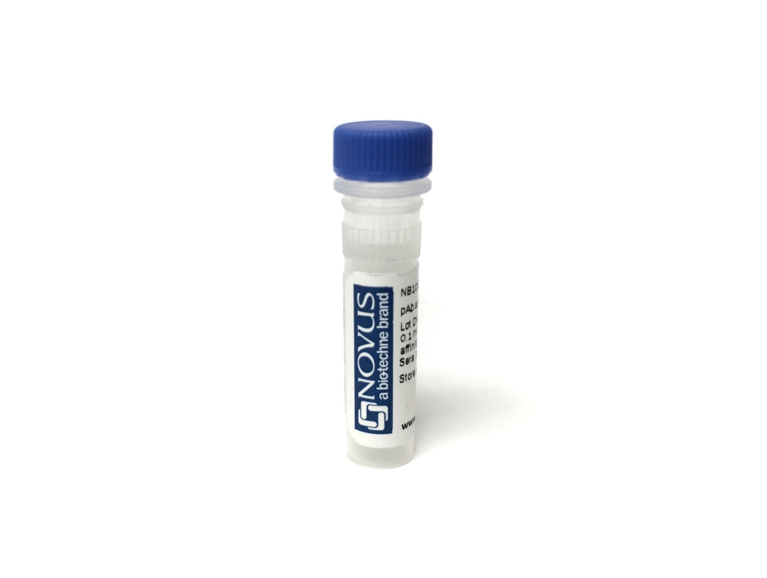
Heparan Sulfate 6-O-Sulfotransferase 3/HS6ST3 Antibody [Biotin]
Novus Biologicals | Catalog # NBP3-78862

Key Product Details
Species Reactivity
Validated:
Predicted:
Applications
Label
Antibody Source
Product Specifications
Immunogen
Clonality
Host
Isotype
Description
This conjugate is made on demand and is not held in inventory. As a result, each order represents a unique lot.
Successful site-specific biotin conjugation is confirmed by reducing SDS-PAGE, revealing a mobility shift of the antibody heavy-chain when biotin is covalently attached. Free biotin from the conjugation process has been removed. The antibody concentration is provided on the individual product label.
For specialized conjugation needs, like large quantities, and specific concentrations, bulk and custom antibody conjugation services are available.
Applications for Heparan Sulfate 6-O-Sulfotransferase 3/HS6ST3 Antibody [Biotin]
Immunocytochemistry/ Immunofluorescence
NBP2-69063
Formulation, Preparation, and Storage
Purification
Formulation
Preservative
Concentration
Shipping
Stability & Storage
Background: Heparan Sulfate 6-O-Sulfotransferase 3/HS6ST3
Alternate Names
Gene Symbol
Additional Heparan Sulfate 6-O-Sulfotransferase 3/HS6ST3 Products
Product Documents for Heparan Sulfate 6-O-Sulfotransferase 3/HS6ST3 Antibody [Biotin]
Certificate of Analysis
To download a Certificate of Analysis, please enter a lot or batch number in the search box below.
Product Specific Notices for Heparan Sulfate 6-O-Sulfotransferase 3/HS6ST3 Antibody [Biotin]
This product is for research use only and is not approved for use in humans or in clinical diagnosis. Primary Antibodies are guaranteed for 1 year from date of receipt.
Related Research Areas
Customer Reviews for Heparan Sulfate 6-O-Sulfotransferase 3/HS6ST3 Antibody [Biotin]
There are currently no reviews for this product. Be the first to review Heparan Sulfate 6-O-Sulfotransferase 3/HS6ST3 Antibody [Biotin] and earn rewards!
Have you used Heparan Sulfate 6-O-Sulfotransferase 3/HS6ST3 Antibody [Biotin]?
Submit a review and receive an Amazon gift card!
$25/€18/£15/$25CAN/¥2500 for a review with an image
$10/€7/£6/$10CAN/¥1110 for a review without an image
Submit a review
Protocols
Find general support by application which include: protocols, troubleshooting, illustrated assays, videos and webinars.
- Appropriate Fixation of IHC/ICC Samples
- Cellular Response to Hypoxia Protocols
- ClariTSA™ Fluorophore Kits
- Detection & Visualization of Antibody Binding
- ICC Cell Smear Protocol for Suspension Cells
- ICC Immunocytochemistry Protocol Videos
- ICC for Adherent Cells
- Immunocytochemistry (ICC) Protocol
- Immunocytochemistry Troubleshooting
- Immunofluorescence of Organoids Embedded in Cultrex Basement Membrane Extract
- Immunohistochemistry (IHC) and Immunocytochemistry (ICC) Protocols
- Preparing Samples for IHC/ICC Experiments
- Preventing Non-Specific Staining (Non-Specific Binding)
- Primary Antibody Selection & Optimization
- Protocol for VisUCyte™ HRP Polymer Detection Reagent
- Protocol for the Fluorescent ICC Staining of Cell Smears - Graphic
- Protocol for the Fluorescent ICC Staining of Cultured Cells on Coverslips - Graphic
- Protocol for the Preparation and Fluorescent ICC Staining of Cells on Coverslips
- Protocol for the Preparation and Fluorescent ICC Staining of Non-adherent Cells
- Protocol for the Preparation and Fluorescent ICC Staining of Stem Cells on Coverslips
- Protocol for the Preparation of a Cell Smear for Non-adherent Cell ICC - Graphic
- TUNEL and Active Caspase-3 Detection by IHC/ICC Protocol
- The Importance of IHC/ICC Controls
- View all Protocols, Troubleshooting, Illustrated assays and Webinars
Associated Pathways
