
Histone H2AX is one of a number of core histone proteins. In the cellular response to genotoxic insults, ATM and related protein kinases phosphorylate the carboxyl-terminal tail of the H2AX protein (gamma-H2AX). gamma-H2AX marks the site of damage and provides a nucleation site for the formation of damage response and repair complexes. BRCA1 carboxyl terminal (BRCT) domain-containing proteins are recruited to gamma-H2AX to create a protein scaffold for further assembly of signaling complexes.
Histone H2AX Antibody (1077751) [CoraFluor™ 1]
Novus Biologicals | Catalog # FAB11529CL1

Scientific images may be resized, cropped, or adjusted for brightness or contrast for presentation purposes. These updates are limited to presentation and do not affect the underlying scientific interpretation of the data. Where available, additional source data may be provided upon request.
Key Product Details
Species Reactivity
Human
Applications
Immunohistochemistry
Label
CoraFluor 1
Antibody Source
Monoclonal Mouse IgG2B Clone # 1077751
Loading...
Product Specifications
Immunogen
E. coli-derived recombinant human H2AX
Gly3-Ser131
Accession # P16104
Gly3-Ser131
Accession # P16104
Specificity
Detects human Histone H2AX.
Clonality
Monoclonal
Host
Mouse
Isotype
IgG2B
Description
This conjugate is made on demand and is not held in inventory. Each order represents a unique lot. All conjugates are generated using validated conjugation protocols and meet strict degree of labeling (F/P) specifications; additional information is available upon request. The antibody concentration is provided on the individual product label.
For specialized conjugation needs, like large quantities, specific concentrations, and defined F/P ratios, bulk and custom antibody conjugation services are available.
For specialized conjugation needs, like large quantities, specific concentrations, and defined F/P ratios, bulk and custom antibody conjugation services are available.
Scientific Data Images for Histone H2AX Antibody (1077751) [CoraFluor™ 1]
Product Feature: CoraFluor Probes for TR-FRET
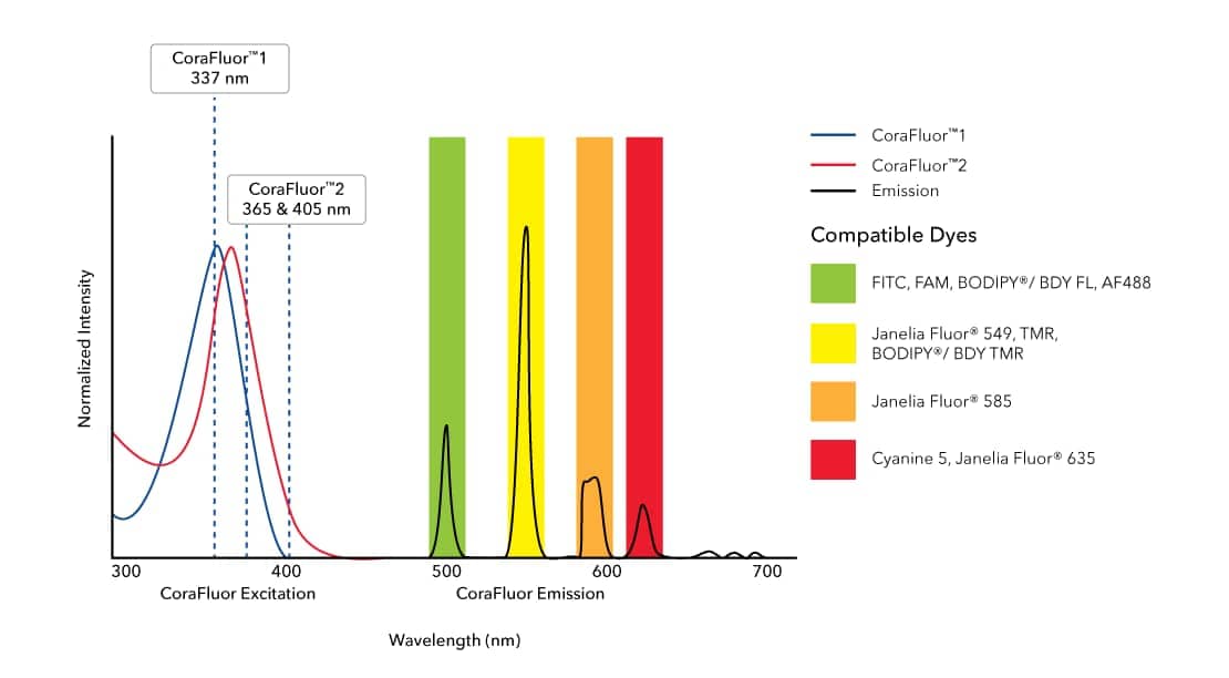
CoraFluor™ 1, amine reactive (Catalog:7920) and CoraFluor™ 2, amine reactive (Catalog # 7950) are terbium-based probes that have been developed for use as TR-FRET donors. They emit wavelengths compatible with commonly used fluorescent acceptor dyes such as BODIPY® (or BDY) and Janelia Fluor® dyes, FITC (Catalog # 5440), TMR and Cyanine 5 (Catalog # 5436). CoraFluor™ fluorescence is brighter and more stable in biological media than existing TR-FRET donors, leading to enhanced sensitivity and improved data generation. CoraFluor™ 1 exhibits excitation upon exposure to a 337 nm UV laser.Applications for Histone H2AX Antibody (1077751) [CoraFluor™ 1]
Application
Recommended Usage
Immunohistochemistry
Optimal dilutions of this antibody should be experimentally determined.
Application Notes
Recommended applications are based on validated applications from the unconjugated base product MAB11529. This conjugated antibody is not kept in inventory and is made to order using validated conjugation protocols.
Formulation, Preparation, and Storage
Purification
Protein A or G purified from hybridoma culture supernatant
Formulation
PBS
Preservative
No Preservative
Concentration
Please see the vial label for concentration. If unlisted please contact technical services.
Shipping
The product is shipped with polar packs. Upon receipt, store it immediately at the temperature recommended below.
Stability & Storage
Store at 4C in the dark. Do not freeze.
Background: Histone H2AX
Alternate Names
H2AFX
Gene Symbol
H2AX
Additional Histone H2AX Products
Product Documents for Histone H2AX Antibody (1077751) [CoraFluor™ 1]
Certificate of Analysis
To download a Certificate of Analysis, please enter a lot or batch number in the search box below.
Product Specific Notices for Histone H2AX Antibody (1077751) [CoraFluor™ 1]
Sold for research purposes only under agreement from Massachusetts General Hospital, US Patent No. 12,134,721. A license is required for development of new commercial assay platforms (e.g., microarrays, microfluidics), diagnostic use or other unlicensed uses. Please contact Technical Support for further details: techsupport@bio-techne.com.
This product is for research use only and is not approved for use in humans or in clinical diagnosis. Primary Antibodies are guaranteed for 1 year from date of receipt.
Related Research Areas
Customer Reviews for Histone H2AX Antibody (1077751) [CoraFluor™ 1]
There are currently no reviews for this product. Be the first to review Histone H2AX Antibody (1077751) [CoraFluor™ 1] and earn rewards!
Have you used Histone H2AX Antibody (1077751) [CoraFluor™ 1]?
Submit a review and receive an Amazon gift card!
$25/€18/£15/$25CAN/¥2500 for a review with an image
$10/€7/£6/$10CAN/¥1110 for a review without an image
Submit a review
Protocols
Find general support by application which include: protocols, troubleshooting, illustrated assays, videos and webinars.
- Antigen Retrieval Protocol (PIER)
- Antigen Retrieval for Frozen Sections Protocol
- Appropriate Fixation of IHC/ICC Samples
- Cellular Response to Hypoxia Protocols
- Chromogenic IHC Staining of Formalin-Fixed Paraffin-Embedded (FFPE) Tissue Protocol
- Chromogenic Immunohistochemistry Staining of Frozen Tissue
- ClariTSA™ Fluorophore Kits
- Detection & Visualization of Antibody Binding
- Fluorescent IHC Staining of Frozen Tissue Protocol
- Graphic Protocol for Heat-induced Epitope Retrieval
- Graphic Protocol for the Preparation and Fluorescent IHC Staining of Frozen Tissue Sections
- Graphic Protocol for the Preparation and Fluorescent IHC Staining of Paraffin-embedded Tissue Sections
- Graphic Protocol for the Preparation of Gelatin-coated Slides for Histological Tissue Sections
- IHC Sample Preparation (Frozen sections vs Paraffin)
- Immunofluorescent IHC Staining of Formalin-Fixed Paraffin-Embedded (FFPE) Tissue Protocol
- Immunohistochemistry (IHC) and Immunocytochemistry (ICC) Protocols
- Immunohistochemistry Frozen Troubleshooting
- Immunohistochemistry Paraffin Troubleshooting
- Preparing Samples for IHC/ICC Experiments
- Preventing Non-Specific Staining (Non-Specific Binding)
- Primary Antibody Selection & Optimization
- Protocol for Heat-Induced Epitope Retrieval (HIER)
- Protocol for Making a 4% Formaldehyde Solution in PBS
- Protocol for VisUCyte™ HRP Polymer Detection Reagent
- Protocol for the Preparation & Fixation of Cells on Coverslips
- Protocol for the Preparation and Chromogenic IHC Staining of Frozen Tissue Sections
- Protocol for the Preparation and Chromogenic IHC Staining of Frozen Tissue Sections - Graphic
- Protocol for the Preparation and Chromogenic IHC Staining of Paraffin-embedded Tissue Sections
- Protocol for the Preparation and Chromogenic IHC Staining of Paraffin-embedded Tissue Sections - Graphic
- Protocol for the Preparation and Fluorescent IHC Staining of Frozen Tissue Sections
- Protocol for the Preparation and Fluorescent IHC Staining of Paraffin-embedded Tissue Sections
- Protocol for the Preparation of Gelatin-coated Slides for Histological Tissue Sections
- TUNEL and Active Caspase-3 Detection by IHC/ICC Protocol
- The Importance of IHC/ICC Controls
- Troubleshooting Guide: Immunohistochemistry
- View all Protocols, Troubleshooting, Illustrated assays and Webinars
Loading...