
Histone H3 [Trimethyl Lys9] Antibody (6F12-H4) - BSA Free
Novus Biologicals | Catalog # NBP1-30141

Scientific images may be resized, cropped, or adjusted for brightness or contrast for presentation purposes. These updates are limited to presentation and do not affect the underlying scientific interpretation of the data. Where available, additional source data may be provided upon request.
Key Product Details
Species Reactivity
Validated:
Human, Mouse, Rat, Porcine, C. elegans, Drosophila, Invertebrate, Mammal, Yeast
Cited:
Human, Mouse, Porcine, Invertebrate, Yeast
Applications
Validated:
Immunohistochemistry, Immunohistochemistry-Paraffin, Western Blot, ELISA, Immunocytochemistry/ Immunofluorescence, Immunoprecipitation, Chromatin Immunoprecipitation (ChIP)
Cited:
Immunohistochemistry-Paraffin, Western Blot, Immunocytochemistry/ Immunofluorescence, Immunoprecipitation, Chemotaxis, Proximity Ligation Assay, IF/IHC
Label
Unconjugated
Antibody Source
Monoclonal Mouse IgG1 kappa Clone # 6F12-H4
Format
BSA Free
Loading...
Product Specifications
Immunogen
This Histone H3 [Trimethyl Lys9] antibody (6F12-H4) was raised against a synthetic peptide made to an N-terminal region of Histone H3 (between amino acids 1-50). [UniProt# P84243]
Reactivity Notes
Predicted to react with most mammalian species. Invertebrate / Blattella germanica (German cockroach) reactivity reported in scientific literature (PMID: 23872316). Porcine reactivity reported in scientific literature (PMID: 25736622)
Modification
Trimethyl Lys9
Localization
Nucleus
Clonality
Monoclonal
Host
Mouse
Isotype
IgG1 kappa
Theoretical MW
15 kDa.
Disclaimer note: The observed molecular weight of the protein may vary from the listed predicted molecular weight due to post translational modifications, post translation cleavages, relative charges, and other experimental factors.
Disclaimer note: The observed molecular weight of the protein may vary from the listed predicted molecular weight due to post translational modifications, post translation cleavages, relative charges, and other experimental factors.
Scientific Data Images for Histone H3 [Trimethyl Lys9] Antibody (6F12-H4) - BSA Free
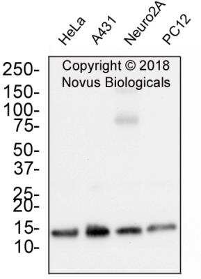
Western Blot: Histone H3 [Trimethyl Lys9] Antibody (6F12-H4) [NBP1-30141] - Whole cell protein from human HeLa, A431, mouse Neuro2A and rat PC12 cells was separated on a 4-20% gel by SDS-PAGE, transferred to 0.2 um PVDF membrane and blocked in 5% non-fat milk in TBST. The membrane was probed with 1.0 ug/ml anti-Histone h3 in block buffer and detected with an anti-mouse HRP secondary antibody using chemiluminescence. Observed molecular weight is ~15 kDa.
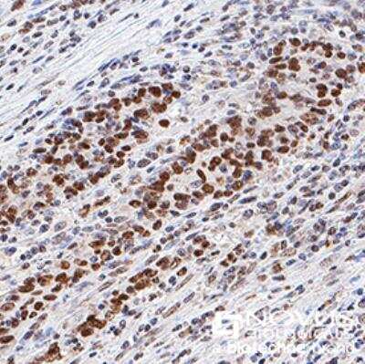
Immunohistochemistry-Paraffin: Histone H3 [Trimethyl Lys9] Antibody (6F12-H4) [NBP1-30141] - Histone 3 was detected in immersion fixed paraffin-embedded sections of human colon cancer using anti-human mouse monoclonal antibody (Catalog # NBP1-30141) at 1:1000 dilution overnight at 4C. Tissue was stained using the VisuCyte anti-mouse HRP polymer detection reagent (Catalog # VC001) with DAB chromogen (brown) and counterstained with hematoxylin (blue). Images may not be copied, printed or otherwise disseminated without express written permission of Novus Biologicals a bio-techne brand.
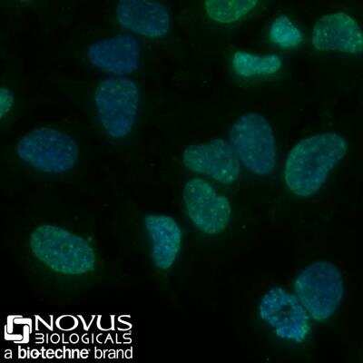
Immunocytochemistry/Immunofluorescence: Histone H3 [Trimethyl Lys9] Antibody (6F12-H4) [NBP1-30141] - HeLa cells were fixed for 10 minutes using 10% formalin and then permeabilized for 5 minutes using 1X PBS + 0.5% Triton X-100. The cells were incubated with anti-Histone H3 [Trymethyl Lys9] (6F12-H4) at 2 ug/ml overnight at 4C and detected with an anti-mouse DyLight 488 (green) at a 1:500 dilution. Nuclei were counterstained with DAPI (blue). Cells were imaged using a 40X objective.
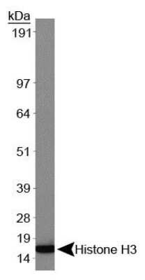
Western Blot: Histone H3 [Trimethyl Lys9] Antibody (6F12-H4) [NBP1-30141] - Analysis of Histone H3 (NBP1-30141) using HeLa nuclear lysate [NB800-PC9]. Observed molecular weight ~15 kDa.
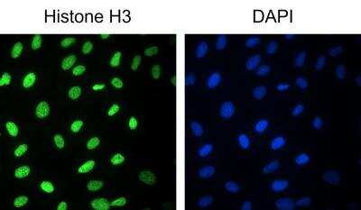
Immunocytochemistry/Immunofluorescence: Histone H3 [Trimethyl Lys9] Antibody (6F12-H4) [NBP1-30141] - Analysis of A549 cells (fixed with methanol) using Histone H3 [Trimethyl Lys9] antibody. Image from verified customer review.
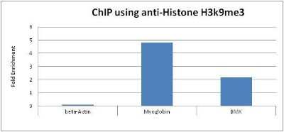
Chromatin Immunoprecipitation: Histone H3 [Trimethyl Lys9] Antibody (6F12-H4) [NBP1-30141] - Used to perform ChIP on HeLa cells.
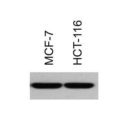
Western Blot: Histone H3 [Trimethyl Lys9] Antibody (6F12-H4) [NBP1-30141] - H3K9Me3 analysis of MCF7 and HCT116. Theorectical molecular weight is ~15 kDa. Image from verified customer review.
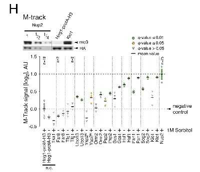
Histone-H3-Trimethyl-Lys9-Antibody-6F12-H4-Western-Blot-NBP1-30141-img0013.jpg
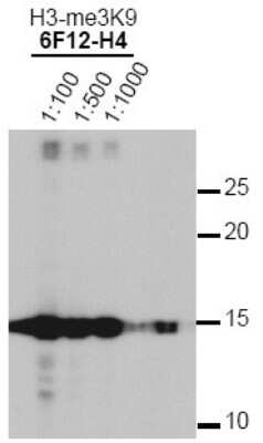
Western Blot: Histone H3 [Trimethyl Lys9] Antibody (6F12-H4) [NBP1-30141] - Analysis of Histone H3 K9-me3 in HeLa histone lysates. Observed molecular weight is ~15 kDa.
![Histone H3 [Trimethyl Lys9] Antibody (6F12-H4)](https://resources.rndsystems.com/images/products/antibody/nbp1-30141_mouse-monoclonal-histone-h3-trimethyl-lys9-antibody-6f12-h4-immunocytochemistry-immunofluorescence-2382024134224..jpg "Histone H3 [Trimethyl Lys9] (6F12-H4) in MCF7 Human Cell Line.")
Histone H3 [Trimethyl Lys9] (6F12-H4) in MCF7 Human Cell Line.
Histone H3 [Trimethyl Lys9] (6F12-H4) was detected in immersion fixed MCF7 human breast cancer cell line using Mouse anti- Histone H3 [Trimethyl Lys9] (6F12-H4) Protein G Purified Monoclonal Antibody conjugated to DyLight 550 (Catalog # NBP1-30141R) (red) at 5 µg/mL overnight at 4C. Cells were counterstained with DAPI (blue). Cells were imaged using a 100X objective and digitally deconvolved.Applications for Histone H3 [Trimethyl Lys9] Antibody (6F12-H4) - BSA Free
Application
Recommended Usage
Chromatin Immunoprecipitation (ChIP)
1:10-1:500
ELISA
1:100-1:2000
Immunocytochemistry/ Immunofluorescence
1:200
Immunohistochemistry
1:1000. Use reported in scientific literature (PMID 23872316)
Immunohistochemistry-Paraffin
1:1000
Immunoprecipitation
1:10-1:500. Use reported in scientific literature (PMID 28270554)
Western Blot
1:2000
Application Notes
In Western blot, a band is seen at ~15 kDa.
Reviewed Applications
Read 2 reviews rated 5 using NBP1-30141 in the following applications:
Formulation, Preparation, and Storage
Purification
Protein G purified
Formulation
Tris-Glycine and 0.15M NaCl
Format
BSA Free
Preservative
0.05% Sodium Azide
Concentration
1 mg/ml
Shipping
The product is shipped with polar packs. Upon receipt, store it immediately at the temperature recommended below.
Stability & Storage
Store at 4C short term. Aliquot and store at -20C long term. Avoid freeze-thaw cycles.
Background: Histone H3
Histones are nuclear proteins responsible for the nucleosome structure of the chromosomal fiber in eukaryotes. Changes in chromatin structure play a large role in the regulation of transcription. The chromatin fibers are compacted through the interaction of a linker histone, H1, with the DNA between the nucleosomes to form higher order chromatin structures.
Common histone modifications include methylation of lysine and arginine, acetylation of lysine, phosphorylation of threonine and serine, and sumoylation, biotinylation, and ubiquitylation of lysine. Posttranslational modifications such as acetylation of core histones regulates gene expression, thus altering protein function and regulation (1). Histone H3 is primarily acetylated at lysines 9, 14, 18, and 23 and have a theoretical molecular weight of 15 kDa. Acetylation at lysine 9 and 14 appears to control histone deposition, chromatin assembly and active transcription. Methylation of arginine residues within histone H3 has also been linked to transcription regulation. Histone H3 has been linked to various types of cancer as a biomarker through the aberrant expression of histone deacetylase (HDAC) enzymes and changes to chromatins (2-4).
References
1. Zhang, Y. X., Akumuo, R. C., Espana, R. A., Yan, C. X., Gao, W. J., & Li, Y. C. (2018). The histone demethylase KDM6B in the medial prefrontal cortex epigenetically regulates cocaine reward memory. Neuropharmacology, 141, 113-125. doi:10.1016/j.neuropharm.2018.08.030
2. Nandakumar, V., Hansen, N., Glenn, H. L., Han, J. H., Helland, S., Hernandez, K,...Meldrum, D. R. (2016). Vorinostat differentially alters 3D nuclear structure of cancer and non-cancerous esophageal cells. Sci Rep, 6, 30593. doi:10.1038/srep30593
3. Zhou, M., Li, Y., Lin, S., Chen, Y., Qian, Y., Zhao, Z., & Fan, H. (2019). H3K9me3, H3K36me3, and H4K20me3 Expression Correlates with Patient Outcome in Esophageal Squamous Cell Carcinoma as Epigenetic Markers. Dig Dis Sci, 64(8), 2147-2157. doi:10.1007/s10620-019-05529-2
4. Li, Y., Guo, D., Sun, R., Chen, P., Qian, Q., & Fan, H. (2019). Methylation Patterns of Lys9 and Lys27 on Histone H3 Correlate with Patient Outcome in Gastric Cancer. Dig Dis Sci, 64(2), 439-446. doi:10.1007/s10620-018-5341-8
Alternate Names
H3F3A, H3K9Me3
Gene Symbol
H3-3A
UniProt
Additional Histone H3 Products
Product Documents for Histone H3 [Trimethyl Lys9] Antibody (6F12-H4) - BSA Free
Certificate of Analysis
To download a Certificate of Analysis, please enter a lot or batch number in the search box below.
Product Specific Notices for Histone H3 [Trimethyl Lys9] Antibody (6F12-H4) - BSA Free
This product is for research use only and is not approved for use in humans or in clinical diagnosis. Primary Antibodies are guaranteed for 1 year from date of receipt.
Related Research Areas
Citations for Histone H3 [Trimethyl Lys9] Antibody (6F12-H4) - BSA Free
 Powered by Bioz
Customer Reviews for Histone H3 [Trimethyl Lys9] Antibody (6F12-H4) - BSA Free (2)
5 out of 5
2 Customer Ratings
Have you used Histone H3 [Trimethyl Lys9] Antibody (6F12-H4) - BSA Free?
Submit a review and receive an Amazon gift card!
$25/€18/£15/$25CAN/¥2500 for a review with an image
$10/€7/£6/$10CAN/¥1110 for a review without an image
Submit a review
Customer Images
![Histone H3 [Trimethyl Lys9] Antibody (6F12-H4) - BSA Free NBP1-30141](https://resources.rndsystems.com/images/reviews/review_nbp1-30141_58626_0_0.jpg)
![Histone H3 [Trimethyl Lys9] Antibody (6F12-H4) - BSA Free NBP1-30141](https://resources.rndsystems.com/images/reviews/Western-Blot_Histone-H3-Antibody-(NBP1-30141)-(01-ml)_NBP1-30141_9976.jpg)
Showing
1
-
2 of
2 reviews
Showing All
Filter By:
-
Application: ImmunocytochemistrySample Tested: A549 cellsSpecies: HumanVerified Customer | Posted 03/11/2022Left: NBP1-30141; Right: DAPI-stained nuclei A549 cells, fixed with methanol.
![Histone H3 [Trimethyl Lys9] Antibody (6F12-H4) - BSA Free NBP1-30141](data:image/png;base64,R0lGODlhAQABAAD/ACwAAAAAAQABAAACADs=)
-
Application: Western BlotSample Tested: H3K9(me)3 immunoblot in MCF7 and HCT116Species: HumanVerified Customer | Posted 09/09/2014H3K9(me)3 immunoblot in MCF7 and HCT116
There are no reviews that match your criteria.
Protocols
View specific protocols for Histone H3 [Trimethyl Lys9] Antibody (6F12-H4) - BSA Free (NBP1-30141):
Western Blot Protocol
1. Perform SDS-PAGE (4-12% MOPS) on samples to be analyzed, loading 40 ug of total protein per lane.
2. Transfer proteins to Nitrocellulose according to the instructions provided by the manufacturer of the transfer
apparatus.
3. Rinse membrane with dH2O and then stain the blot using Ponceau S for 1-2 minutes to access the transfer of
proteins onto the nitrocellulose membrane. Rinse the blot in water to remove excess stain and mark the lane locations
and locations of molecular weight markers using a pencil.
4. Rinse the blot in TBS for approximately 5 minutes.
5. Block the membrane using 5% BSA in TBS + Tween, 1 hour at RT.
6. Rinse the membrane in dH2O and then wash the membrane in wash buffer [TBS + 0.1% Tween] 3 times for 10
minutes each.
7. Dilute the rabbit anti-Histone H4 [K20-me1]primary antibody (NBP1-30091) in blocking buffer and incubate 1 hour at room
temperature.
8. Rinse the membrane in dH2O and then wash the membrane in wash buffer [TBS + 0.1% Tween] 3 times for 10
minutes each.
9. Apply the diluted rabbit-IgG HRP-conjugated secondary antibody in blocking buffer (as per manufacturers
instructions) and incubate 1 hour at room temperature.
10. Wash the blot in wash buffer [TBS + 0.1% Tween] 3 times for 10 minutes each (this step can be repeated as
required to reduce background).
11. Apply the detection reagent of choice in accordance with the manufacturers instructions (Pierce ECL).
Note: Tween-20 can be added to the blocking or antibody dilution buffer at a final concentration of 0.05-0.2%, provided
it does not interfere with antibody-antigen binding.
Find general support by application which include: protocols, troubleshooting, illustrated assays, videos and webinars.
- Antigen Retrieval Protocol (PIER)
- Antigen Retrieval for Frozen Sections Protocol
- Appropriate Fixation of IHC/ICC Samples
- Cellular Response to Hypoxia Protocols
- ChIP Protocol Video
- Chromatin Immunoprecipitation (ChIP) Protocol
- Chromatin Immunoprecipitation Protocol
- Chromogenic IHC Staining of Formalin-Fixed Paraffin-Embedded (FFPE) Tissue Protocol
- Chromogenic Immunohistochemistry Staining of Frozen Tissue
- ClariTSA™ Fluorophore Kits
- Detection & Visualization of Antibody Binding
- ELISA Sample Preparation & Collection Guide
- ELISA Troubleshooting Guide
- Fluorescent IHC Staining of Frozen Tissue Protocol
- Graphic Protocol for Heat-induced Epitope Retrieval
- Graphic Protocol for the Preparation and Fluorescent IHC Staining of Frozen Tissue Sections
- Graphic Protocol for the Preparation and Fluorescent IHC Staining of Paraffin-embedded Tissue Sections
- Graphic Protocol for the Preparation of Gelatin-coated Slides for Histological Tissue Sections
- How to Run an R&D Systems DuoSet ELISA
- How to Run an R&D Systems Quantikine ELISA
- How to Run an R&D Systems Quantikine™ QuicKit™ ELISA
- ICC Cell Smear Protocol for Suspension Cells
- ICC Immunocytochemistry Protocol Videos
- ICC for Adherent Cells
- IHC Sample Preparation (Frozen sections vs Paraffin)
- Immunocytochemistry (ICC) Protocol
- Immunocytochemistry Troubleshooting
- Immunofluorescence of Organoids Embedded in Cultrex Basement Membrane Extract
- Immunofluorescent IHC Staining of Formalin-Fixed Paraffin-Embedded (FFPE) Tissue Protocol
- Immunohistochemistry (IHC) and Immunocytochemistry (ICC) Protocols
- Immunohistochemistry Frozen Troubleshooting
- Immunohistochemistry Paraffin Troubleshooting
- Immunoprecipitation Protocol
- Preparing Samples for IHC/ICC Experiments
- Preventing Non-Specific Staining (Non-Specific Binding)
- Primary Antibody Selection & Optimization
- Protocol for Heat-Induced Epitope Retrieval (HIER)
- Protocol for Making a 4% Formaldehyde Solution in PBS
- Protocol for VisUCyte™ HRP Polymer Detection Reagent
- Protocol for the Fluorescent ICC Staining of Cell Smears - Graphic
- Protocol for the Fluorescent ICC Staining of Cultured Cells on Coverslips - Graphic
- Protocol for the Preparation & Fixation of Cells on Coverslips
- Protocol for the Preparation and Chromogenic IHC Staining of Frozen Tissue Sections
- Protocol for the Preparation and Chromogenic IHC Staining of Frozen Tissue Sections - Graphic
- Protocol for the Preparation and Chromogenic IHC Staining of Paraffin-embedded Tissue Sections
- Protocol for the Preparation and Chromogenic IHC Staining of Paraffin-embedded Tissue Sections - Graphic
- Protocol for the Preparation and Fluorescent ICC Staining of Cells on Coverslips
- Protocol for the Preparation and Fluorescent ICC Staining of Non-adherent Cells
- Protocol for the Preparation and Fluorescent ICC Staining of Stem Cells on Coverslips
- Protocol for the Preparation and Fluorescent IHC Staining of Frozen Tissue Sections
- Protocol for the Preparation and Fluorescent IHC Staining of Paraffin-embedded Tissue Sections
- Protocol for the Preparation of Gelatin-coated Slides for Histological Tissue Sections
- Protocol for the Preparation of a Cell Smear for Non-adherent Cell ICC - Graphic
- Quantikine HS ELISA Kit Assay Principle, Alkaline Phosphatase
- Quantikine HS ELISA Kit Principle, Streptavidin-HRP Polymer
- R&D Systems Quality Control Western Blot Protocol
- Sandwich ELISA (Colorimetric) – Biotin/Streptavidin Detection Protocol
- Sandwich ELISA (Colorimetric) – Direct Detection Protocol
- TUNEL and Active Caspase-3 Detection by IHC/ICC Protocol
- The Importance of IHC/ICC Controls
- Troubleshooting Guide: ELISA
- Troubleshooting Guide: Immunohistochemistry
- Troubleshooting Guide: Western Blot Figures
- Western Blot Conditions
- Western Blot Protocol
- Western Blot Protocol for Cell Lysates
- Western Blot Troubleshooting
- Western Blot Troubleshooting Guide
- View all Protocols, Troubleshooting, Illustrated assays and Webinars
Loading...