
HLA G Antibody (MEM-G/1) - BSA Free
Novus Biologicals | Catalog # NB500-302

![Immunohistochemistry: HLA G Antibody (MEM-G/1) - BSA Free [NB500-302]](https://resources.rndsystems.com/images/products/HLA-G-Antibody-MEM-G-1-Immunohistochemistry-NB500-302-img0005.jpg "Immunohistochemistry: HLA G Antibody (MEM-G/1) - BSA Free [NB500-302]")
Scientific images may be resized, cropped, or adjusted for brightness or contrast for presentation purposes. These updates are limited to presentation and do not affect the underlying scientific interpretation of the data. Where available, additional source data may be provided upon request.
Key Product Details
Validated by
Biological Validation
Species Reactivity
Validated:
Human
Cited:
Human
Applications
Validated:
Immunohistochemistry, Immunohistochemistry-Paraffin, Immunohistochemistry-Frozen, Western Blot, Immunocytochemistry/ Immunofluorescence
Cited:
Immunohistochemistry-Paraffin, Western Blot, Immunocytochemistry/ Immunofluorescence, IF/IHC
Label
Unconjugated
Antibody Source
Monoclonal Mouse IgG1 Clone # MEM-G/1
Format
BSA Free
Loading...
Product Specifications
Immunogen
HLA-A2.1/human beta2-microglobulin double transgenic mice were immunized with murine L cells transfected with both human beta2-microglobulin and HLA G Antibody (G233)
Localization
Cell Membrane
Specificity
The antibody MEM-G/1 reacts with denaturated HLA-G heavy chain. HLA-G belongs to the MHC Class I molecules (MHC Class Ib; nonclassical) and it is expressed on the surface of trophoblast cells.
Clonality
Monoclonal
Host
Mouse
Isotype
IgG1
Scientific Data Images for HLA G Antibody (MEM-G/1) - BSA Free
![Western Blot: HLA G Antibody (MEM-G/1)BSA Free [NB500-302]](https://resources.rndsystems.com/images/products/HLA-G-Antibody-MEM-G-1-Western-Blot-NB500-302-img0003.jpg "Western Blot: HLA G Antibody (MEM-G/1)BSA Free [NB500-302]")
Western Blot: HLA G Antibody (MEM-G/1)BSA Free [NB500-302]
Western Blot: HLA G Antibody (MEM-G/1) [NB500-302] - Using the Biotin direct conjugate analysis (reducing conditions) of HLA-G1 in HLA-G1 transfectants using the antibody MEM-G/1 biotin.![Immunohistochemistry: HLA G Antibody (MEM-G/1) - BSA Free [NB500-302]](https://resources.rndsystems.com/images/products/HLA-G-Antibody-MEM-G-1-Immunohistochemistry-NB500-302-img0001.jpg "Immunohistochemistry: HLA G Antibody (MEM-G/1) - BSA Free [NB500-302]")
Immunohistochemistry: HLA G Antibody (MEM-G/1) - BSA Free [NB500-302]
Immunohistochemistry: HLA G Antibody (MEM-G/1) [NB500-302] - Fig. 1B - first-trimester placenta (paraffin-embedded sections)![Immunohistochemistry: HLA G Antibody (MEM-G/1) - BSA Free [NB500-302]](https://resources.rndsystems.com/images/products/HLA-G-Antibody-MEM-G-1-Immunohistochemistry-NB500-302-img0002.jpg "Immunohistochemistry: HLA G Antibody (MEM-G/1) - BSA Free [NB500-302]")
Immunohistochemistry: HLA G Antibody (MEM-G/1) - BSA Free [NB500-302]
Immunohistochemistry: HLA G Antibody (MEM-G/1) [NB500-302] - Fig. 1. Immunohistochemistry staining with anti-human HLA-G (MEM-G/1). Fig. 1A - pulmonary disseases (paraffin-embedded sections) The antibody MEM-G/1 stains infiltrating macrophages in pulmonary diseases. In the top left corner see the detail of macrophage.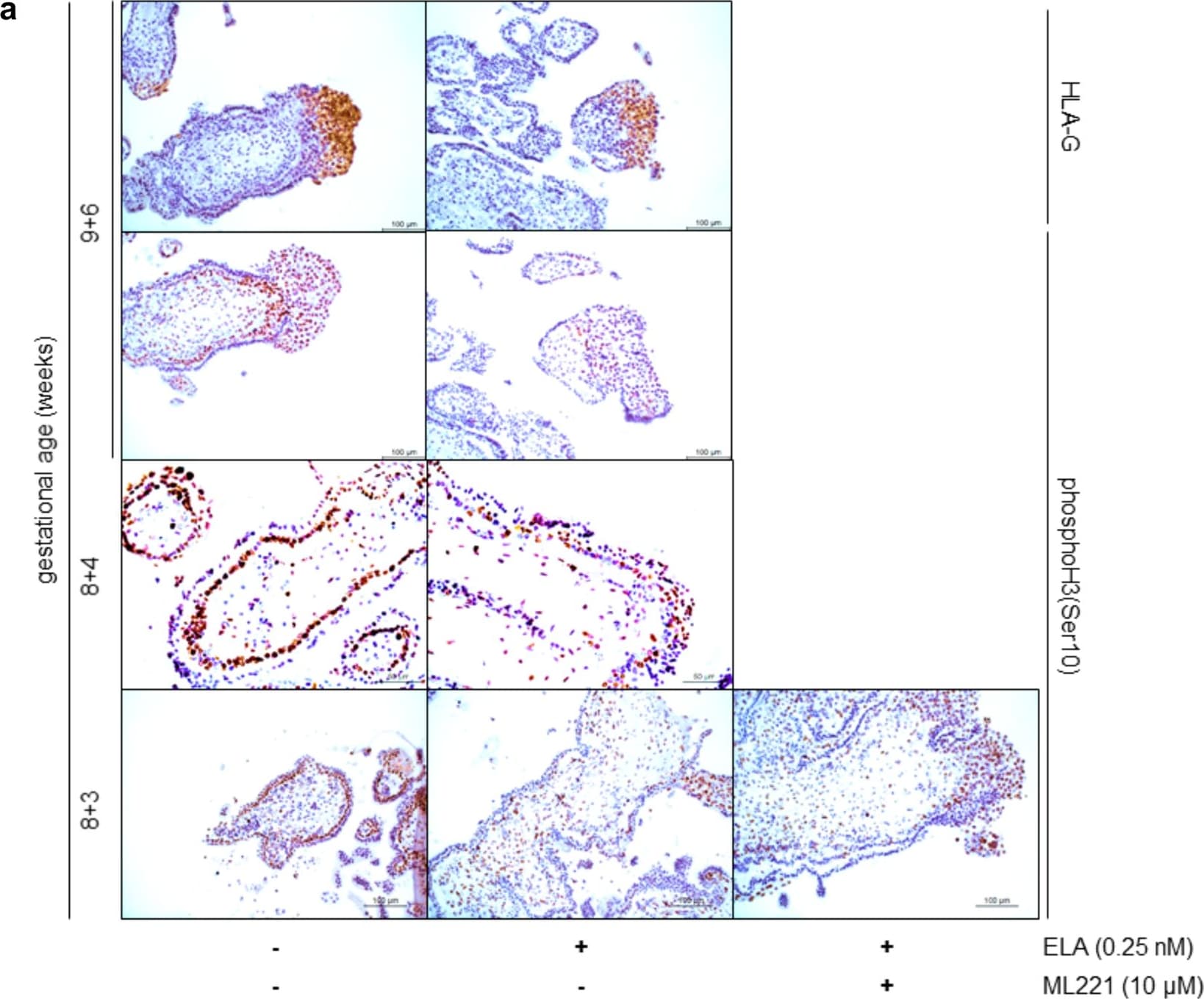
Immunohistochemistry: HLA G Antibody (MEM-G/1) - BSA Free [NB500-302] -
Immunohistochemistry: HLA G Antibody (MEM-G/1) - BSA Free [NB500-302] - ELA affects first trimester placental explant trophoblast proliferation. (a) HLA-G immunostaining (brown color top panel) indicates the location of extravillous trophoblasts. PhosphoH3(Ser10) immunostaining (brown color lower 3 panels) identifies proliferating cells undergoing mitosis. In extravillous trophoblasts of explants treated with ELA proliferation appears to decrease. In control explants almost all villous cytotrophoblasts proliferate, while this is much less apparent in explants treated with ELA. Treatment with ELA in combination with ML221 does not return villous cytotrophoblast proliferation to the level as seen in control explants. (b) Quantifying the percentage of proliferating extravillous trophoblasts observed by immunohistochemistry shows a significant decrease upon treatment with ELA which cannot be rescued by the addition of ML221. P = 0.08 between controls & explants treated with ELA & ML221. n = 12 placentas, 5 explants per treatment. Bars represent mean ± SEM & data was tested with one-way ANOVA followed by Bonferroni multiple comparisons test. *Indicates p < 0.05. (c) Treatment of explants with an ELA neutralizing antibody shows a significant increase of extravillous trophoblast proliferation upon quantifying immunohistochemistry stainings. n = 4 placentas, 5 explants per treatment. Bars represent mean ± SEM & data was tested with a Student’s t-test. *Indicates p < 0.05. Image collected & cropped by CiteAb from the following publication (https://pubmed.ncbi.nlm.nih.gov/31836787), licensed under a CC-BY license. Not internally tested by Novus Biologicals.Applications for HLA G Antibody (MEM-G/1) - BSA Free
Application
Recommended Usage
Immunohistochemistry
1:60 - 1:100
Immunohistochemistry-Paraffin
1:60 - 1:100
Western Blot
1-2 ug/ml
Application Notes
Immunohistochemistry (paraffin sections) incubation: 1 h at RT; positive tissue: human placenta - extravillous cytotrophoblast, heat retrieval in 0.01M citrate buffer (4x2 min. in microwave oven). Use in ICC/IF reported in scientific literature (PMID: 27849611).
Formulation, Preparation, and Storage
Purification
Protein A purified
Formulation
Phosphate buffered saline (PBS), pH 7.4
Format
BSA Free
Preservative
15mM Sodium Azide
Concentration
1 mg/ml
Shipping
The product is shipped with polar packs. Upon receipt, store it immediately at the temperature recommended below.
Stability & Storage
Store at 4C. Do not freeze.
Background: HLA-G
Discovered in cytotrophoblasts, HLA-G is involved in fetal maternal immune tolerance and some studies have linked its downregulation with severe preeclampsia (3). HLA-G mediated immune suppression works by impeding cell proliferation, differentiation, cytotoxicity, cytokine secretion and chemotaxis; and activation of regulatory cells and MDSCs or M2 type macrophage. HLA-G is constitutively expressed in immune-privileged sites such as pancreatic islets, mesenchymal stem cells (MSCs) and the cornea. Upregulation of HLA-G occurs in pathological states including cancer, allo-transplantations, viral infections, autoimmune and inflammatory diseases (4). In cancer, HLA-G expression is induced by hypoxia and has been correlated with advanced tumor stage, tumor metastasis, and poor therapeutic response and survival. HLA-G is an attractive tumor associated-antigen (TAA) for immunotherapy and is considered a major immune checkpoint (ICP).
References
1. Loustau, M., Anna, F., Drean, R., Lecomte, M., Langlade-Demoyen, P., & Caumartin, J. (2020). HLA-G Neo-Expression on Tumors. Front Immunol, 11:1685. PMID: 32922387
2. Lin A, Yan WH. (2018) Heterogeneity of HLA-G Expression in Cancers: Facing the Challenges. Front Immunol. 9:2164. PMID: 30319626
3. Djurisic S, Hviid TV. (2014) HLA Class Ib Molecules and Immune Cells in Pregnancy and Preeclampsia. Front Immunol. 5:652. PMID: 25566263
4. Lila N, Rouas-Freiss N, Dausset J, Carpentier A, Carosella ED. (2001) Soluble HLA-G protein secreted by allo-specific CD4+ T cells suppresses the allo-proliferative response: a CD4+ T cell regulatory mechanism. Proc Natl Acad Sci U S A. 98(21):12150-12155. PMID: 11572934
Long Name
Major Histocompatibility Complex, Class I, G
Alternate Names
HLA G, HLA-6.0, HLAG, MHC Class I Antigen G, MHC-G
Gene Symbol
HLA-G
Additional HLA-G Products
Product Documents for HLA G Antibody (MEM-G/1) - BSA Free
Certificate of Analysis
To download a Certificate of Analysis, please enter a lot or batch number in the search box below.
Product Specific Notices for HLA G Antibody (MEM-G/1) - BSA Free
This product is for research use only and is not approved for use in humans or in clinical diagnosis. Primary Antibodies are guaranteed for 1 year from date of receipt.
Related Research Areas
Citations for HLA G Antibody (MEM-G/1) - BSA Free
 Powered by Bioz
Customer Reviews for HLA G Antibody (MEM-G/1) - BSA Free
There are currently no reviews for this product. Be the first to review HLA G Antibody (MEM-G/1) - BSA Free and earn rewards!
Have you used HLA G Antibody (MEM-G/1) - BSA Free?
Submit a review and receive an Amazon gift card!
$25/€18/£15/$25CAN/¥2500 for a review with an image
$10/€7/£6/$10CAN/¥1110 for a review without an image
Submit a review
Protocols
Find general support by application which include: protocols, troubleshooting, illustrated assays, videos and webinars.
- Antigen Retrieval Protocol (PIER)
- Antigen Retrieval for Frozen Sections Protocol
- Appropriate Fixation of IHC/ICC Samples
- Cellular Response to Hypoxia Protocols
- Chromogenic IHC Staining of Formalin-Fixed Paraffin-Embedded (FFPE) Tissue Protocol
- Chromogenic Immunohistochemistry Staining of Frozen Tissue
- ClariTSA™ Fluorophore Kits
- Detection & Visualization of Antibody Binding
- Fluorescent IHC Staining of Frozen Tissue Protocol
- Graphic Protocol for Heat-induced Epitope Retrieval
- Graphic Protocol for the Preparation and Fluorescent IHC Staining of Frozen Tissue Sections
- Graphic Protocol for the Preparation and Fluorescent IHC Staining of Paraffin-embedded Tissue Sections
- Graphic Protocol for the Preparation of Gelatin-coated Slides for Histological Tissue Sections
- ICC Cell Smear Protocol for Suspension Cells
- ICC Immunocytochemistry Protocol Videos
- ICC for Adherent Cells
- IHC Sample Preparation (Frozen sections vs Paraffin)
- Immunocytochemistry (ICC) Protocol
- Immunocytochemistry Troubleshooting
- Immunofluorescence of Organoids Embedded in Cultrex Basement Membrane Extract
- Immunofluorescent IHC Staining of Formalin-Fixed Paraffin-Embedded (FFPE) Tissue Protocol
- Immunohistochemistry (IHC) and Immunocytochemistry (ICC) Protocols
- Immunohistochemistry Frozen Troubleshooting
- Immunohistochemistry Paraffin Troubleshooting
- Preparing Samples for IHC/ICC Experiments
- Preventing Non-Specific Staining (Non-Specific Binding)
- Primary Antibody Selection & Optimization
- Protocol for Heat-Induced Epitope Retrieval (HIER)
- Protocol for Making a 4% Formaldehyde Solution in PBS
- Protocol for VisUCyte™ HRP Polymer Detection Reagent
- Protocol for the Fluorescent ICC Staining of Cell Smears - Graphic
- Protocol for the Fluorescent ICC Staining of Cultured Cells on Coverslips - Graphic
- Protocol for the Preparation & Fixation of Cells on Coverslips
- Protocol for the Preparation and Chromogenic IHC Staining of Frozen Tissue Sections
- Protocol for the Preparation and Chromogenic IHC Staining of Frozen Tissue Sections - Graphic
- Protocol for the Preparation and Chromogenic IHC Staining of Paraffin-embedded Tissue Sections
- Protocol for the Preparation and Chromogenic IHC Staining of Paraffin-embedded Tissue Sections - Graphic
- Protocol for the Preparation and Fluorescent ICC Staining of Cells on Coverslips
- Protocol for the Preparation and Fluorescent ICC Staining of Non-adherent Cells
- Protocol for the Preparation and Fluorescent ICC Staining of Stem Cells on Coverslips
- Protocol for the Preparation and Fluorescent IHC Staining of Frozen Tissue Sections
- Protocol for the Preparation and Fluorescent IHC Staining of Paraffin-embedded Tissue Sections
- Protocol for the Preparation of Gelatin-coated Slides for Histological Tissue Sections
- Protocol for the Preparation of a Cell Smear for Non-adherent Cell ICC - Graphic
- R&D Systems Quality Control Western Blot Protocol
- TUNEL and Active Caspase-3 Detection by IHC/ICC Protocol
- The Importance of IHC/ICC Controls
- Troubleshooting Guide: Immunohistochemistry
- Troubleshooting Guide: Western Blot Figures
- Western Blot Conditions
- Western Blot Protocol
- Western Blot Protocol for Cell Lysates
- Western Blot Troubleshooting
- Western Blot Troubleshooting Guide
- View all Protocols, Troubleshooting, Illustrated assays and Webinars
Loading...