
Hormone-sensitive Lipase/HSL [p Ser660] Antibody - Azide and BSA Free
Novus Biologicals | Catalog # NBP3-05459

Scientific images may be resized, cropped, or adjusted for brightness or contrast for presentation purposes. These updates are limited to presentation and do not affect the underlying scientific interpretation of the data. Where available, additional source data may be provided upon request.
Loading...
Key Product Details
Validated by
Biological Validation
Species Reactivity
Human, Mouse
Applications
Western Blot
Label
Unconjugated
Antibody Source
Polyclonal Rabbit IgG
Format
Azide and BSA Free
Loading...
Product Specifications
Immunogen
A synthetic phosphorylated peptide around S660 of human Hormone-sensitive Lipase/HSL (NP_005348.2). QTSRS
Modification
p Ser660
Clonality
Polyclonal
Host
Rabbit
Isotype
IgG
Scientific Data Images for Hormone-sensitive Lipase/HSL [p Ser660] Antibody - Azide and BSA Free
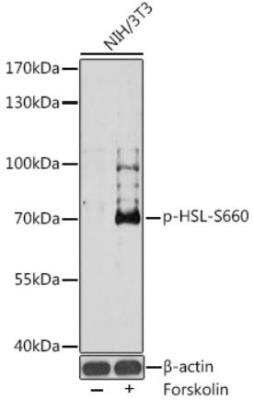
Western Blot: Hormone-sensitive Lipase/HSL [p Ser660] AntibodyAzide and BSA Free [NBP3-05459]
Western Blot: Hormone-sensitive Lipase/HSL [p Ser660] Antibody [NBP3-05459] - Analysis of extracts of NIH/3T3 cells, using Phospho-HSL-S660 antibody at 1:1000 dilution.NIH/3T3 cells were treated by Forskolin (10 uM) at 37c for 30 minutes after serum-starvation overnight. Secondary antibody: HRP Goat Anti-Rabbit IgG (H+L) at 1:10000 dilution. Lysates/proteins: 25ug per lane. Blocking buffer: 3% BSA.Detection: ECL Basic Kit. Exposure time: 180s.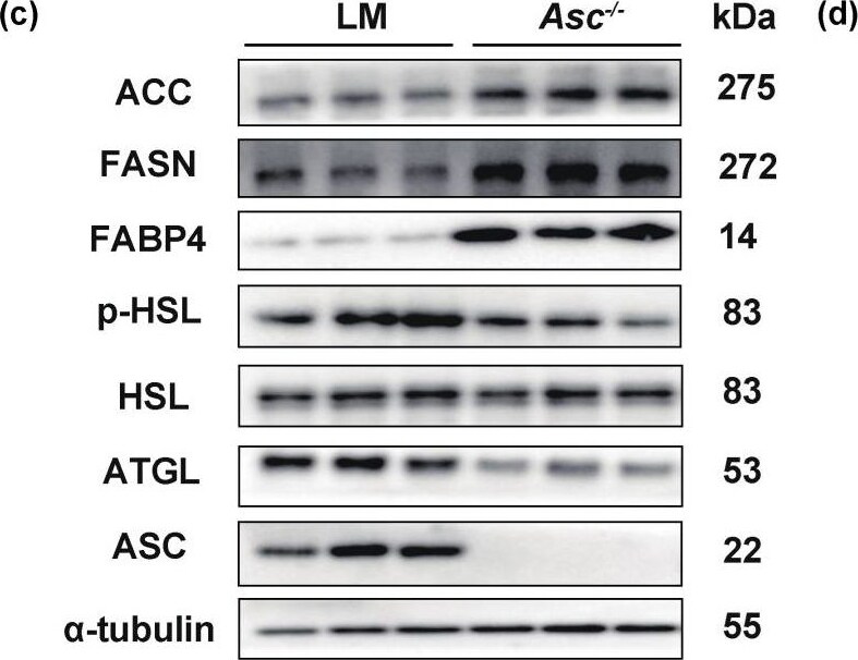
Western Blot: Hormone-sensitive Lipase/HSL [p Ser660] Antibody - Azide and BSA Free [NBP3-05459] -
Ablation of ASC in stromal vascular fractions (SVF) cells improved the lipogenesis but impaired lipolysis. (a–d) Primary SAT SVF cells of LM and Asc−/− mice (Red-LM mice, Blue-Asc−/− mice). (a) Oil Red O staining of the 6th day under lipogenesis inducement treated primary SAT SVF cells, separated from LM and Asc−/− mice, scale bar 200 um. (b) The expression of lipogenesis genes was analyzed by RT-qPCR. (c) Expression of lipogenesis (ACC, FASN, FABP4) and lipolysis (p-HSL, HSL, ATGL) protein in indicated cells. (d–h) Primary SAT SVF cells separated from WT mice were transfected with siRNA targeting for ASC on the 4th day (Blue-NC, Yellow-si Asc). (d,e) mRNA and protein level of ASC in indicated cells. (f) Oil Red O staining of the 6th day under lipogenesis inducement treated in indicated cells, scale bar 200 um. (g) Expression of lipogenesis genes in indicated cells. (h) Expression of lipogenesis (ACC, FASN, FABP4) and lipolysis (p-HSL, HSL, ATGL) protein in indicated cells. Data are presented as means +/- SD from three independent experiments. * p < 0.05 and ** p < 0.01. Image collected and cropped by CiteAb from the following open publication (https://pubmed.ncbi.nlm.nih.gov/36077447), licensed under a CC-BY license. Not internally tested by Novus Biologicals.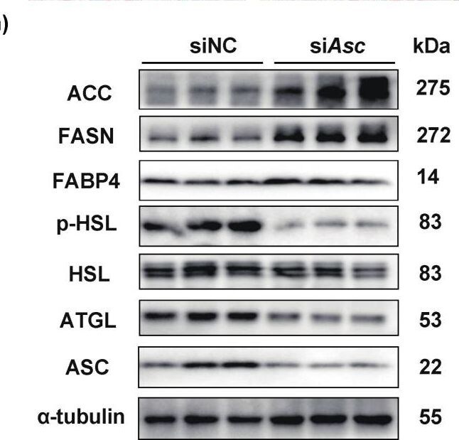
Western Blot: Hormone-sensitive Lipase/HSL [p Ser660] Antibody - Azide and BSA Free [NBP3-05459] -
Ablation of ASC in stromal vascular fractions (SVF) cells improved the lipogenesis but impaired lipolysis. (a–d) Primary SAT SVF cells of LM and Asc−/− mice (Red-LM mice, Blue-Asc−/− mice). (a) Oil Red O staining of the 6th day under lipogenesis inducement treated primary SAT SVF cells, separated from LM and Asc−/− mice, scale bar 200 um. (b) The expression of lipogenesis genes was analyzed by RT-qPCR. (c) Expression of lipogenesis (ACC, FASN, FABP4) and lipolysis (p-HSL, HSL, ATGL) protein in indicated cells. (d–h) Primary SAT SVF cells separated from WT mice were transfected with siRNA targeting for ASC on the 4th day (Blue-NC, Yellow-si Asc). (d,e) mRNA and protein level of ASC in indicated cells. (f) Oil Red O staining of the 6th day under lipogenesis inducement treated in indicated cells, scale bar 200 um. (g) Expression of lipogenesis genes in indicated cells. (h) Expression of lipogenesis (ACC, FASN, FABP4) and lipolysis (p-HSL, HSL, ATGL) protein in indicated cells. Data are presented as means +/- SD from three independent experiments. * p < 0.05 and ** p < 0.01. Image collected and cropped by CiteAb from the following open publication (https://pubmed.ncbi.nlm.nih.gov/36077447), licensed under a CC-BY license. Not internally tested by Novus Biologicals.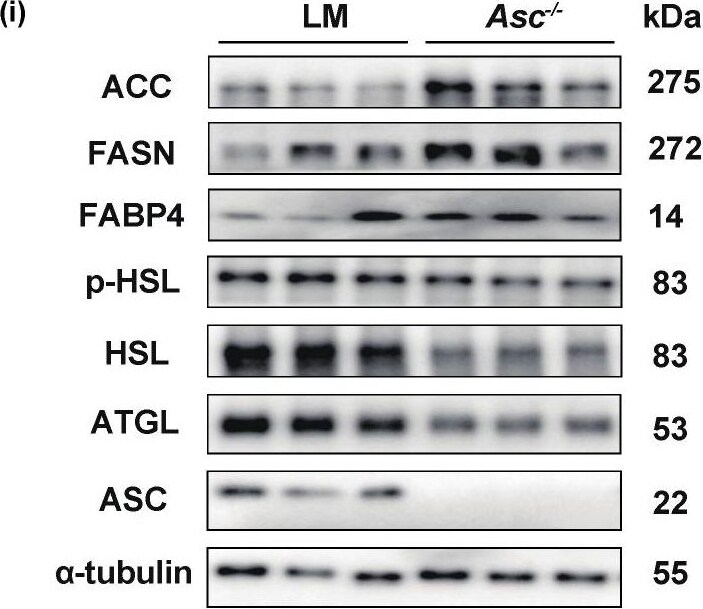
Western Blot: Hormone-sensitive Lipase/HSL [p Ser660] Antibody - Azide and BSA Free [NBP3-05459] -
Increased lipogenesis and decreased lipolysis were observed in subcutaneous adipose tissue of Asc−/− mice fed with HFD. (a) Body weight change of LM (n = 8/group) and Asc knockout (Asc−/−) mice (n = 10/group), fed with HFD (12 weeks). (b) SAT groups are as described in (a). (c) Fat index (ratio of SAT weight to whole body weight of indicated mice. (d) H&E and quantification of SAT cell size, scale bar 50 um. (e,f) GTT and ITT analysis of indicated mice. (g) In indicated mice, plasma concentrations of triglyceride (TG), total cholesterol (TC), and free fatty acids (NEFA) at baseline. (h) mRNA expression of lipogenesis genes in SAT from indicated mice. (i) Western blot analysis of lipogenesis (ACC, FASN, FABP4) and lipolysis (p-HSL, HSL, ATGL) proteins in SAT. All the mice were male and fed with HFD for 12 weeks if not indicated otherwise. (n = 8–10/group) (Red-LM mice, Blue-Asc−/− mice). * p < 0.05, ** p < 0.01, *** p < 0.001 and **** p < 0.0001. Image collected and cropped by CiteAb from the following open publication (https://pubmed.ncbi.nlm.nih.gov/36077447), licensed under a CC-BY license. Not internally tested by Novus Biologicals.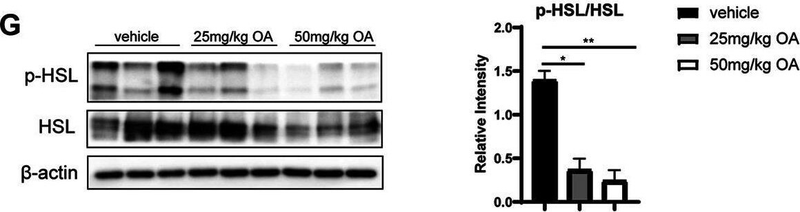
Western Blot: Hormone-sensitive Lipase/HSL [p Ser660] Antibody - Azide and BSA Free [NBP3-05459] -
OA improves glucose and lipid metabolism and alleviates diet-induced IR. (A) Body weight of mice in vehicle, 25 mg/kg OA, 50 mg/kg OA group at the end of the experiment (n = 7). (B) Glucose tolerance test (GTT) in mice after 4 weeks of OA treatment (n = 7). (C) Fasting plasma insulin levels (n = 7). (D) HOMA-IR index (n = 7). (E) Western blots of phospho-Ser473 Akt (p-Akt), and Akt in eWAT of mice. (F) Plasma concentrations of triglyceride, total cholesterol, FFA at baseline (fasted), and Adipo-IR index (n = 5–7). (G) Western blots of phospho-Ser660 HSL (p-HSL), and HSL in the eWAT of mice. *p < 0.05, **p < 0.01, ***p < 0.001, ****p < 0.0001. Image collected and cropped by CiteAb from the following open publication (https://pubmed.ncbi.nlm.nih.gov/34393781), licensed under a CC-BY license. Not internally tested by Novus Biologicals.Applications for Hormone-sensitive Lipase/HSL [p Ser660] Antibody - Azide and BSA Free
Application
Recommended Usage
Western Blot
1:500 - 1:1000
Formulation, Preparation, and Storage
Purification
Affinity purified
Formulation
PBS (pH 7.3), 50% glycerol
Format
Azide and BSA Free
Preservative
0.02% Sodium Azide
Concentration
Please see the vial label for concentration. If unlisted please contact technical services.
Shipping
The product is shipped with polar packs. Upon receipt, store it immediately at the temperature recommended below.
Stability & Storage
Store at -20C. Avoid freeze-thaw cycles.
Background: Hormone-sensitive Lipase/HSL
HSL is the major enzyme responsible for the mobilization of free fatty acids from adopise tissue, and HSL antibodies are useful tools for lipid and metabolism research.
Alternate Names
Hormonesensitive Lipase, HSL, LHS, LIPE
Gene Symbol
LIPE
Additional Hormone-sensitive Lipase/HSL Products
Product Documents for Hormone-sensitive Lipase/HSL [p Ser660] Antibody - Azide and BSA Free
Certificate of Analysis
To download a Certificate of Analysis, please enter a lot or batch number in the search box below.
Product Specific Notices for Hormone-sensitive Lipase/HSL [p Ser660] Antibody - Azide and BSA Free
This product is for research use only and is not approved for use in humans or in clinical diagnosis. Primary Antibodies are guaranteed for 1 year from date of receipt.
Related Research Areas
Customer Reviews for Hormone-sensitive Lipase/HSL [p Ser660] Antibody - Azide and BSA Free
There are currently no reviews for this product. Be the first to review Hormone-sensitive Lipase/HSL [p Ser660] Antibody - Azide and BSA Free and earn rewards!
Have you used Hormone-sensitive Lipase/HSL [p Ser660] Antibody - Azide and BSA Free?
Submit a review and receive an Amazon gift card!
$25/€18/£15/$25CAN/¥2500 for a review with an image
$10/€7/£6/$10CAN/¥1110 for a review without an image
Submit a review
Protocols
Find general support by application which include: protocols, troubleshooting, illustrated assays, videos and webinars.
- Cellular Response to Hypoxia Protocols
- R&D Systems Quality Control Western Blot Protocol
- Troubleshooting Guide: Western Blot Figures
- Western Blot Conditions
- Western Blot Protocol
- Western Blot Protocol for Cell Lysates
- Western Blot Troubleshooting
- Western Blot Troubleshooting Guide
- View all Protocols, Troubleshooting, Illustrated assays and Webinars
Loading...