

Scientific images may be resized, cropped, or adjusted for brightness or contrast for presentation purposes. These updates are limited to presentation and do not affect the underlying scientific interpretation of the data. Where available, additional source data may be provided upon request.
Key Product Details
Species Reactivity
Human
Applications
Western Blot, Immunocytochemistry
Label
Unconjugated
Antibody Source
Polyclonal Goat IgG
Loading...
Product Specifications
Immunogen
Human embryonic kidney cell line HEK293-derived human B7-H7/HHLA2
Met1-Asn344
Accession # Q9UM44
Met1-Asn344
Accession # Q9UM44
Specificity
Detects human B7-H7/HHLA2 in direct ELISAs.
Clonality
Polyclonal
Host
Goat
Isotype
IgG
Scientific Data Images for Human B7-H7/HHLA2 Antibody
Detection of Human B7-H7/HHLA2 by Western Blot.
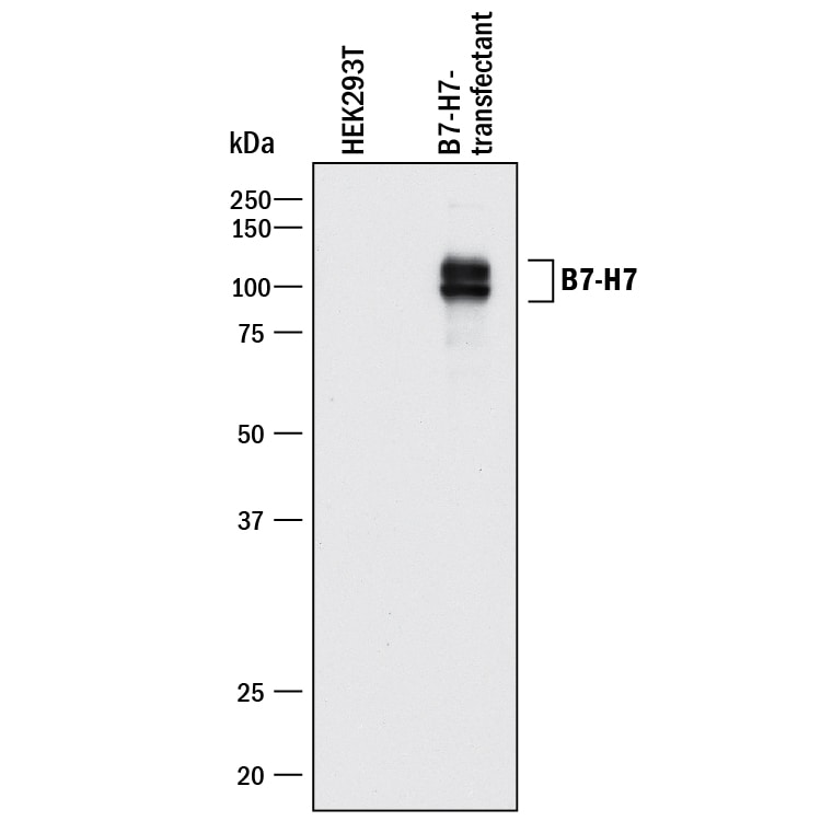
Western blot shows lysates of HEK293T human embryonic kidny cell line transfected with human B7-H7. PVDF membrane was probed with 1 µg/mL of Goat Anti-Human B7-H7/HHLA2 Antigen Affinity-purified Polyclonal Antibody (Catalog # AF8084) followed by HRP-conjugated Anti-Goat IgG Secondary Antibody (Catalog # HAF017). Specific bands were detected for B7-H7/HHLA2 at approximately 90-110 kDa (as indicated). This experiment was conducted under reducing conditions and using Immunoblot Buffer Group 1.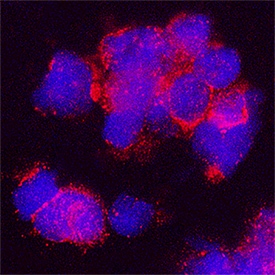
B7-H7/HHLA2 in Human Monocytes.
B7-H7/HHLA2 was detected in immersion fixed human monocytes using Goat Anti-Human B7-H7/HHLA2 Antigen Affinity-purified Polyclonal Antibody (Catalog # AF8084) at 15 µg/mL for 3 hours at room temperature. Cells were stained using the NorthernLights™ 557-conjugated Anti-Goat IgG Secondary Antibody (red; Catalog # NL001) and counterstained with DAPI (blue). Specific staining was localized to cytoplasm. View our protocol for Fluorescent ICC Staining of Non-adherent Cells.Applications for Human B7-H7/HHLA2 Antibody
Application
Recommended Usage
Immunocytochemistry
1-15 µg/mL
Sample: Immersion fixed human monocytes
Sample: Immersion fixed human monocytes
Western Blot
1 µg/mL
Sample: HEK293T human embryonic kidny cell line transfected with human B7-H7
Sample: HEK293T human embryonic kidny cell line transfected with human B7-H7
Formulation, Preparation, and Storage
Purification
Antigen Affinity-purified
Reconstitution
Reconstitute at 0.2 mg/mL in sterile PBS. For liquid material, refer to CoA for concentration.
Loading...
Formulation
Lyophilized from a 0.2 μm filtered solution in PBS with Trehalose. *Small pack size (SP) is supplied either lyophilized or as a 0.2 µm filtered solution in PBS.
Shipping
Lyophilized product is shipped at ambient temperature. Liquid small pack size (-SP) is shipped with polar packs. Upon receipt, store immediately at the temperature recommended below.
Stability & Storage
Use a manual defrost freezer and avoid repeated freeze-thaw cycles.
- 12 months from date of receipt, -20 to -70 °C as supplied.
- 1 month, 2 to 8 °C under sterile conditions after reconstitution.
- 6 months, -20 to -70 °C under sterile conditions after reconstitution.
Calculators
Background: B7-H7/HHLA2
B7‑H7 is constitutively expressed on monocytes and is up-regulated by LPS and IFN-gamma stimulation. It is expressed on LPS/IFN-gamma treated B cells but not on resting B cells (5). B7-H7 binds to cell surface determinants on resting and mature T cells, B cells, and monocytes as well as on immature and mature dendritic cells (5). Soluble B7-H7 inhibits the proliferation of activated CD4+ and CD8+ T cells and their production of IFN-gamma, TNF-alpha, IL-5, IL-10, IL-13, IL-17A, and IL-22 (5).
References
- Zou, W. and L. Chen (2008) Nat. Rev. Immunol. 8:467.
- Bour-Jordan, H. et al. (2011) Immunol. Rev. 241:180.
- Mager, D.L. et al. (1999) Genomics 59:255.
- Flajnik, M.M. et al. (2012) Immunogenetics 64:571.
- Zhao, R. et al. (2013) Proc. Natl. Acad. Sci. USA 110:9879.
Long Name
B7 Homolog 7
Alternate Names
B7H7, HHLA2
Gene Symbol
HHLA2
UniProt
Additional B7-H7/HHLA2 Products
Product Documents for Human B7-H7/HHLA2 Antibody
Certificate of Analysis
To download a Certificate of Analysis, please enter a lot or batch number in the search box below.
Note: Certificate of Analysis not available for kit components.
Product Specific Notices for Human B7-H7/HHLA2 Antibody
For research use only
Related Research Areas
Customer Reviews for Human B7-H7/HHLA2 Antibody
There are currently no reviews for this product. Be the first to review Human B7-H7/HHLA2 Antibody and earn rewards!
Have you used Human B7-H7/HHLA2 Antibody?
Submit a review and receive an Amazon gift card!
$25/€18/£15/$25CAN/¥2500 for a review with an image
$10/€7/£6/$10CAN/¥1110 for a review without an image
Submit a review
Protocols
Find general support by application which include: protocols, troubleshooting, illustrated assays, videos and webinars.
- Appropriate Fixation of IHC/ICC Samples
- Cellular Response to Hypoxia Protocols
- ClariTSA™ Fluorophore Kits
- Detection & Visualization of Antibody Binding
- ICC Cell Smear Protocol for Suspension Cells
- ICC Immunocytochemistry Protocol Videos
- ICC for Adherent Cells
- Immunocytochemistry (ICC) Protocol
- Immunocytochemistry Troubleshooting
- Immunofluorescence of Organoids Embedded in Cultrex Basement Membrane Extract
- Immunohistochemistry (IHC) and Immunocytochemistry (ICC) Protocols
- Preparing Samples for IHC/ICC Experiments
- Preventing Non-Specific Staining (Non-Specific Binding)
- Primary Antibody Selection & Optimization
- Protocol for VisUCyte™ HRP Polymer Detection Reagent
- Protocol for the Fluorescent ICC Staining of Cell Smears - Graphic
- Protocol for the Fluorescent ICC Staining of Cultured Cells on Coverslips - Graphic
- Protocol for the Preparation and Fluorescent ICC Staining of Cells on Coverslips
- Protocol for the Preparation and Fluorescent ICC Staining of Non-adherent Cells
- Protocol for the Preparation and Fluorescent ICC Staining of Stem Cells on Coverslips
- Protocol for the Preparation of a Cell Smear for Non-adherent Cell ICC - Graphic
- R&D Systems Quality Control Western Blot Protocol
- TUNEL and Active Caspase-3 Detection by IHC/ICC Protocol
- The Importance of IHC/ICC Controls
- Troubleshooting Guide: Western Blot Figures
- Western Blot Conditions
- Western Blot Protocol
- Western Blot Protocol for Cell Lysates
- Western Blot Troubleshooting
- Western Blot Troubleshooting Guide
- View all Protocols, Troubleshooting, Illustrated assays and Webinars
Loading...