
Cellular responses to bone morphogenetic proteins (BMPs) have been shown to be mediated by the formation of hetero-oligomeric complexes of the type I and type II serine/threonine kinase receptors. BMP receptor IB (BMPR-IB), also known as activin receptor-like kinase (ALK)-6, is one of seven known type I serine/threonine kinases that are required for the signal transduction of TGF-beta family cytokines. In contrast to the TGF-beta receptor system in which the type I receptor does not bind TGF-beta in the absence of the type II receptor, type I receptors involved in BMP signaling (including BMPR-IA, BMPR-IB/ALK-6, and ActR-I/ALK-2) can independently bind the various BMP family proteins in the absence of type II receptors. Recombinant soluble BMPR-IB binds BMP-4 with high-affinity in solution and is a potent BMP-4 antagonist in vitro. BMPR-IB is expressed in various tissues during embryogenesis. In adult tissues, BMPR-IB is only found in the brain. The extracellular domain of BMPR-IB shares little amino acid sequence identity with the other mammalian ALK type I receptor kinases, but the cysteine residues are conserved. Human and mouse BMPR-IB are highly conserved and share 98% amino acid sequence identity.
Human BMPR-IB/ALK-6 Antibody (477914)
R&D Systems | Catalog # MAB5051


Scientific images may be resized, cropped, or adjusted for brightness or contrast for presentation purposes. These updates are limited to presentation and do not affect the underlying scientific interpretation of the data. Where available, additional source data may be provided upon request.
Key Product Details
Species Reactivity
Validated:
Cited:
Applications
Validated:
Cited:
Label
Antibody Source
Product Specifications
Immunogen
Lys14-Arg126
Accession # O00238
Specificity
Clonality
Host
Isotype
Scientific Data Images for Human BMPR-IB/ALK-6 Antibody (477914)
Detection of BMPR‑IB/ALK‑6 in PC‑3 Human Cell Line by Flow Cytometry.
PC-3 human prostate cancer cell line was stained with Mouse Anti-Human BMPR-IB/ ALK-6 Monoclonal Antibody (Catalog # MAB5051, filled histogram) or isotype control antibody (Catalog # MAB0041, open histogram), followed by Allophycocyanin-conjugated Anti-Mouse IgG F(ab')2Secondary Antibody (Catalog # F0101B).
Detection of BMPR-IB/ALK-6 in Human iPS cells differentiated to Mesoderm by Flow Cytometry.
Human iPS cells differentiated to mesoderm (using Catalog # SC030B) were stained with Mouse Anti-Human BMPR-IB/ALK-6 Monoclonal Antibody (Catalog # MAB5051, filled histogram) or isotype control antibody (Catalog # MAB0041, open histogram) followed by anti-Mouse IgG PE-conjugated secondary antibosy (Catalog # F0101B). View our protocol for Staining Membrane-associated Proteins.
BMPR‑IB/ALK‑6 in PC‑3 Human Cell Line.
BMPR-IB/ALK-6 was detected in immersion fixed PC-3 human prostate cancer cell line using Mouse Anti-Human BMPR-IB/ALK-6 Monoclonal Antibody (Catalog # MAB5051) at 10 µg/mL for 3 hours at room temperature. Cells were stained using the NorthernLights™ 557-conjugated Anti-Mouse IgG Secondary Antibody (red; Catalog # NL007) and counterstained with DAPI (blue). Specific staining was localized to the cytoplasm and cell surface. View our protocol for Fluorescent ICC Staining of Cells on Coverslips.Applications for Human BMPR-IB/ALK-6 Antibody (477914)
CyTOF-ready
Flow Cytometry
Sample: PC‑3 human prostate cancer cell line and human iPS cells differentiated to mesoderm (using Catalog # SC030B)
Immunocytochemistry
Sample: Immersion fixed PC-3 human prostate cancer cell line
Reviewed Applications
Read 1 review rated 5 using MAB5051 in the following applications:
Flow Cytometry Panel Builder
Bio-Techne Knows Flow Cytometry
Save time and reduce costly mistakes by quickly finding compatible reagents using the Panel Builder Tool.
Advanced Features
- Spectra Viewer - Custom analysis of spectra from multiple fluorochromes
- Spillover Popups - Visualize the spectra of individual fluorochromes
- Antigen Density Selector - Match fluorochrome brightness with antigen density

Formulation, Preparation, and Storage
Purification
Reconstitution
Reconstitute at 0.5 mg/mL in sterile PBS. For liquid material, refer to CoA for concentration.
Formulation
Shipping
Stability & Storage
- 12 months from date of receipt, -20 to -70 °C as supplied.
- 1 month, 2 to 8 °C under sterile conditions after reconstitution.
- 6 months, -20 to -70 °C under sterile conditions after reconstitution.
Calculators
Background: BMPR-IB/ALK-6
References
- Kawabata, M. et al. (1998) Cytokine and Growth Factor Reviews 9:49.
- Ebendal, T. et al. (1998) J. Neuroscience Research 51:139.
Long Name
Alternate Names
Gene Symbol
UniProt
Additional BMPR-IB/ALK-6 Products
Product Documents for Human BMPR-IB/ALK-6 Antibody (477914)
Certificate of Analysis
To download a Certificate of Analysis, please enter a lot or batch number in the search box below.
Note: Certificate of Analysis not available for kit components.
Product Specific Notices for Human BMPR-IB/ALK-6 Antibody (477914)
For research use only
Related Research Areas
Citations for Human BMPR-IB/ALK-6 Antibody (477914)
 Powered by Bioz
Customer Reviews for Human BMPR-IB/ALK-6 Antibody (477914) (1)
Have you used Human BMPR-IB/ALK-6 Antibody (477914)?
Submit a review and receive an Amazon gift card!
$25/€18/£15/$25CAN/¥2500 for a review with an image
$10/€7/£6/$10CAN/¥1110 for a review without an image
Submit a review
Customer Images
-
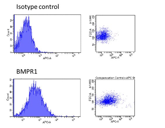
Application: Flow CytometrySample Tested: Cells from neural differentiation of hiPSCsSpecies: HumanVerified Customer | Posted 10/20/2017The cells were stained with 1µl of anti-BMPR1 antibody in 100 µl FACS staining buffer for 30 min at 4C. After washing, the cells were incubated with 1µl of anti-mouse IgG2b-APC antibody in 100 µl FACS staining buffer for 20 min at 4C.

There are no reviews that match your criteria.
Protocols
Find general support by application which include: protocols, troubleshooting, illustrated assays, videos and webinars.
- 7-Amino Actinomycin D (7-AAD) Cell Viability Flow Cytometry Protocol
- Appropriate Fixation of IHC/ICC Samples
- Cellular Response to Hypoxia Protocols
- ClariTSA™ Fluorophore Kits
- Detection & Visualization of Antibody Binding
- Extracellular Membrane Flow Cytometry Protocol
- Flow Cytometry Protocol for Cell Surface Markers
- Flow Cytometry Protocol for Staining Membrane Associated Proteins
- Flow Cytometry Staining Protocols
- Flow Cytometry Troubleshooting Guide
- ICC Cell Smear Protocol for Suspension Cells
- ICC Immunocytochemistry Protocol Videos
- ICC for Adherent Cells
- Immunocytochemistry (ICC) Protocol
- Immunocytochemistry Troubleshooting
- Immunofluorescence of Organoids Embedded in Cultrex Basement Membrane Extract
- Immunohistochemistry (IHC) and Immunocytochemistry (ICC) Protocols
- Intracellular Flow Cytometry Protocol Using Alcohol (Methanol)
- Intracellular Flow Cytometry Protocol Using Detergents
- Intracellular Nuclear Staining Flow Cytometry Protocol Using Detergents
- Intracellular Staining Flow Cytometry Protocol Using Alcohol Permeabilization
- Intracellular Staining Flow Cytometry Protocol Using Detergents to Permeabilize Cells
- Preparing Samples for IHC/ICC Experiments
- Preventing Non-Specific Staining (Non-Specific Binding)
- Primary Antibody Selection & Optimization
- Propidium Iodide Cell Viability Flow Cytometry Protocol
- Protocol for Liperfluo
- Protocol for VisUCyte™ HRP Polymer Detection Reagent
- Protocol for the Characterization of Human Th22 Cells
- Protocol for the Characterization of Human Th9 Cells
- Protocol for the Fluorescent ICC Staining of Cell Smears - Graphic
- Protocol for the Fluorescent ICC Staining of Cultured Cells on Coverslips - Graphic
- Protocol for the Preparation and Fluorescent ICC Staining of Cells on Coverslips
- Protocol for the Preparation and Fluorescent ICC Staining of Non-adherent Cells
- Protocol for the Preparation and Fluorescent ICC Staining of Stem Cells on Coverslips
- Protocol for the Preparation of a Cell Smear for Non-adherent Cell ICC - Graphic
- Protocol: Annexin V and PI Staining by Flow Cytometry
- Protocol: Annexin V and PI Staining for Apoptosis by Flow Cytometry
- TUNEL and Active Caspase-3 Detection by IHC/ICC Protocol
- The Importance of IHC/ICC Controls
- Troubleshooting Guide: Fluorokine Flow Cytometry Kits
- View all Protocols, Troubleshooting, Illustrated assays and Webinars