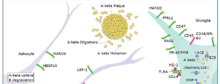
CD36, alternatively known as platelet membrane glycoprotein IV (GPIV), GPIIIb, thrombospondin receptor, collagen receptor, fatty acid translocase (FAT), and scavenger receptor class B, member 3 (SR-B3), is an integral membrane glycoprotein that has multiple physiological functions (1). It is broadly expressed on a variety of cell types including microvascular endothelium, adipocytes, skeletal muscle, epithelial cells of the retina, breast, and intestine, smooth muscle cells, erythroid precursors, platelets, megakaryocytes, dendritic cells, monocytes/macrophages, and microglia (1, 2). As a member of the scavenger receptor family, CD36 is a multiligand pattern recognition receptor that interacts with a large number of structurally dissimilar ligands, including long chain fatty acid (LCFA), advanced glycation end products (AGE), thrombospondin-1, oxidized low-density lipoproteins (oxLDLs), high density lipoprotein (HDL), phosphatidylserine, apoptotic cells, beta -amyloid fibrils (fA beta ), collagens I and IV, and Plasmodium falciparum-infected erythrocytes (3). CD36 is required for the anti-angiogenic effects of thrombospondin-1 in the corneal neovascularization assay (4). It plays a role in lipid metabolism and has been identified as a fatty acid translocase necessary for the binding and transport of LCFA in cells and tissues (5). CD36 has been implicated in the clearance of apoptotic cells and cell debris and has also been shown to mediate the internalization and degradation of a variety of its ligands such as oxLDL, AGE and fA beta (3). Upon ligand binding, CD36 transduces signals that mediate a wide range of pro-inflammatory cellular responses (2). CD36 plays a significant role in the initiation and pathogenesis of chronic inflammatory diseases such as Alzheimer’s disease and atherosclerosis (2, 3). The human CD36 gene encodes a single-chain 472 amino acid residue protein containing both an N- and a C-terminal cytoplasmic tail and an extracellular loop.
Human CD36/SR-B3 Antibody
R&D Systems | Catalog # AF1955

Scientific images may be resized, cropped, or adjusted for brightness or contrast for presentation purposes. These updates are limited to presentation and do not affect the underlying scientific interpretation of the data. Where available, additional source data may be provided upon request.
Key Product Details
Species Reactivity
Validated:
Cited:
Applications
Validated:
Cited:
Label
Antibody Source
Product Specifications
Immunogen
Gly30-Asn439
Accession # P16671
Specificity
Clonality
Host
Isotype
Scientific Data Images for Human CD36/SR-B3 Antibody
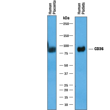
Detection of Human CD36/SR‑B3 by Western Blot.
Western blot shows lysates of human placenta tissue and human platelets. PVDF membrane was probed with 1 µg/mL of Goat Anti-Human CD36/SR-B3 Antigen Affinity-purified Polyclonal Antibody (Catalog # AF1955) followed by HRP-conjugated Anti-Goat IgG Secondary Antibody (Catalog # HAF019). A specific band was detected for CD36/SR-B3 at approximately 85-90 kDa (as indicated). This experiment was conducted under reducing conditions and using Immunoblot Buffer Group 1.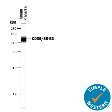
Detection of Human CD36/SR‑B3 by Simple WesternTM.
Simple Western lane view shows lysates of human placenta tissue, loaded at 0.2 mg/mL. A specific band was detected for CD36/SR-B3 at approximately 140 kDa (as indicated) using 10 µg/mL of Goat Anti-Human CD36/SR-B3 Antigen Affinity-purified Polyclonal Antibody (Catalog # AF1955) followed by 1:50 dilution of HRP-conjugated Anti-Goat IgG Secondary Antibody (Catalog # HAF109). This experiment was conducted under reducing conditions and using the 12-230 kDa separation system.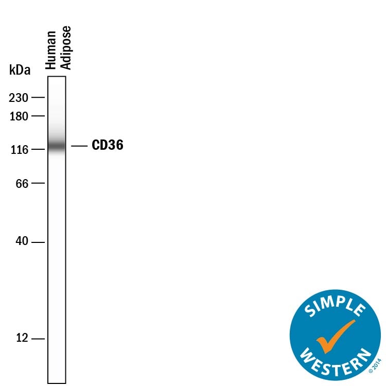
Detection of Human CD36/SR‑B3 by Simple WesternTM.
Simple Western lane view shows lysates of human adipose tissue, loaded at 0.2 mg/mL. A specific band was detected for CD36/SR-B3 at approximately 125 kDa (as indicated) using 50 µg/mL of Goat Anti-Human CD36/SR-B3 Antigen Affinity-purified Polyclonal Antibody (Catalog # AF1955) followed by 1:50 dilution of HRP-conjugated Anti-Goat IgG Secondary Antibody (Catalog # HAF109). This experiment was conducted under reducing conditions and using the 12-230 kDa separation system.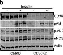
Detection of CD36/SR-B3 by Western Blot
CD36 regulates insulin signalling in primary derived hMEC. (a) Alterations in genes related to PI3K–insulin signalling in control hMECs (CtrlKD, hMECs treated with scrambled siRNA) and CD36KD (KD, hMECs treated with anti-CD36 siRNA), n=5 per group. (b) Western blot of CtrlKD and CD36KD hMECs, showing effect of insulin (100 nmol/l, 5 min) on phosphorylation of Akt and eNOS. (c, d) Densitometry analysis of pAkt/total Akt and of p-eNOS/total eNOS (n=3 p<0.05). (e) Effect of CD36 knockdown on insulin-dependent interaction of Cav-1 and eNOS. (f) Effect of CD36 on regulation of NO production. CtrlKD and CD36KD hMECs were serum starved and subjected to insulin stimulation (100 nmol/l, 5 min). n=4 assays. Western blots are representative of three independent experiments. Data are shown as means ± SD. *p<0.05, ***p<0.005. Ins, insulin Image collected and cropped by CiteAb from the following open publication (https://pubmed.ncbi.nlm.nih.gov/39503770), licensed under a CC-BY license. Not internally tested by R&D Systems.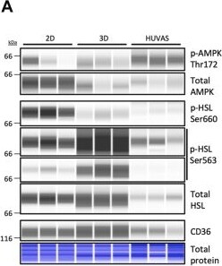
Detection of CD36/SR-B3 by Simple Western
HUVAS adipocytes are characterized by AMPK activation and high fatty acid uptake. A, B: Capillary Western blot analysis of p-AMPK T172, total AMPK, p-HSLS660, p-HSL S563 (shown with 2 contrasts), total HSL, and CD36 as well as quantification of the p-AMPK/AMPK and p-HSL/HSL ratios after normalization to total protein levels (Fig. S6A, S7A) across HUVAS, 2D, and 3D cultures from 3 technical replicates of the same individual on d30. C, D, isoprenaline-induced (C, shown as induced/basal) and basal (D) lipolysis measured by glycerol release across cultures from 8 technical replicates of the same individual. Similar results were obtained from cells from >three different cell donors. E, F: Quantification of the uptake of a BODIPY-labelled long-chain fatty acid tracer (BODIPY-FA) across HUVAS, 3D, and 2D cultures (E), and in HUVAS and 3D cultures in the absence or presence of the CD36 inhibitor SMS121 or vehicle (F). N ≥ 5 throughout; values in (F) are expressed as relative to the mean of HUVAS control to combine data from several experiments and cell donors. G–H: Quantification of CD36 mRNA expression expressed as normalized counts (G) and protein from capillary Western blot analysis (H) after normalization to total protein levels (supplemental Fig. S6A) from 3 technical replicates of the same individual on d30. I–K: Effects of chronic treatment of HUVAS with CD36 inhibitor SMS121 d20-d30 shown as representative confocal images at d30 (I), adipocyte lipid droplet diameter (J), and unilocularity (K). n = 40 cells per spheroid and 2 spheroids per individual. Scale bars: 50 μm. Image collected and cropped by CiteAb from the following open publication (https://pubmed.ncbi.nlm.nih.gov/41850682), licensed under a CC-BY license. Not internally tested by R&D Systems.Applications for Human CD36/SR-B3 Antibody
Simple Western
Sample: Human placenta tissue and Human adipose tissue
Western Blot
Sample: human placenta tissue and human platelets
Formulation, Preparation, and Storage
Purification
Reconstitution
Reconstitute at 0.2 mg/mL in sterile PBS. For liquid material, refer to CoA for concentration.
Formulation
Shipping
Stability & Storage
- 12 months from date of receipt, -20 to -70 °C as supplied.
- 1 month, 2 to 8 °C under sterile conditions after reconstitution.
- 6 months, -20 to -70 °C under sterile conditions after reconstitution.
Calculators
Background: CD36/SR-B3
References
- Febbraio, M. et al. (2001) J. Clin. Invest. 108:785.
- Khoury, J. et al. (2003) J. Exp. Med. 197:1657.
- Husemann, J. et al. (2002) Glia 40:195.
- Armstrong, L and P. Bornstein (2003) Matrix. Biol. 22:63.
- Febbraio M. et al. (1999) J. Biol. Chem. 274:19055.
Long Name
Alternate Names
Gene Symbol
UniProt
Additional CD36/SR-B3 Products
Product Documents for Human CD36/SR-B3 Antibody
Certificate of Analysis
To download a Certificate of Analysis, please enter a lot or batch number in the search box below.
Note: Certificate of Analysis not available for kit components.
Product Specific Notices for Human CD36/SR-B3 Antibody
For research use only
Citations for Human CD36/SR-B3 Antibody
 Powered by Bioz
Customer Reviews for Human CD36/SR-B3 Antibody
There are currently no reviews for this product. Be the first to review Human CD36/SR-B3 Antibody and earn rewards!
Have you used Human CD36/SR-B3 Antibody?
Submit a review and receive an Amazon gift card!
$25/€18/£15/$25CAN/¥2500 for a review with an image
$10/€7/£6/$10CAN/¥1110 for a review without an image
Submit a review
Protocols
Find general support by application which include: protocols, troubleshooting, illustrated assays, videos and webinars.
- Cellular Response to Hypoxia Protocols
- R&D Systems Quality Control Western Blot Protocol
- Troubleshooting Guide: Western Blot Figures
- Western Blot Conditions
- Western Blot Protocol
- Western Blot Protocol for Cell Lysates
- Western Blot Troubleshooting
- Western Blot Troubleshooting Guide
- View all Protocols, Troubleshooting, Illustrated assays and Webinars