
Human CLEC-2/CLEC1B Antibody (219150)
R&D Systems | Catalog # MAB1718

Scientific images may be resized, cropped, or adjusted for brightness or contrast for presentation purposes. These updates are limited to presentation and do not affect the underlying scientific interpretation of the data. Where available, additional source data may be provided upon request.
Key Product Details
Species Reactivity
Validated:
Human
Cited:
Human
Applications
Validated:
Western Blot
Cited:
Western Blot, Neutralization, Flow Cytometry
Label
Unconjugated
Antibody Source
Monoclonal Mouse IgG2B Clone # 219150
Loading...
Product Specifications
Immunogen
NS0-derived recombinant human CLEC-2 extracellular domain
Gln58-Pro229
Accession # AAF36777
Gln58-Pro229
Accession # AAF36777
Specificity
Detects human CLEC-2/CLEC1B in direct ELISAs and Western blots. In direct ELISAs, no cross-reactivity with recombinant humanCLEC-1 is observed.
Clonality
Monoclonal
Host
Mouse
Isotype
IgG2B
Scientific Data Images for Human CLEC-2/CLEC1B Antibody (219150)
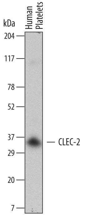
Detection of Human CLEC-2/CLEC1B by Western Blot.
Western blot shows lysates of human platelets. PVDF Membrane was probed with 2 µg/mL of Mouse Anti-Human CLEC-2/CLEC1B Monoclonal Antibody (Catalog # MAB1718) followed by HRP-conjugated Anti-Mouse IgG Secondary Antibody (Catalog # HAF007). A specific band was detected for CLEC-2/CLEC1B at approximately 35 kDa (as indicated). This experiment was conducted under reducing conditions and using Immunoblot Buffer Group 5.Applications for Human CLEC-2/CLEC1B Antibody (219150)
Application
Recommended Usage
Western Blot
2 µg/mL
Sample: Human platelets
Sample: Human platelets
Formulation, Preparation, and Storage
Purification
Protein A or G purified from hybridoma culture supernatant
Reconstitution
Reconstitute at 0.5 mg/mL in sterile PBS. For liquid material, refer to CoA for concentration.
Loading...
Formulation
Lyophilized from a 0.2 μm filtered solution in PBS with Trehalose. *Small pack size (SP) is supplied either lyophilized or as a 0.2 µm filtered solution in PBS.
Shipping
Lyophilized product is shipped at ambient temperature. Liquid small pack size (-SP) is shipped with polar packs. Upon receipt, store immediately at the temperature recommended below.
Stability & Storage
Use a manual defrost freezer and avoid repeated freeze-thaw cycles.
- 12 months from date of receipt, -20 to -70 °C as supplied.
- 1 month, 2 to 8 °C under sterile conditions after reconstitution.
- 6 months, -20 to -70 °C under sterile conditions after reconstitution.
Calculators
Background: CLEC-2/CLEC1B
C‑type lectin-like family of receptors, CLEC-2's CRD lacks the amino acid residues that are crucial for Ca2+-dependent carbohydrate binding, making it a
non‑classical C-type lectin receptor (1, 6). A splicing variant at aa 22-55 produces two isoforms for CLEC-2. Isoform 1 is the longer protein, and in isoform 2, an alanine residue is substituted for aa 22-55. Human CLEC-2 shares 63% aa sequence identity with mouse CLEC-2. CLEC-2 is expressed preferentially in liver, and is also detected in myeloid cells (monocytes, dendritic cells, and granulocytes) (1), platelets, and megakaryocytes (4). CLEC-2 is the receptor for the
platelet-aggregating snake venom protein rhodocytin (3-4) and the molecule podoplanin, a transmembrane sialoglycoprotein that, when bound to CLEC-2, is involved in platelet aggregation, tumor metastasis, and lymphatic vessel formation (2, 7). CLEC-2 has also been shown to enhance infectivity of HIV-1 by mediating HIV-1 attachment and transfer by CLEC-2 transfected cells and platelets (8).
References
-
Colonna, M. et al. (2000) Eur. J. Immunol. 30:697.
- Christou, C.M. et al. (2008) Biochem. J. 411:133.
- Watson, A.A. et al. (2007) J. Biol. Chem. 282:3165.
- Suzuki-Inoue, K. et al. (2006) Blood 107:542.
- Fuller, G.L. et al. (2007) J. Biol. Chem. 282:12397.
- Weis, W.I. et al. (1998) Immunol. Rev. 163:19.
- Suzuki-Inoue, K. et al. (2007) J. Biol. Chem. 282:25993.
- Chaipan, C. et al. (2006) J. Virol. 80:8951.
Long Name
C-type Lectin-like Receptor 2
Alternate Names
CLEC1B, CLEC2
Gene Symbol
CLEC1B
UniProt
Additional CLEC-2/CLEC1B Products
Product Documents for Human CLEC-2/CLEC1B Antibody (219150)
Certificate of Analysis
To download a Certificate of Analysis, please enter a lot or batch number in the search box below.
Note: Certificate of Analysis not available for kit components.
Product Specific Notices for Human CLEC-2/CLEC1B Antibody (219150)
For research use only
Citations for Human CLEC-2/CLEC1B Antibody (219150)
 Powered by Bioz
Customer Reviews for Human CLEC-2/CLEC1B Antibody (219150)
There are currently no reviews for this product. Be the first to review Human CLEC-2/CLEC1B Antibody (219150) and earn rewards!
Have you used Human CLEC-2/CLEC1B Antibody (219150)?
Submit a review and receive an Amazon gift card!
$25/€18/£15/$25CAN/¥2500 for a review with an image
$10/€7/£6/$10CAN/¥1110 for a review without an image
Submit a review
Protocols
Find general support by application which include: protocols, troubleshooting, illustrated assays, videos and webinars.
- Cellular Response to Hypoxia Protocols
- R&D Systems Quality Control Western Blot Protocol
- Troubleshooting Guide: Western Blot Figures
- Western Blot Conditions
- Western Blot Protocol
- Western Blot Protocol for Cell Lysates
- Western Blot Troubleshooting
- Western Blot Troubleshooting Guide
- View all Protocols, Troubleshooting, Illustrated assays and Webinars
Loading...