
CXCL9, a member of the alpha subfamily of chemokines that lack the ELR domain, was initially identified as a lymphokine-activated gene in mouse macrophages. Human CXCL9 was subsequently cloned using mouse MIG cDNA as a probe. The CXCL9 gene is induced in macrophages and in primary glial cells of the central nervous system specifically in response to IFN-gamma. CXCL9 has been shown to be a chemoattractant for activated T-lymphocytes and TIL but not for neutrophils or monocytes. The human CXCL9 cDNA encodes a 125 amino acid (aa) residue precursor protein with a 22 aa residue signal peptide that is cleaved to yield a 103 aa residue mature protein. CXCL9 has an extended carboxy-terminus containing greater than 50% basic aa residues and is larger than most other chemokines. The carboxy-terminal residues of CXCL9 are prone to proteolytic cleavage resulting in size heterogeneity of natural and recombinant CXCL9. CXCL9 with large carboxy-terminal deletions have been shown to have diminished activity in the calcium flux assay. A chemokine receptor (CXCR3) specific for CXCL9 and IP-10 has been cloned and shown to be highly expressed in IL-2-activated T-lymphocytes. The E. coli-expressed CXCL9 preparations produced at R&D Systems have been shown to contain greater than 80% full length CXCL9.
Human CXCL9/MIG Antibody (49106)
R&D Systems | Catalog # MAB392


Scientific images may be resized, cropped, or adjusted for brightness or contrast for presentation purposes. These updates are limited to presentation and do not affect the underlying scientific interpretation of the data. Where available, additional source data may be provided upon request.
Key Product Details
Validated by
Species Reactivity
Validated:
Cited:
Applications
Validated:
Cited:
Label
Antibody Source
Product Specifications
Immunogen
Thr23-Thr125
Accession # Q07325
Specificity
Clonality
Host
Isotype
Endotoxin Level
Scientific Data Images for Human CXCL9/MIG Antibody (49106)
Chemotaxis Induced by CXCL9/MIG and Neutralization by Human CXCL9/MIG Antibody.
Recombinant Human CXCL9/MIG (Catalog # 392-MG) chemoattracts the BaF3 mouse pro-B cell line transfected with mouse CXCR3 in a dose-dependent manner (orange line). The amount of cells that migrated through to the lower chemotaxis chamber was measured by Resazurin (Catalog # AR002). Chemotaxis elicited by Recombinant Human CXCL9/MIG (0.25 µg/mL) is neutralized (green line) by increasing concentrations of Mouse Anti-Human CXCL9/MIG Monoclonal Antibody (Catalog # MAB392). The ND50 is typically 0.5-4 µg/mL.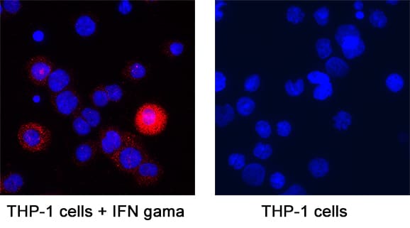
CXCL9/MIG in THP‑1 Human Cell Line.
CXCL9/MIG was detected in immersion fixed THP‑1 human acute monocytic leukemia cell line treated with IFN gamma (positive staining) and THP‑1 human acute monocytic leukemia cell line (untreated; negative staining) using Mouse Anti-Human CXCL9/MIG Monoclonal Antibody (Catalog # MAB392) at 25 µg/mL for 3 hours at room temperature. Cells were stained using the NorthernLights™ 557-conjugated Anti-Mouse IgG Secondary Antibody (red; NL007) and counterstained with DAPI (blue). Specific staining was localized to cytoplasm. Staining was performed using our protocol for Fluorescent ICC Staining of Non-adherent Cells.
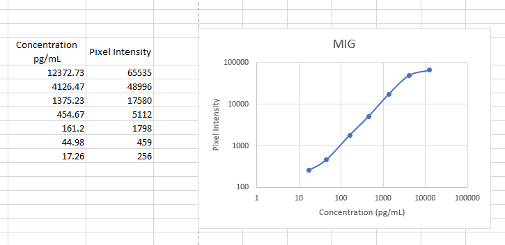
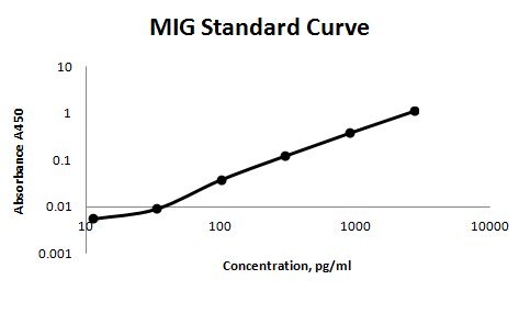
Human CXCL9 / MIG ELISA Standard Curve
Recombinant Human CXCL9/MIG (Catalog # 392-MG) was serially diluted and captured by Mouse Anti-Human CXCL9/MIG Monoclonal Antibody (Catalog # MAB392) coated on a Clear Polystyrene Microplate (Catalog # DY990). Goat Anti-Human CXCL9/MIG Antigen Affinity-purified Polyclonal Antibody (Catalog # AF392) was biotinylated and incubated with the protein captured on the plate. Detection of the standard curve was achieved by incubating Streptavidin-HRP (Catalog # DY998)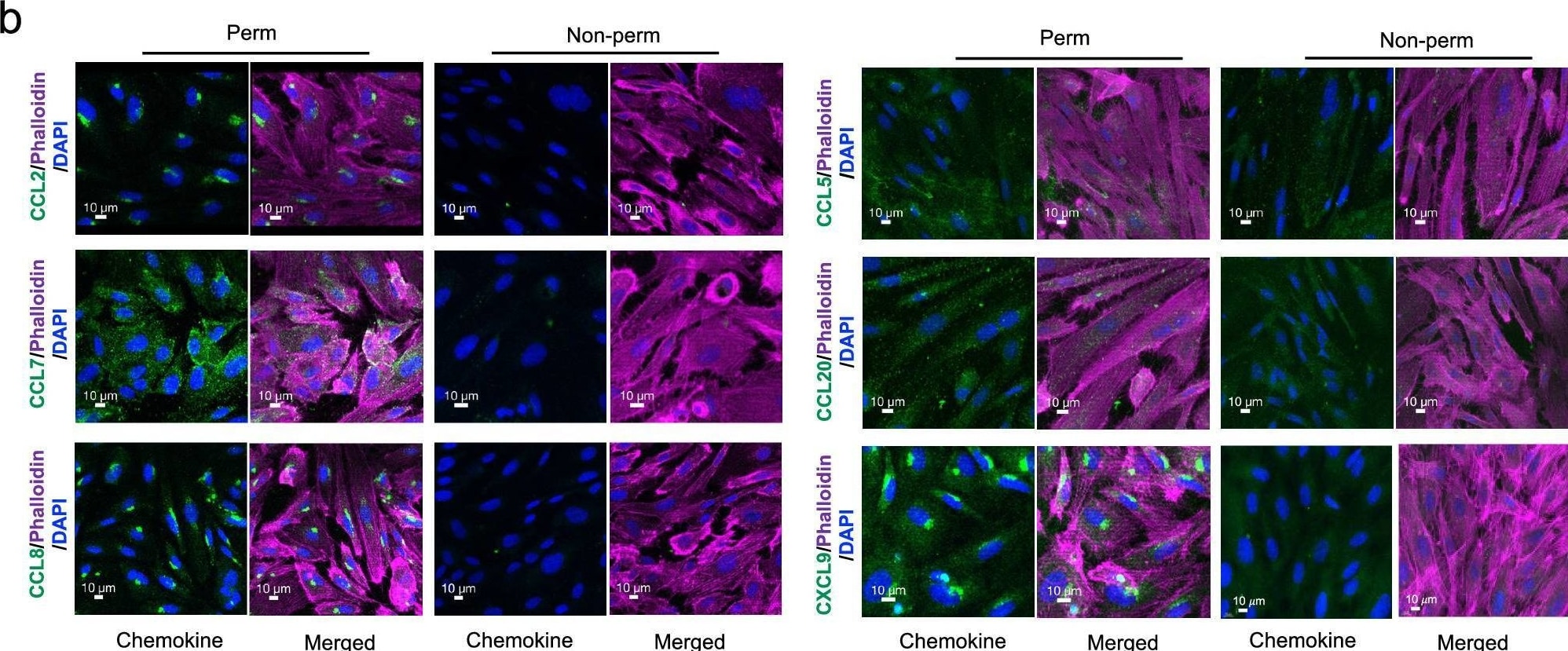
Detection of CXCL9/MIG by Immunohistochemistry
CCR2 ligands are secreted but do not bind to HUVECs.a Culture medium collected from the upper and lower wells of transwells with HUVEC monolayers either not stimulated (NS) or stimulated with TNF-alpha + IFN-gamma were assayed for CCL2, CCL7, and CCL8 by ELISA (n = 3 separate experiments). Each symbol shows data from one experiment, with assays done in duplicate, and bars indicate means ± SEM. b Confocal microscopy images at × 40 magnification of permeabilized and non-permeabilized TNF-alpha + IFN-gamma -stimulated HUVECs immunostained for CCL2, CCL7, CCL8, CCL5, CCL20, and CXCL9 (green) and stained using phalloidin for polymerized actin (magenta) and DAPI for nuclei (blue). The scale bars indicate 10 µm. Images are representative of three experiments. c Biotinylated CCL5 (0.1 μM) and CCL2 (1 μM) were incubated with CHO-K1 cells and the heparan-sulfate deficient cell line, D-677. Chemokine binding was detected using streptavidin-phycoerythrin (PE) and flow cytometry. Data shown are representative of three separate experiments. Image collected and cropped by CiteAb from the following open publication (https://www.nature.com/articles/s41467-025-57002-6), licensed under a CC-BY license. Not internally tested by R&D Systems.Applications for Human CXCL9/MIG Antibody (49106)
CyTOF-ready
Immunocytochemistry
Sample: Immersion fixed THP‑1 human acute monocytic leukemia cell line treated with IFN gamma
Intracellular Staining by Flow Cytometry
Sample: THP-1 cells treated with Recombinant Human IFN‑ gamma (Catalog # 285‑IF), fixed with paraformaldehyde, and permeabilized with saponin
Western Blot
Sample: Recombinant Human CXCL9/MIG (Catalog # 392-MG)
under non-reducing conditions only
Neutralization
Human CXCL9/MIG Sandwich Immunoassay
Reviewed Applications
Read 3 reviews rated 5 using MAB392 in the following applications:
Flow Cytometry Panel Builder
Bio-Techne Knows Flow Cytometry
Save time and reduce costly mistakes by quickly finding compatible reagents using the Panel Builder Tool.
Advanced Features
- Spectra Viewer - Custom analysis of spectra from multiple fluorochromes
- Spillover Popups - Visualize the spectra of individual fluorochromes
- Antigen Density Selector - Match fluorochrome brightness with antigen density

Formulation, Preparation, and Storage
Purification
Reconstitution
Reconstitute at 0.5 mg/mL in sterile PBS. For liquid material, refer to CoA for concentration.
Formulation
Shipping
Stability & Storage
- 12 months from date of receipt, -20 to -70 °C as supplied.
- 1 month, 2 to 8 °C under sterile conditions after reconstitution.
- 6 months, -20 to -70 °C under sterile conditions after reconstitution.
Calculators
Background: CXCL9/MIG
References
- Loetscher, M. et al. (1996) J. Exp. Med. 184:963.
- Liao, F. et al. (1995) J. Exp. Med. 182:1301.
- Vanguri, P. (1995) J. Neuroimmunol. 56:35.
Alternate Names
Gene Symbol
UniProt
Additional CXCL9/MIG Products
Product Documents for Human CXCL9/MIG Antibody (49106)
Certificate of Analysis
To download a Certificate of Analysis, please enter a lot or batch number in the search box below.
Note: Certificate of Analysis not available for kit components.
Product Specific Notices for Human CXCL9/MIG Antibody (49106)
For research use only
Citations for Human CXCL9/MIG Antibody (49106)
 Powered by Bioz
Customer Reviews for Human CXCL9/MIG Antibody (49106) (3)
Have you used Human CXCL9/MIG Antibody (49106)?
Submit a review and receive an Amazon gift card!
$25/€18/£15/$25CAN/¥2500 for a review with an image
$10/€7/£6/$10CAN/¥1110 for a review without an image
Submit a review
Customer Images
-
Application: ELISASample Tested: SerumSpecies: PrimateVerified Customer | Posted 11/14/2022Worked as a capture antibody in our non-human primate ELISA

-
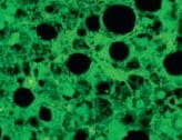
Application: Immunocytochemistry/ImmunofluorescenceSample Tested: macrophagesSpecies: HumanVerified Customer | Posted 09/04/2021CXCL9 antibody showed positive macrophages in hepatitis liver tissue.
-
Application: ELISASample Tested: Serum and PlasmaSpecies: HumanVerified Customer | Posted 12/06/2017This antibody was used as a capture in a sandwich ELISA for MIG. The detection antibody was BAM392. The standard was 392-MG-010. Sensitivity was around 5 pg/ml.
There are no reviews that match your criteria.
Protocols
Find general support by application which include: protocols, troubleshooting, illustrated assays, videos and webinars.
- 7-Amino Actinomycin D (7-AAD) Cell Viability Flow Cytometry Protocol
- Appropriate Fixation of IHC/ICC Samples
- Cellular Response to Hypoxia Protocols
- ClariTSA™ Fluorophore Kits
- Detection & Visualization of Antibody Binding
- Extracellular Membrane Flow Cytometry Protocol
- Flow Cytometry Protocol for Cell Surface Markers
- Flow Cytometry Protocol for Staining Membrane Associated Proteins
- Flow Cytometry Staining Protocols
- Flow Cytometry Troubleshooting Guide
- ICC Cell Smear Protocol for Suspension Cells
- ICC Immunocytochemistry Protocol Videos
- ICC for Adherent Cells
- Immunocytochemistry (ICC) Protocol
- Immunocytochemistry Troubleshooting
- Immunofluorescence of Organoids Embedded in Cultrex Basement Membrane Extract
- Immunohistochemistry (IHC) and Immunocytochemistry (ICC) Protocols
- Intracellular Flow Cytometry Protocol Using Alcohol (Methanol)
- Intracellular Flow Cytometry Protocol Using Detergents
- Intracellular Nuclear Staining Flow Cytometry Protocol Using Detergents
- Intracellular Staining Flow Cytometry Protocol Using Alcohol Permeabilization
- Intracellular Staining Flow Cytometry Protocol Using Detergents to Permeabilize Cells
- Preparing Samples for IHC/ICC Experiments
- Preventing Non-Specific Staining (Non-Specific Binding)
- Primary Antibody Selection & Optimization
- Propidium Iodide Cell Viability Flow Cytometry Protocol
- Protocol for Liperfluo
- Protocol for VisUCyte™ HRP Polymer Detection Reagent
- Protocol for the Characterization of Human Th22 Cells
- Protocol for the Characterization of Human Th9 Cells
- Protocol for the Fluorescent ICC Staining of Cell Smears - Graphic
- Protocol for the Fluorescent ICC Staining of Cultured Cells on Coverslips - Graphic
- Protocol for the Preparation and Fluorescent ICC Staining of Cells on Coverslips
- Protocol for the Preparation and Fluorescent ICC Staining of Non-adherent Cells
- Protocol for the Preparation and Fluorescent ICC Staining of Stem Cells on Coverslips
- Protocol for the Preparation of a Cell Smear for Non-adherent Cell ICC - Graphic
- Protocol: Annexin V and PI Staining by Flow Cytometry
- Protocol: Annexin V and PI Staining for Apoptosis by Flow Cytometry
- R&D Systems Quality Control Western Blot Protocol
- TUNEL and Active Caspase-3 Detection by IHC/ICC Protocol
- The Importance of IHC/ICC Controls
- Troubleshooting Guide: Fluorokine Flow Cytometry Kits
- Troubleshooting Guide: Western Blot Figures
- Western Blot Conditions
- Western Blot Protocol
- Western Blot Protocol for Cell Lysates
- Western Blot Troubleshooting
- Western Blot Troubleshooting Guide
- View all Protocols, Troubleshooting, Illustrated assays and Webinars
Associated Pathways
