
Dectin-2, also known as CLEC6A, CLECSF10, and NKCL, belongs to the C-type lectin family of transmembrane immune regulatory glycoproteins. Dectin-2, plus CLEC4A-E constitute a subgroup of molecules that exhibit approximately 40% amino acid (aa) sequence identity in their extracellular domains (ECD), and have a conserved cysteine spacing in their carbohydrate recognition domains (CRD) (1, 2). Mature human Dectin-2 is a type II transmembrane protein with a short cytoplasmic tail, a transmembrane segment, and a 168 aa ECD with a stalk region and one CRD (3, 4). Within the ECD, human Dectin-2 shares 71% and 75% aa sequence identity with bovine and mouse Dectin-2, respectively. An alternately spliced beta isoform has a deletion of portions of the transmembrane and cytoplasmic regions (5). Full length Dectin-2 is a 27 kDa molecule that is expressed on monocytes, tissue macrophages, and activated CD4+ T cells (4‑6). The CRD of Dectin-2 contains an EPN motif which is characteristic of calcium-dependent mannose-binding lectins. Dectin-2 selectively interacts with high mannose structures in the Man9GlcNAc2 configuration (7). It mediates the recognition of a variety of microorganisms, particularly the filamentous forms of yeast and fungii (7, 8). The short cytoplasmic tail does not contain signaling motifs but mediates association with the ITAM-containing Fc receptor gamma subunit on macrophages (8). Ligation of Dectin-2 induces tyrosine phosphorylation of the gamma subunit, activation of NF kappa B, and enhanced release of TNF-alpha and IL-1ra (8). Macrophage Dectin-2 is up‑regulated in vivo by inflammatory stimuli and UV-B irradiation (5, 6, 9). Dectin-2 is known to participate in UV-induced immunosuppression by interacting with CD4+CD25+ regulatory T cells, which then induce dendritic cells to release IL-4, IL-10, and TGF-beta (10).
Human Dectin-2/CLEC6A Antibody
R&D Systems | Catalog # AF3114

Scientific images may be resized, cropped, or adjusted for brightness or contrast for presentation purposes. These updates are limited to presentation and do not affect the underlying scientific interpretation of the data. Where available, additional source data may be provided upon request.
Key Product Details
Species Reactivity
Validated:
Cited:
Applications
Validated:
Cited:
Label
Antibody Source
Product Specifications
Immunogen
Thr46-Leu209
Accession # Q6EIG7
Specificity
Clonality
Host
Isotype
Scientific Data Images for Human Dectin-2/CLEC6A Antibody
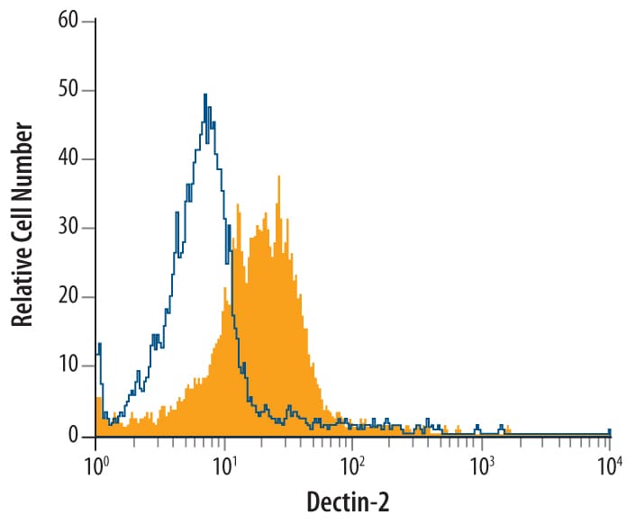
Detection of Dectin‑2/CLEC6A in Human Monocytes by Flow Cytometry.
Human blood-derived monocytes were stained with Goat Anti-Human Dectin-2/CLEC6A Antigen Affinity-purified Polyclonal Antibody (Catalog # AF3114, filled histogram) or control antibody (Catalog # AB-108-C, open histogram), followed by Allophycocyanin-conjugated Anti-Goat IgG Secondary Antibody (Catalog # F0108).Applications for Human Dectin-2/CLEC6A Antibody
CyTOF-ready
Flow Cytometry
Sample: Human blood-derived monocytes
Western Blot
Sample: Recombinant Human Dectin‑2/CLEC6A (Catalog # 3114-DC)
Flow Cytometry Panel Builder
Bio-Techne Knows Flow Cytometry
Save time and reduce costly mistakes by quickly finding compatible reagents using the Panel Builder Tool.
Advanced Features
- Spectra Viewer - Custom analysis of spectra from multiple fluorochromes
- Spillover Popups - Visualize the spectra of individual fluorochromes
- Antigen Density Selector - Match fluorochrome brightness with antigen density

Formulation, Preparation, and Storage
Purification
Reconstitution
Reconstitute at 0.2 mg/mL in sterile PBS. For liquid material, refer to CoA for concentration.
Formulation
Shipping
Stability & Storage
- 12 months from date of receipt, -20 to -70 °C as supplied.
- 1 month, 2 to 8 °C under sterile conditions after reconstitution.
- 6 months, -20 to -70 °C under sterile conditions after reconstitution.
Calculators
Background: Dectin-2/CLEC6A
References
- Kanazawa, N. (2007) J. Dermatol. Sci. 45:77.
- Kanazawa, N. et al. (2004) Immunobiology 209:179.
- Flornes, L.M. et al. (2004) Immunogenetics 56:506.
- Kanazawa, N. et al. (2004) J. Invest. Dermatol. 122:1522.
- Gavino, A.C. et al. (2005) Exp. Dermatol. 14:281.
- Taylor, P.R. et al. (2005) Eur. J. Immunol. 35:2163.
- McGreal, E.P. et al. (2006) Glycobiology 16:422.
- Sato, K. et al. (2006) J. Biol. Chem. 281:38854.
- Bonkobara, M. et al. (2005) Photochem. Photobiol. 81:944.
- Aragane, Y. et al. (2003) J. Immunol. 171:3801.
Alternate Names
Gene Symbol
UniProt
Additional Dectin-2/CLEC6A Products
Product Documents for Human Dectin-2/CLEC6A Antibody
Certificate of Analysis
To download a Certificate of Analysis, please enter a lot or batch number in the search box below.
Note: Certificate of Analysis not available for kit components.
Product Specific Notices for Human Dectin-2/CLEC6A Antibody
For research use only
Related Research Areas
Citations for Human Dectin-2/CLEC6A Antibody
 Powered by Bioz
Customer Reviews for Human Dectin-2/CLEC6A Antibody
There are currently no reviews for this product. Be the first to review Human Dectin-2/CLEC6A Antibody and earn rewards!
Have you used Human Dectin-2/CLEC6A Antibody?
Submit a review and receive an Amazon gift card!
$25/€18/£15/$25CAN/¥2500 for a review with an image
$10/€7/£6/$10CAN/¥1110 for a review without an image
Submit a review
Protocols
Find general support by application which include: protocols, troubleshooting, illustrated assays, videos and webinars.
- 7-Amino Actinomycin D (7-AAD) Cell Viability Flow Cytometry Protocol
- Cellular Response to Hypoxia Protocols
- Extracellular Membrane Flow Cytometry Protocol
- Flow Cytometry Protocol for Cell Surface Markers
- Flow Cytometry Protocol for Staining Membrane Associated Proteins
- Flow Cytometry Staining Protocols
- Flow Cytometry Troubleshooting Guide
- Intracellular Flow Cytometry Protocol Using Alcohol (Methanol)
- Intracellular Flow Cytometry Protocol Using Detergents
- Intracellular Nuclear Staining Flow Cytometry Protocol Using Detergents
- Intracellular Staining Flow Cytometry Protocol Using Alcohol Permeabilization
- Intracellular Staining Flow Cytometry Protocol Using Detergents to Permeabilize Cells
- Propidium Iodide Cell Viability Flow Cytometry Protocol
- Protocol for Liperfluo
- Protocol for the Characterization of Human Th22 Cells
- Protocol for the Characterization of Human Th9 Cells
- Protocol: Annexin V and PI Staining by Flow Cytometry
- Protocol: Annexin V and PI Staining for Apoptosis by Flow Cytometry
- R&D Systems Quality Control Western Blot Protocol
- Troubleshooting Guide: Fluorokine Flow Cytometry Kits
- Troubleshooting Guide: Western Blot Figures
- Western Blot Conditions
- Western Blot Protocol
- Western Blot Protocol for Cell Lysates
- Western Blot Troubleshooting
- Western Blot Troubleshooting Guide
- View all Protocols, Troubleshooting, Illustrated assays and Webinars
Associated Pathways
