
Dkk-3, also known as REIC (Reduced Expansion in Immortalized Cells), is one of four numbered members of the Dickkopf family of Wnt antagonists (1). Dkk-3 is a secreted monomer expressed in many normal human tissues, most strongly in heart, brain and spinal cord (1, 2), and during early embryonic development in the mouse (3). N-glycosylation at up to four sites preceding or between two conserved cysteine-rich motifs results in expression of a 45-65 kDa glycoprotein (1, 4). The cysteine-rich motifs contain 10 cysteines each, with prokineticin and colipase families containing sequences similar to those of the second motif (1, 5). Human Dkk-3 shows 82%, 88%, 85%, and 53% amino acid (aa) identity with mouse, bovine, canine, and chick Dkk-3, respectively, and 37-45% aa identity with other human Dkk family members. Several lines of evidence implicate Dkk-3 as a negative growth regulator. Dkk-3 is downregulated in many tumors as compared to normal cells, sometimes by loss of heterozygosity (4, 6). Downregulation by CpG hypermethylation in acute lymphoblastic leukemia is correlated with faster progression and shorter survival (7). Release of cultured cells from serum starvation results in downregulation of Dkk-3 in late G1 phase of the cell cycle (6). Over-expression of Dkk‑3 results in tumor cell-line-specific growth inhibition, induction of apoptosis, and decreased tumorigenicity in nude mice (2, 4, 6). The prototype Dickkopf member, Dkk-1, antagonizes Wnt family signaling by binding to Wnt receptors LRP5 and LRP6 (low-density lipoprotein receptor-related proteins) and promoting their internalization (1, 9, 10). Results are less straightforward for Dkk-3, where some studies show binding to LRP5/6 while others do not. These effects appear to be dependent on the cells and conditions used (1, 6-10).
Human Dkk-3 Antibody [Biotin]
R&D Systems | Catalog # BAF1118

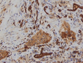
Scientific images may be resized, cropped, or adjusted for brightness or contrast for presentation purposes. These updates are limited to presentation and do not affect the underlying scientific interpretation of the data. Where available, additional source data may be provided upon request.
Key Product Details
Species Reactivity
Validated:
Cited:
Applications
Validated:
Cited:
Label
Antibody Source
Product Specifications
Immunogen
Ala22-Ile350
Accession # Q9UBP4
Specificity
Clonality
Host
Isotype
Scientific Data Images for Human Dkk-3 Antibody [Biotin]
Dkk‑3 in Human Lung.
Dkk‑3 was detected in immersion fixed paraffin-embedded sections of human lung using Goat Anti-Human Dkk‑3 Biotinylated Antigen Affinity-purified Polyclonal Antibody (Catalog # BAF1118) at 15 µg/mL overnight at 4 °C. Tissue was stained using the Anti-Goat HRP-DAB Cell & Tissue Staining Kit (brown; CTS008) and counterstained with hematoxylin (blue). View our protocol for Chromogenic IHC Staining of Paraffin-embedded Tissue Sections.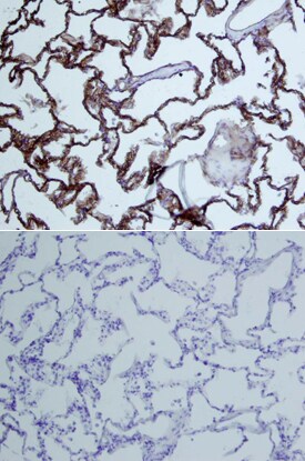
Dkk‑3 in Human Lung.
Dkk-3 was detected in immersion fixed paraffin-embedded sections of human lung array using Goat Anti-Human Dkk-3 Biotinylated Antigen Affinity-purified Polyclonal Antibody (Catalog # BAF1118) at 15 µg/mL overnight at 4 °C. Tissue was stained using the Anti-Goat HRP-DAB Cell & Tissue Staining Kit (brown; CTS008) and counterstained with hematoxylin (blue). Lower panel shows a lack of labeling if primary antibodies are omitted and tissue is stained only with secondary antibody followed by incubation with detection reagents. View our protocol for Chromogenic IHC Staining of Paraffin-embedded Tissue Sections.
Human Dkk-3 ELISA Standard Curve
Recombinant Human Dkk‑3 (Catalog # 1118-DK) was serially diluted and captured by Mouse Anti-Human Dkk‑3 Monoclonal Antibody (Catalog # MAB11181) coated on a Clear Polystyrene Microplate (Catalog # DY990). Goat Anti-Human Dkk‑3 Antigen Affinity-purified Polyclonal Antibody (Catalog # AF1118) was biotinylated and incubated with the protein captured on the plate. Detection of the standard curve was achieved by incubating Streptavidin-HRP (Catalog # DY998)Applications for Human Dkk-3 Antibody [Biotin]
Immunohistochemistry
Sample: Immersion fixed paraffin-embedded sections of human lung
Human Dkk-3 Sandwich Immunoassay
Formulation, Preparation, and Storage
Purification
Reconstitution
Reconstitute at 0.2 mg/mL in sterile PBS.
Formulation
Shipping
Stability & Storage
- 12 months from date of receipt, -20 to -70 °C as supplied.
- 1 month, 2 to 8 °C under sterile conditions after reconstitution.
- 6 months, -20 to -70 °C under sterile conditions after reconstitution.
Calculators
Background: Dkk-3
References
- Krupnik, V.E. et al. (1999) Gene 238:301.
- Tsuji, T. et al. (2000) Biochem. Biophys. Res. Comm. 268:20.
- Kemp, C. et al. (2005) Dev. Dyn. 233:1064.
- Hsieh, S.-Y. et al. (2004) Oncogene 23:9183.
- Bullock, C.M. et al. (2004) Mol. Pharmacol. 65:582.
- Tsuji, T. et al. (2001) Biochem. Biophys. Res. Comm. 289:257.
- Roman-Gomez, J. et al. (2004) Br. J. Cancer 91:707.
- Hoang, B.H. et al. (2004) Cancer Res. 64:2734.
- Caricasole, A. et al. (2003) J. Biol. Chem. 278:37024.
- Mao, B. et al. (2001) Nature 411:321.
Long Name
Alternate Names
Gene Symbol
UniProt
Additional Dkk-3 Products
Product Documents for Human Dkk-3 Antibody [Biotin]
Certificate of Analysis
To download a Certificate of Analysis, please enter a lot or batch number in the search box below.
Note: Certificate of Analysis not available for kit components.
Product Specific Notices for Human Dkk-3 Antibody [Biotin]
For research use only
Related Research Areas
Citations for Human Dkk-3 Antibody [Biotin]
 Powered by Bioz
Customer Reviews for Human Dkk-3 Antibody [Biotin]
There are currently no reviews for this product. Be the first to review Human Dkk-3 Antibody [Biotin] and earn rewards!
Have you used Human Dkk-3 Antibody [Biotin]?
Submit a review and receive an Amazon gift card!
$25/€18/£15/$25CAN/¥2500 for a review with an image
$10/€7/£6/$10CAN/¥1110 for a review without an image
Submit a review
Protocols
Find general support by application which include: protocols, troubleshooting, illustrated assays, videos and webinars.
- Antigen Retrieval Protocol (PIER)
- Antigen Retrieval for Frozen Sections Protocol
- Appropriate Fixation of IHC/ICC Samples
- Cellular Response to Hypoxia Protocols
- Chromogenic IHC Staining of Formalin-Fixed Paraffin-Embedded (FFPE) Tissue Protocol
- Chromogenic Immunohistochemistry Staining of Frozen Tissue
- ClariTSA™ Fluorophore Kits
- Detection & Visualization of Antibody Binding
- Fluorescent IHC Staining of Frozen Tissue Protocol
- Graphic Protocol for Heat-induced Epitope Retrieval
- Graphic Protocol for the Preparation and Fluorescent IHC Staining of Frozen Tissue Sections
- Graphic Protocol for the Preparation and Fluorescent IHC Staining of Paraffin-embedded Tissue Sections
- Graphic Protocol for the Preparation of Gelatin-coated Slides for Histological Tissue Sections
- IHC Sample Preparation (Frozen sections vs Paraffin)
- Immunofluorescent IHC Staining of Formalin-Fixed Paraffin-Embedded (FFPE) Tissue Protocol
- Immunohistochemistry (IHC) and Immunocytochemistry (ICC) Protocols
- Immunohistochemistry Frozen Troubleshooting
- Immunohistochemistry Paraffin Troubleshooting
- Preparing Samples for IHC/ICC Experiments
- Preventing Non-Specific Staining (Non-Specific Binding)
- Primary Antibody Selection & Optimization
- Protocol for Heat-Induced Epitope Retrieval (HIER)
- Protocol for Making a 4% Formaldehyde Solution in PBS
- Protocol for VisUCyte™ HRP Polymer Detection Reagent
- Protocol for the Preparation & Fixation of Cells on Coverslips
- Protocol for the Preparation and Chromogenic IHC Staining of Frozen Tissue Sections
- Protocol for the Preparation and Chromogenic IHC Staining of Frozen Tissue Sections - Graphic
- Protocol for the Preparation and Chromogenic IHC Staining of Paraffin-embedded Tissue Sections
- Protocol for the Preparation and Chromogenic IHC Staining of Paraffin-embedded Tissue Sections - Graphic
- Protocol for the Preparation and Fluorescent IHC Staining of Frozen Tissue Sections
- Protocol for the Preparation and Fluorescent IHC Staining of Paraffin-embedded Tissue Sections
- Protocol for the Preparation of Gelatin-coated Slides for Histological Tissue Sections
- TUNEL and Active Caspase-3 Detection by IHC/ICC Protocol
- The Importance of IHC/ICC Controls
- Troubleshooting Guide: Immunohistochemistry
- View all Protocols, Troubleshooting, Illustrated assays and Webinars