
EMMPRIN is expressed in areas of tissue remodeling, including tumors, endometrium, placenta, skin, and regions undergoing angiogenesis (1, 2, 6-9). It is also expressed in cells with high metabolic activity, such as lymphoblasts, macrophages and tumor cells (2, 10). On cells with elevated metabolic rates, EMMPRIN is often co-expressed with the amino acid transporter CD98h (11). EMMPRIN also interacts with caveolin-1 (via its C2-like domain), and this reduces the level of EMMPRIN glycosylation and subsequent EMMPRIN multimerization and activity (12). EMMPRIN’s TM sequence contains a Glu and a Pro which are important for intracellular interactions with cyclophilins (CyP) (3, 13, 14). CyPA (cyclosporin A receptor) and CyP60 interactions with the TM segment promote leukocyte inflammatory chemotaxis and surface expression of EMMPRIN, respectively (13, 14). An active 22 kDa fragment can be shed from tumor cells by MT1-MMP (1). Tumor cells can also release active, full-length EMMPRIN in microvesicles (15, 16). Functionally, EMMPRIN is known to induce urokinase-type plasminogen activator (uPA), VEGF, hyaluronan, and multiple MMPs (1, 2, 7, 8, 9). Human EMMPRIN (269 aa) shows 58%, 58%, 62%, and 52% aa identity with mouse, rat, bovine, and chicken EMMPRIN, respectively. It also shows 25% and 38% aa identity with the related proteins, embigin and neuroplastin (SDR-1), respectively (4).
Human EMMPRIN/CD147 Antibody [Biotin]
R&D Systems | Catalog # BAF972

Scientific images may be resized, cropped, or adjusted for brightness or contrast for presentation purposes. These updates are limited to presentation and do not affect the underlying scientific interpretation of the data. Where available, additional source data may be provided upon request.
Key Product Details
Species Reactivity
Validated:
Cited:
Applications
Validated:
Cited:
Label
Antibody Source
Product Specifications
Immunogen
Thr25-His205
Accession # Q54A51
Specificity
Clonality
Host
Isotype
Scientific Data Images for Human EMMPRIN/CD147 Antibody [Biotin]
Detection of Human EMMPRIN/CD147 by Immunohistochemistry
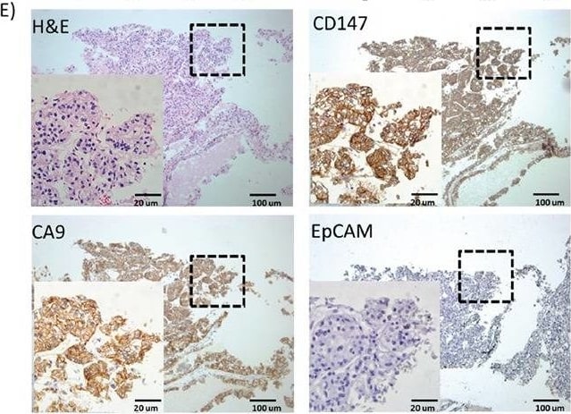
IHC analysis of EpCAM, CA9 and CD147 expression in RCC patient cohortA. Positive expression percentages of EpCAM, CA9 and CD147 in RCC patient samples of different clinical stages by IHC analysis (I, n=29; II, n=19; III, n=16; IV, n=6). B. IHC scores of EpCAM, CA9 and CD147 in RCC samples of (A). *** p<0.001. C. Positive expression percentages of CA9+ and/or CD147+ in EpCAM+ (I, n=8; II, n=3; III, n=1; IV, n=1) or EpCAM- (I, n=21; II, n=16; III, n=15; IV, n=5) RCC patient samples of different clinical stages by IHC analysis. D. IHC scores of EpCAM, CA9 and CD147 in RCC samples of (C). E. H&E and IHC staining of EpCAM, CA9 and CD147 in representative EpCAM- RCC patient tissues. The selected enlarged areas are indicated in dashed rectangles. Original magnification, 400× (enlarged) and 100× (original); scale bars: 20 μm (enlarged) and 100 μm (original). Image collected and cropped by CiteAb from the following publication (https://www.oncotarget.com/lookup/doi/10.18632/oncotarget.10979), licensed under a CC-BY license. Not internally tested by R&D Systems.
Human EMMPRIN / CD147 ELISA Standard Curve
Recombinant Human EMMPRIN/CD147 Fc Chimera (Catalog # 972-EMN) was serially diluted and captured by Mouse Anti-Human EMMPRIN/CD147 Monoclonal Antibody (Catalog # MAB972) coated on a Clear Polystyrene Microplate (Catalog # DY990). Goat Anti-Human EMMPRIN/CD147 Antigen Affinity-purified Polyclonal Antibody (Catalog # AF972) was biotinylated and incubated with the protein captured on the plate. Detection of the standard curve was achieved by incubating Streptavidin-HRP (Catalog # DY998)Applications for Human EMMPRIN/CD147 Antibody [Biotin]
Western Blot
Sample: Recombinant Human EMMPRIN/CD147 Fc Chimera (Catalog # 972-EMN)
Reviewed Applications
Read 1 review rated 4 using BAF972 in the following applications:
Formulation, Preparation, and Storage
Purification
Reconstitution
Reconstitute at 0.2 mg/mL in sterile PBS.
Formulation
Shipping
Stability & Storage
- 12 months from date of receipt, -20 to -70 °C as supplied.
- 1 month, 2 to 8 °C under sterile conditions after reconstitution.
- 6 months, -20 to -70 °C under sterile conditions after reconstitution.
Calculators
Background: EMMPRIN/CD147
References
- Gabison, E.E. et al. (2005) Biochimie 87:361.
- Yurchenko, V. et al. (2006) Immunology 117:301.
- Kasinrerk, W. et al. (1992) J. Immunol. 149:847.
- Miyauchi, T. et al. (1991) J. Biochem. 110:770.
- Hanna, S.M. et al. (2003) BMC Biochem. 4:17.
- Riethdorf, S. et al. (2006) Int. J. Cancer 119:1800.
- Braundmeier, A.G. et al. (2006) J. Clin. Endocrinol. Metab. 91:2358.
- Tang, Y. et al. (2006) Mol. Cancer Res. 4:371.
- Quemener, C. et al. (2007) Cancer Res. 67:9.
- Wilson, M.C. et al. (2005) J. Biol. Chem. 280:27213.
- Xu, D. and M.E. Hemler (2005) Mol. Cell. Proteomics 4:1061.
- Tang, W. et al. (2004) Mol. Biol. Cell 15:4043.
- Arora, K. et al. (2005) J. Immunol. 175:517.
- Pushkarsky, T. et al. (2005) J. Biol. Chem. 280:27866.
- Egawa, N. et al. (2006) J. Biol. Chem. 281:37576.
- Sidhu, S.S. et al. (2004) Oncogene 23:956.
Long Name
Alternate Names
Gene Symbol
UniProt
Additional EMMPRIN/CD147 Products
Product Documents for Human EMMPRIN/CD147 Antibody [Biotin]
Certificate of Analysis
To download a Certificate of Analysis, please enter a lot or batch number in the search box below.
Note: Certificate of Analysis not available for kit components.
Product Specific Notices for Human EMMPRIN/CD147 Antibody [Biotin]
For research use only
Citations for Human EMMPRIN/CD147 Antibody [Biotin]
 Powered by Bioz
Customer Reviews for Human EMMPRIN/CD147 Antibody [Biotin] (1)
Have you used Human EMMPRIN/CD147 Antibody [Biotin]?
Submit a review and receive an Amazon gift card!
$25/€18/£15/$25CAN/¥2500 for a review with an image
$10/€7/£6/$10CAN/¥1110 for a review without an image
Submit a review
-
Application: ELISASample Tested: See PMID: 23598007Species: HumanVerified Customer | Posted 01/15/2015
There are no reviews that match your criteria.
Protocols
Find general support by application which include: protocols, troubleshooting, illustrated assays, videos and webinars.
- Cellular Response to Hypoxia Protocols
- R&D Systems Quality Control Western Blot Protocol
- Troubleshooting Guide: Western Blot Figures
- Western Blot Conditions
- Western Blot Protocol
- Western Blot Protocol for Cell Lysates
- Western Blot Troubleshooting
- Western Blot Troubleshooting Guide
- View all Protocols, Troubleshooting, Illustrated assays and Webinars