
Ephrin-A5, also known as AL-1, RAGS, LERK-7, and EFL-5 (1), is a member of the ephrin ligand family which binds members of the Eph receptor family. All ligands share a conserved extracellular sequence, which most likely corresponds to the receptor binding domain. This conserved sequence consists of approximately 125 amino acids and includes four invariant cysteines. The A-class ligands have a GPI anchor following the conserved sequence. Ephrin-A5 has been shown to bind EphA2, EphA3, EphA4, EphA5, EphA6, EphA7, and EphA8 (2, 3). The extracellular domains of murine and human Ephrin-A5 share 99% amino acid identity. Only membrane-bound or Fc-clustered ligands are capable of activating the receptor in vitro. While soluble monomeric ligands bind the receptor, they do not induce receptor autophosphorylation and activation (2). In vivo, the ligands and receptors display reciprocal expression (3). It has been found that nearly all the receptors and ligands are expressed in developing and adult neural tissue (3). The ephrin/Eph families also appear to play a role in angiogenesis (3).

Scientific images may be resized, cropped, or adjusted for brightness or contrast for presentation purposes. These updates are limited to presentation and do not affect the underlying scientific interpretation of the data. Where available, additional source data may be provided upon request.
Key Product Details
Species Reactivity
Validated:
Cited:
Applications
Validated:
Cited:
Label
Antibody Source
Product Specifications
Immunogen
Gln21-Asn203
Accession # P52803
Specificity
Clonality
Host
Isotype
Scientific Data Images for Human Ephrin-A5 Antibody
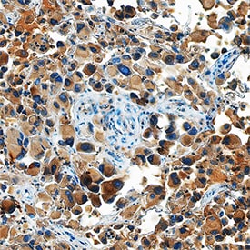
Ephrin‑A5 in Human Melanoma.
Ephrin-A5 was detected in immersion fixed paraffin-embedded sections of human melanoma using Goat Anti-Human Ephrin-A5 Antigen Affinity-purified Polyclonal Antibody (Catalog # AF3743) at 15 µg/mL overnight at 4 °C. Tissue was stained using the Anti-Goat HRP-DAB Cell & Tissue Staining Kit (brown; Catalog # CTS008) and counterstained with hematoxylin (blue). Specific staining was localized to cytoplasm. View our protocol for Chromogenic IHC Staining of Paraffin-embedded Tissue Sections.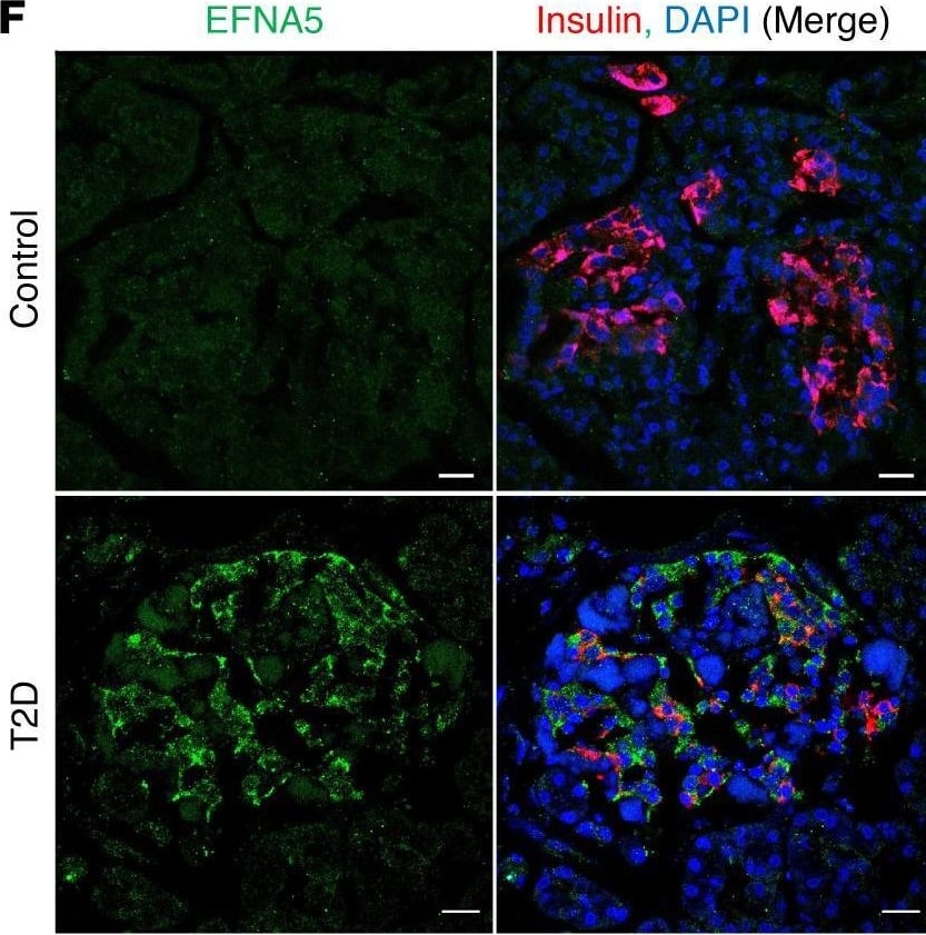
Detection of Ephrin-A5 by Immunohistochemistry
Increased Notch and Ephrin signaling in metabolically inflexible human beta cells.(A) scRNA-Seq of islets from nondiabetic and T2D patients, with beta cells subclustered by metabolic pathways to define healthy (H beta ) and metabolically inflexible (MI beta ) as per a previous report (48). tSNE, t-distributed stochastic neighbor embedding. (B) Violin plots of RBPJ activity in H beta and MI beta cell populations. (C) GSEA of EPH-Ephrin signaling in DEGs of MI beta versus H beta populations. NES, normalized enrichment score. (D) Average activity of EPH-Ephrin signaling components in H beta and MI beta populations. (E) RBPJ activity as compared with EPH-Ephrin signaling components, with each dot representing a single H beta (black) and MI beta (red) cell. Extended figure with all EPH-Ephrin signaling components and Pearson’s r values in Supplemental Figure 11A. (F) Representative images of pancreatic sections stained with antibodies against EFNA5 and insulin from nondiabetic and T2D patients. Extended figure with additional staining in Supplemental Figure 12B. Scale bars: 20 μm. Image collected and cropped by CiteAb from the following open publication (https://insight.jci.org/articles/view/157694), licensed under a CC-BY license. Not internally tested by R&D Systems.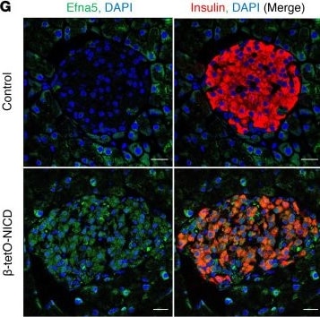
Detection of Ephrin-A5 by Immunohistochemistry
RNA-Seq of beta -tetO-NICD islets reveals potential morphogenetic effectors.(A) Volcano plot showing differentially expressed genes (DEGs, adjusted P < 0.05) in islets from beta -tetO-NICD and Cre+ controls after 8 weeks’ Dox. (B) Downregulated and (C) upregulated biological processes in beta -tetO-NICD islets, after Gene Ontology enrichment analysis using Enrichr. (D) Heatmap of representative DEGs across samples, with log2-normalized gene expression value shown. Efna5, Ephrin A5. (E) STRING network analysis of upregulated DEGs in beta -tetO-NICD islets showing enrichment analysis of processes associated with the network, highlighting processes associated with tissue morphogenesis. (F) Absolute quantitation, relative to invariant control Ppia, of genes encoding Ephrin ligands in islets isolated from beta -tetO-NICD and control mice after 8 weeks’ Dox (n = 5–6 mice/group). (G) Representative images of pancreatic sections stained with antibodies against Efna5 and insulin from beta -tetO-NICD and Cre– controls after 8 weeks’ Dox. Scale bars: 20 μm. All data are shown with group means; *, P < 0.05, **, P < 0.01, ***, P < 0.001, ****, P < 0.0001 by 2-tailed t test. Image collected and cropped by CiteAb from the following open publication (https://insight.jci.org/articles/view/157694), licensed under a CC-BY license. Not internally tested by R&D Systems.Applications for Human Ephrin-A5 Antibody
Immunohistochemistry
Sample: Immersion fixed paraffin-embedded sections of human melanoma tissue
Western Blot
Sample: Recombinant Human Ephrin-A5 Fc Chimera (Catalog # 374-EA)
Reviewed Applications
Read 3 reviews rated 4 using AF3743 in the following applications:
Formulation, Preparation, and Storage
Purification
Reconstitution
Reconstitute at 0.2 mg/mL in sterile PBS. For liquid material, refer to CoA for concentration.
Formulation
Shipping
Stability & Storage
- 12 months from date of receipt, -20 to -70 °C as supplied.
- 1 month, 2 to 8 °C under sterile conditions after reconstitution.
- 6 months, -20 to -70 °C under sterile conditions after reconstitution.
Calculators
Background: Ephrin-A5
References
- Eph Nomenclature Committee [letter] (1997) Cell 90:403.
- Flanagan, J.G. and P. Vanderhaeghen (1998) Annu. Rev. Neurosci. 21:309.
- Pasquale, E.B. (1997) Curr. Opin. Cell Biol. 9:608.
Alternate Names
Gene Symbol
UniProt
Additional Ephrin-A5 Products
Product Documents for Human Ephrin-A5 Antibody
Certificate of Analysis
To download a Certificate of Analysis, please enter a lot or batch number in the search box below.
Note: Certificate of Analysis not available for kit components.
Product Specific Notices for Human Ephrin-A5 Antibody
For research use only
Related Research Areas
Citations for Human Ephrin-A5 Antibody
 Powered by Bioz
Customer Reviews for Human Ephrin-A5 Antibody (3)
Have you used Human Ephrin-A5 Antibody?
Submit a review and receive an Amazon gift card!
$25/€18/£15/$25CAN/¥2500 for a review with an image
$10/€7/£6/$10CAN/¥1110 for a review without an image
Submit a review
Customer Images


-
Application: Western BlotSample Tested: HT-29 colon cancer cell line and T84 human colon carcinoma cell lineSpecies: HumanVerified Customer | Posted 04/04/2020

-
Application: Western BlotSample Tested: DA3 mouse myeloma cell lineSpecies: MouseVerified Customer | Posted 02/18/2019
-
Application: Western BlotSample Tested: Capan-1 human pancreatic adenocarcinoma cell line and AR42J human acinar pancreatic cancer cell lineSpecies: HumanVerified Customer | Posted 10/17/2018
There are no reviews that match your criteria.
Protocols
Find general support by application which include: protocols, troubleshooting, illustrated assays, videos and webinars.
- Antigen Retrieval Protocol (PIER)
- Antigen Retrieval for Frozen Sections Protocol
- Appropriate Fixation of IHC/ICC Samples
- Cellular Response to Hypoxia Protocols
- Chromogenic IHC Staining of Formalin-Fixed Paraffin-Embedded (FFPE) Tissue Protocol
- Chromogenic Immunohistochemistry Staining of Frozen Tissue
- ClariTSA™ Fluorophore Kits
- Detection & Visualization of Antibody Binding
- Fluorescent IHC Staining of Frozen Tissue Protocol
- Graphic Protocol for Heat-induced Epitope Retrieval
- Graphic Protocol for the Preparation and Fluorescent IHC Staining of Frozen Tissue Sections
- Graphic Protocol for the Preparation and Fluorescent IHC Staining of Paraffin-embedded Tissue Sections
- Graphic Protocol for the Preparation of Gelatin-coated Slides for Histological Tissue Sections
- IHC Sample Preparation (Frozen sections vs Paraffin)
- Immunofluorescent IHC Staining of Formalin-Fixed Paraffin-Embedded (FFPE) Tissue Protocol
- Immunohistochemistry (IHC) and Immunocytochemistry (ICC) Protocols
- Immunohistochemistry Frozen Troubleshooting
- Immunohistochemistry Paraffin Troubleshooting
- Preparing Samples for IHC/ICC Experiments
- Preventing Non-Specific Staining (Non-Specific Binding)
- Primary Antibody Selection & Optimization
- Protocol for Heat-Induced Epitope Retrieval (HIER)
- Protocol for Making a 4% Formaldehyde Solution in PBS
- Protocol for VisUCyte™ HRP Polymer Detection Reagent
- Protocol for the Preparation & Fixation of Cells on Coverslips
- Protocol for the Preparation and Chromogenic IHC Staining of Frozen Tissue Sections
- Protocol for the Preparation and Chromogenic IHC Staining of Frozen Tissue Sections - Graphic
- Protocol for the Preparation and Chromogenic IHC Staining of Paraffin-embedded Tissue Sections
- Protocol for the Preparation and Chromogenic IHC Staining of Paraffin-embedded Tissue Sections - Graphic
- Protocol for the Preparation and Fluorescent IHC Staining of Frozen Tissue Sections
- Protocol for the Preparation and Fluorescent IHC Staining of Paraffin-embedded Tissue Sections
- Protocol for the Preparation of Gelatin-coated Slides for Histological Tissue Sections
- R&D Systems Quality Control Western Blot Protocol
- TUNEL and Active Caspase-3 Detection by IHC/ICC Protocol
- The Importance of IHC/ICC Controls
- Troubleshooting Guide: Immunohistochemistry
- Troubleshooting Guide: Western Blot Figures
- Western Blot Conditions
- Western Blot Protocol
- Western Blot Protocol for Cell Lysates
- Western Blot Troubleshooting
- Western Blot Troubleshooting Guide
- View all Protocols, Troubleshooting, Illustrated assays and Webinars