Estrogen Receptor beta (ERβ,NR3A2) is a member of the steroid receptor family. The natural ligand for ER is the classical estrogenic compound 17β-estradiol. ERβ is expressed in the granulosa cell layer of primary, secondary and mature follicles in the ovary, in bone, bladder, uterus, testis, epididymis, gastrointestinal tract, kidney, breast, heart, vessel wall, immune system, lung, pituitary, hippocampus and hypothalamus. Roles for ERβ in the reproductive and cardiovascular systems have been reported, although these are the subject of conflicting reports. ERβ has been postulated to act primarily as a modulator of ERα function. ER&beta has been shown to form homodimers as well as heterodimers with ERα. Both ERα and ERβ can give rise to numerous isoforms.
Human ER beta/NR3A2 Antibody (PPZ0506)
R&D Systems | Catalog # PP-PPZ0506-00

Scientific images may be resized, cropped, or adjusted for brightness or contrast for presentation purposes. These updates are limited to presentation and do not affect the underlying scientific interpretation of the data. Where available, additional source data may be provided upon request.
Key Product Details
Species Reactivity
Validated:
Cited:
Applications
Validated:
Cited:
Label
Antibody Source
Product Specifications
Immunogen
Clonality
Host
Isotype
Scientific Data Images for Human ER beta/NR3A2 Antibody (PPZ0506)
Detection of ER beta/NR3A2 by Immunohistochemistry
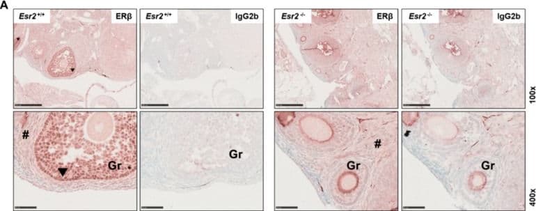
Female reproductive tissues of Esr2-depleted mice are negative for estrogen receptor beta (ER beta ) expression. Ovaries (A) and uterine tissue (B) of Esr2 knockout (Esr2−/−) and wild-type (Esr2+/+) animals from the same mouse line were stained with PPZ0506 antibody or with isotype-specific IgG2b negative control. Only granulosa cells (Gr) in Esr2+/+ ovaries strongly express ER beta, whereas in Esr2−/− ovary and uterine tissue no specific nuclear staining was observed. Specific staining is shown by arrowheads in Esr2+/+ and # indicates diffuse non-specific background staining. Scale bar 250 µm (100×) or 50 µm (400×). Image collected and cropped by CiteAb from the following open publication (https://www.mdpi.com/2227-9059/10/12/3100), licensed under a CC-BY license. Not internally tested by R&D Systems.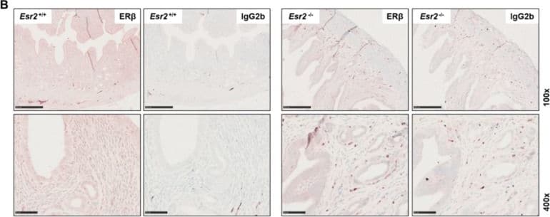
Detection of ER beta/NR3A2 by Immunohistochemistry
Female reproductive tissues of Esr2-depleted mice are negative for estrogen receptor beta (ER beta ) expression. Ovaries (A) and uterine tissue (B) of Esr2 knockout (Esr2−/−) and wild-type (Esr2+/+) animals from the same mouse line were stained with PPZ0506 antibody or with isotype-specific IgG2b negative control. Only granulosa cells (Gr) in Esr2+/+ ovaries strongly express ER beta, whereas in Esr2−/− ovary and uterine tissue no specific nuclear staining was observed. Specific staining is shown by arrowheads in Esr2+/+ and # indicates diffuse non-specific background staining. Scale bar 250 µm (100×) or 50 µm (400×). Image collected and cropped by CiteAb from the following open publication (https://www.mdpi.com/2227-9059/10/12/3100), licensed under a CC-BY license. Not internally tested by R&D Systems.Applications for Human ER beta/NR3A2 Antibody (PPZ0506)
Immunoprecipitation
Western Blot
Reviewed Applications
Read 2 reviews rated 5 using PP-PPZ0506-00 in the following applications:
Formulation, Preparation, and Storage
Formulation
Shipping
Stability & Storage
Background: ER beta/NR3A2
Long Name
Alternate Names
Entrez Gene IDs
Gene Symbol
Additional ER beta/NR3A2 Products
Product Documents for Human ER beta/NR3A2 Antibody (PPZ0506)
Certificate of Analysis
To download a Certificate of Analysis, please enter a lot or batch number in the search box below.
Note: Certificate of Analysis not available for kit components.
Product Specific Notices for Human ER beta/NR3A2 Antibody (PPZ0506)
For research use only
Related Research Areas
Citations for Human ER beta/NR3A2 Antibody (PPZ0506)
 Powered by Bioz
Customer Reviews for Human ER beta/NR3A2 Antibody (PPZ0506) (2)
Have you used Human ER beta/NR3A2 Antibody (PPZ0506)?
Submit a review and receive an Amazon gift card!
$25/€18/£15/$25CAN/¥2500 for a review with an image
$10/€7/£6/$10CAN/¥1110 for a review without an image
Submit a review
Customer Images


-
Application: ImmunohistochemistrySample Tested: Ovary tissueSpecies: HumanVerified Customer | Posted 09/16/2025heating pretreatment, x200 primary antibody
 Bio-Techne ResponseThis review reflects a new species or application tested on a primary antibody.
-
Application: ImmunohistochemistrySample Tested: Cancer Tissue and Normal tissueSpecies: HumanVerified Customer | Posted 08/15/2017The present antibody proved excellent for IHC (1:600 dilution) of formalin-fixed paraffin-embedded tissues and cells, Western blotting (1:1000) and IP as determined by comparing results from ERbeta-positive cells and ERbeta-negative cells, and corroborated by mass spectrometry. It was tested on a wide range of human normal tissues and cancer, and correlated to mRNA levels (RNA-seq). It stained the nucleus of granulosa cells of the normal ovary, testis, adrenal gland, tonsil, and lymphocytes in various tissues; tumor cells of granulosa cell tumors and a subset of malignant melanoma. See full article at Nature Communications (Andersson et al, 2017): https://www.nature.com/articles/ncomms15840
There are no reviews that match your criteria.
Protocols
Find general support by application which include: protocols, troubleshooting, illustrated assays, videos and webinars.
- Cellular Response to Hypoxia Protocols
- Immunoprecipitation Protocol
- R&D Systems Quality Control Western Blot Protocol
- Troubleshooting Guide: Western Blot Figures
- Western Blot Conditions
- Western Blot Protocol
- Western Blot Protocol for Cell Lysates
- Western Blot Troubleshooting
- Western Blot Troubleshooting Guide
- View all Protocols, Troubleshooting, Illustrated assays and Webinars
Associated Pathways