


Scientific images may be resized, cropped, or adjusted for brightness or contrast for presentation purposes. These updates are limited to presentation and do not affect the underlying scientific interpretation of the data. Where available, additional source data may be provided upon request.
Key Product Details
Species Reactivity
Validated:
Cited:
Applications
Validated:
Cited:
Label
Antibody Source
Product Specifications
Immunogen
Met1-Ala132
Accession # O15540
Specificity
Clonality
Host
Isotype
Scientific Data Images for Human FABP7/B-FABP Antibody
FABP7 in Human Melanoma.
FABP7 was detected in immersion fixed paraffin-embedded sections of human melanoma using 5 µg/mL Goat Anti-Human FABP7 Antigen Affinity-purified Polyclonal Antibody (Catalog # AF3166) overnight at 4 °C. Tissue was stained with the Anti-Goat HRP-DAB Cell & Tissue Staining Kit (brown; Catalog # CTS008) and counterstained with hematoxylin (blue). View our protocol for Chromogenic IHC Staining of Paraffin-embedded Tissue Sections.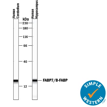
Detection of Human FABP7/B‑FABP by Simple WesternTM.
Simple Western lane view shows lysates of human cerebellum tissue and human hippocampus tissue, loaded at 0.2 mg/mL. A specific band was detected for FABP7/B-FABP at approximately 18 kDa (as indicated) using 50 µg/mL of Goat Anti-Human FABP7/B-FABP Antigen Affinity-purified Polyclonal Antibody (Catalog # AF3166) followed by 1:50 dilution of HRP-conjugated Anti-Goat IgG Secondary Antibody (Catalog # HAF109). This experiment was conducted under reducing conditions and using the 12-230 kDa separation system.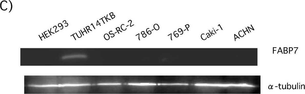
Detection of Human FABP7/B-FABP by Western Blot
FABP7 expression in RCC cell lines. A) Q-PCR analysis was performed using total RNA extracted from a human embryonic kidney (HEK293) and RCC (TUHR14TKB, OS-RC-2, 786-O, 769-P, Caki-1, and ACHN) cell lines. FABP7 expression was determined with Q-PCR by the primers listed in Table 2. B) HEK293 and RCC (TUHR14TKB, OS-RC-2, 786-O, 769-P, Caki-1, and ACHN) cells were cotransfected with the -1122+89 pGL4-FABP7 promoter construct, and cultured for one day. Extracts prepared from transfected cells were assayed for luciferase activity. FABP7 promoter activities were normalized to the control's Renilla luciferase activity. The results shown are an average of four independent experiments with standard deviations indicated by the error bars. C) Western blot analysis was performed using cytoplasmic extracts from HEK293 and RCC (TUHR14TKB, OS-RC-2, 786-O, 769-P, Caki-1, and ACHN) cell lines. Image collected and cropped by CiteAb from the following open publication (https://pubmed.ncbi.nlm.nih.gov/21771320), licensed under a CC-BY license. Not internally tested by R&D Systems.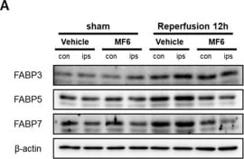
Detection of FABP7/B-FABP by Western Blot
Effects of MF6 administration on FABP3, FABP5 and FABP7 expression levels in I/R mice. Mice were subjected to right tMCAO for 2 h and administered 3 mg/kg MF6 at 30 min after reperfusion. At 12 h after reperfusion, the second slice of the right (ipsilateral) and left (contralateral) brain areas, including the cortex and striatum, were used for Western blot analysis of FABP levels. (A) Representative images of Western blots. Quantitative analyses of FABP3 (B), FABP5 (C) and FABP7 (D) proteins expression levels in the contralateral (black) and ipsilateral (blue) regions of the brains. ** p < 0.01 vs. the sham-treated group, administered the vehicle (contralateral/ipsilateral); #p < 0.05, ##p < 0.01 vs. the I/R-treated group, administered the vehicle (contralateral/ipsilateral) (n = 6 per group). Differences were statistically analyzed using two-way analysis of variance (ANOVA) followed by Student–Newman–Keuls test. Image collected and cropped by CiteAb from the following open publication (https://pubmed.ncbi.nlm.nih.gov/34068550), licensed under a CC-BY license. Not internally tested by R&D Systems.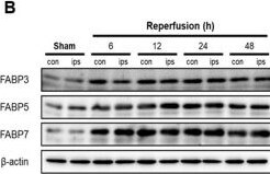
Detection of FABP7/B-FABP by Western Blot
Effects of I/R injury on FABP3, FABP5 and FABP7 expression levels in the brain. (A) The locations of samples. Brains were cut into four slices (2-mm thick) from the front of the cortex. The second slices (designated as con and ips slices) of the brain, including the cortex and striatum, were used for Western blot analysis and PGE2-content analysis in the following experiments. (B–E) Mice were subjected to right tMCAO for 2 h. At 6, 12, 24 and 48 h after reperfusion, the second slice of the right brain (ipsilateral) and the left brain (contralateral) areas were collected for Western blot analysis of FABPs levels. (B) Representative images of Western blots. Quantitative analyses of FABP3 (C), FABP5 (D) and FABP7 (E) expression levels in contralateral (black) and ipsilateral (blue) areas of the brains. #p < 0.05, ##p < 0.01 vs. the sham group (contralateral); * p < 0.05, ** p < 0.01 vs. the sham group (ipsilateral) (n = 6 per group). Differences were statistically analyzed using one-way analysis of variance (ANOVA) followed by Dunnett’s test. Image collected and cropped by CiteAb from the following open publication (https://pubmed.ncbi.nlm.nih.gov/34068550), licensed under a CC-BY license. Not internally tested by R&D Systems.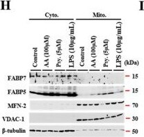
Detection of FABP7/B-FABP by Western Blot
Abnormal accumulation of FABP5 in mitochondria under psychosine exposure. (A) Confocal images of actin (cyan) and Mito-tracker (red), KG-1C cells were treated with AA (100 μM), psychosine (5 μM), or LPS (10 μg/mL), respectively. (B) Microscopy image of KG-1C cells, stained with Mito-tracker. Inverted image was processed by ImageJ software. (C,D) Quantification of dendrite length (n > 40) (C) and mitochondria numbers in the cell body and dendrites (n > 26) (D). KG-1C cells were treated with AA (100 μM), psychosine (5 μM), or LPS (10 μg/mL), respectively. (E) Confocal images of TOM20 (green), FABP5 (red), and DAPI (blue). KG-1C cells were treated with psychosine (5 μM) for 0, 6, 12, or 24 h, respectively. Mitochondrial fission occurs during psychosine exposure and FABP5 shifts to mitochondria during mitochondrial fission. (F) Amplified images of mitochondria in (E), noise was processed by waifu2x-caffe software. (G) Confocal images of Mito-tracker (green), FABP5 (red), and actin (cyan). KG-1C cells treated with psychosine (5 μM) for 24 h, show co-localization of FABP5 and mitochondria after psychosine treatment. (H) Western blot analysis showing that FABP5, but not FABP7, is elevated in the mitochondrial fraction in psychosine-treated KG-1C cells (n = 4). (I,J) Quantification of FABP7 in the cytosolic fraction and FABP5 in both the cytosolic fraction and mitochondrial fraction. (K) Schematic diagram of FABP5 shifting to mitochondria during psychosine exposure. The data are shown as mean ± standard error of the mean and were obtained using a one-way analysis of variance. ** p < 0.01. AA, arachidonic acid; psy, psychosine; LPS, lipopolysaccharide; Cyto., cytosolic fraction; Mito., mitochondrial fraction; NS, not statistically significant. Scale bar = 50 μm. Image collected and cropped by CiteAb from the following open publication (https://pubmed.ncbi.nlm.nih.gov/33419250), licensed under a CC-BY license. Not internally tested by R&D Systems.Applications for Human FABP7/B-FABP Antibody
Immunohistochemistry
Sample: Immersion fixed paraffin-embedded sections of human melanoma and astrocytoma
Simple Western
Sample: Human cerebellum tissue and human hippocampus tissue
Western Blot
Sample: Recombinant Human FABP7
Reviewed Applications
Read 2 reviews rated 4 using AF3166 in the following applications:
Formulation, Preparation, and Storage
Purification
Reconstitution
Reconstitute at 0.2 mg/mL in sterile PBS. For liquid material, refer to CoA for concentration.
Formulation
Shipping
Stability & Storage
- 12 months from date of receipt, -20 to -70 °C as supplied.
- 1 month, 2 to 8 °C under sterile conditions after reconstitution.
- 6 months, -20 to -70 °C under sterile conditions after reconstitution.
Calculators
Background: FABP7/B-FABP
Long Name
Alternate Names
Gene Symbol
UniProt
Additional FABP7/B-FABP Products
Product Documents for Human FABP7/B-FABP Antibody
Certificate of Analysis
To download a Certificate of Analysis, please enter a lot or batch number in the search box below.
Note: Certificate of Analysis not available for kit components.
Product Specific Notices for Human FABP7/B-FABP Antibody
For research use only
Related Research Areas
Citations for Human FABP7/B-FABP Antibody
 Powered by Bioz
Customer Reviews for Human FABP7/B-FABP Antibody (2)
Have you used Human FABP7/B-FABP Antibody?
Submit a review and receive an Amazon gift card!
$25/€18/£15/$25CAN/¥2500 for a review with an image
$10/€7/£6/$10CAN/¥1110 for a review without an image
Submit a review
-
Application: Western BlotSample Tested: See PMID 21771320Species: RatVerified Customer | Posted 01/07/2015
-
Application: ELISASample Tested: See PMID 22646866Species: HumanVerified Customer | Posted 01/07/2015
There are no reviews that match your criteria.
Protocols
Find general support by application which include: protocols, troubleshooting, illustrated assays, videos and webinars.
- Antigen Retrieval Protocol (PIER)
- Antigen Retrieval for Frozen Sections Protocol
- Appropriate Fixation of IHC/ICC Samples
- Cellular Response to Hypoxia Protocols
- Chromogenic IHC Staining of Formalin-Fixed Paraffin-Embedded (FFPE) Tissue Protocol
- Chromogenic Immunohistochemistry Staining of Frozen Tissue
- ClariTSA™ Fluorophore Kits
- Detection & Visualization of Antibody Binding
- Fluorescent IHC Staining of Frozen Tissue Protocol
- Graphic Protocol for Heat-induced Epitope Retrieval
- Graphic Protocol for the Preparation and Fluorescent IHC Staining of Frozen Tissue Sections
- Graphic Protocol for the Preparation and Fluorescent IHC Staining of Paraffin-embedded Tissue Sections
- Graphic Protocol for the Preparation of Gelatin-coated Slides for Histological Tissue Sections
- IHC Sample Preparation (Frozen sections vs Paraffin)
- Immunofluorescent IHC Staining of Formalin-Fixed Paraffin-Embedded (FFPE) Tissue Protocol
- Immunohistochemistry (IHC) and Immunocytochemistry (ICC) Protocols
- Immunohistochemistry Frozen Troubleshooting
- Immunohistochemistry Paraffin Troubleshooting
- Preparing Samples for IHC/ICC Experiments
- Preventing Non-Specific Staining (Non-Specific Binding)
- Primary Antibody Selection & Optimization
- Protocol for Heat-Induced Epitope Retrieval (HIER)
- Protocol for Making a 4% Formaldehyde Solution in PBS
- Protocol for VisUCyte™ HRP Polymer Detection Reagent
- Protocol for the Preparation & Fixation of Cells on Coverslips
- Protocol for the Preparation and Chromogenic IHC Staining of Frozen Tissue Sections
- Protocol for the Preparation and Chromogenic IHC Staining of Frozen Tissue Sections - Graphic
- Protocol for the Preparation and Chromogenic IHC Staining of Paraffin-embedded Tissue Sections
- Protocol for the Preparation and Chromogenic IHC Staining of Paraffin-embedded Tissue Sections - Graphic
- Protocol for the Preparation and Fluorescent IHC Staining of Frozen Tissue Sections
- Protocol for the Preparation and Fluorescent IHC Staining of Paraffin-embedded Tissue Sections
- Protocol for the Preparation of Gelatin-coated Slides for Histological Tissue Sections
- R&D Systems Quality Control Western Blot Protocol
- TUNEL and Active Caspase-3 Detection by IHC/ICC Protocol
- The Importance of IHC/ICC Controls
- Troubleshooting Guide: Immunohistochemistry
- Troubleshooting Guide: Western Blot Figures
- Western Blot Conditions
- Western Blot Protocol
- Western Blot Protocol for Cell Lysates
- Western Blot Troubleshooting
- Western Blot Troubleshooting Guide
- View all Protocols, Troubleshooting, Illustrated assays and Webinars
Associated Pathways
