
Human FGFR2 (IIIb) Antibody (98707)
R&D Systems | Catalog # MAB665


Scientific images may be resized, cropped, or adjusted for brightness or contrast for presentation purposes. These updates are limited to presentation and do not affect the underlying scientific interpretation of the data. Where available, additional source data may be provided upon request.
Key Product Details
Species Reactivity
Validated:
Cited:
Applications
Validated:
Cited:
Label
Antibody Source
Product Specifications
Immunogen
Specificity
Clonality
Host
Isotype
Endotoxin Level
Scientific Data Images for Human FGFR2 (IIIb) Antibody (98707)
FGF R2 alpha Inhibition of FGF acidic-dependent Cell Proliferation and Neutralization by Human FGF R2 Antibody.
Recombinant Human FGF R2a (IIIb) Fc Chimera (Catalog # 663-FR) inhibits Recombinant Human FGF acidic (Catalog # 232-FA) induced proliferation in the NR6R‑3T3 mouse fibroblast cell line in a dose-dependent manner (orange line). Inhibition of Recombinant Human FGF acidic (0.3 ng/mL) activity elicited by Recombinant Human FGF R2a (IIIb) Fc Chimera (6 ng/mL) is neutralized (green line) by increasing concentrations of Human FGF R2 (IIIb) Monoclonal Antibody (Catalog # MAB665). The ND50 is typically 2-8 µg/mL in the presence of heparin (10 µg/mL).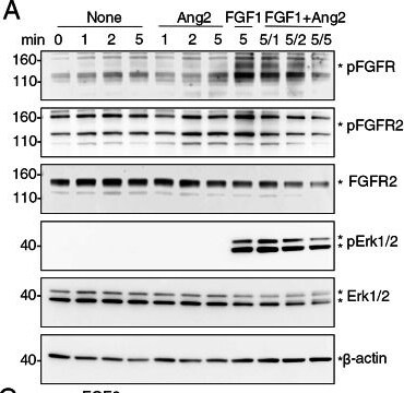
Detection of FGFR2 (IIIb) by Western Blot
Ang2 reduces p-FGFR, p-Erk1/2, p-STAT3 and p-AKT in HEK293T cells. A, B HEK293T cells were incubated in starvation medium only (None), with Ang2 (3 nM), with FGF1 (3 nM), or with Ang2 (3 nM) + FGF1 (3 nM) for the indicated times (min). Cell lysates were immunoblotted with the indicated antibodies. Representative results from 3 experiments. When FGF1 + Ang2 were added to the cells, the incubation time of FGF1 was 5 min and the incubation time of Ang2 is either 1, 2 or 5 minutes (displayed as 5/1, 5/2, 5/5). C HEK293T cells were incubated in starvation medium with FGF2 (3 nM) alone (2 or 5 min), or with Ang2 (3 nM), 5 min. The results show Ang2 + FGF2 for 5 min reduce FGF2-induced Erk1/2 activity (5 min). D Left panel: HEK293T cells were incubated in starvation medium only (None), FGF1 only (5 nM; 5 min), or with Ang2 (5 nM; 5 min) without or with AMG386 (AMG, 5 ng/ml; 1, 2, or 5 min). Right panel: HEK293T cells were incubated with medium only (None), FGF1 only (5 nM; 5 min), or FGF1 (5 nM; 5 min) + Ang2 (5 nM; 5 min), with or without AMG386 (5 ng/ml; 1, 2, or 5 min). Relative band intensity (p-Erk/total Erk) is shown in the bar graph. Representative of 3 experiments Image collected and cropped by CiteAb from the following open publication (https://link.springer.com/10.1007/s10456-025-09988-2), licensed under a CC-BY license. Not internally tested by R&D Systems.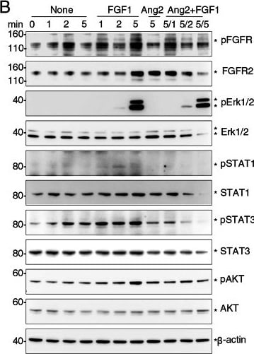
Detection of FGFR2 (IIIb) by Western Blot
Ang2 reduces p-FGFR, p-Erk1/2, p-STAT3 and p-AKT in HEK293T cells. A, B HEK293T cells were incubated in starvation medium only (None), with Ang2 (3 nM), with FGF1 (3 nM), or with Ang2 (3 nM) + FGF1 (3 nM) for the indicated times (min). Cell lysates were immunoblotted with the indicated antibodies. Representative results from 3 experiments. When FGF1 + Ang2 were added to the cells, the incubation time of FGF1 was 5 min and the incubation time of Ang2 is either 1, 2 or 5 minutes (displayed as 5/1, 5/2, 5/5). C HEK293T cells were incubated in starvation medium with FGF2 (3 nM) alone (2 or 5 min), or with Ang2 (3 nM), 5 min. The results show Ang2 + FGF2 for 5 min reduce FGF2-induced Erk1/2 activity (5 min). D Left panel: HEK293T cells were incubated in starvation medium only (None), FGF1 only (5 nM; 5 min), or with Ang2 (5 nM; 5 min) without or with AMG386 (AMG, 5 ng/ml; 1, 2, or 5 min). Right panel: HEK293T cells were incubated with medium only (None), FGF1 only (5 nM; 5 min), or FGF1 (5 nM; 5 min) + Ang2 (5 nM; 5 min), with or without AMG386 (5 ng/ml; 1, 2, or 5 min). Relative band intensity (p-Erk/total Erk) is shown in the bar graph. Representative of 3 experiments Image collected and cropped by CiteAb from the following open publication (https://link.springer.com/10.1007/s10456-025-09988-2), licensed under a CC-BY license. Not internally tested by R&D Systems.Applications for Human FGFR2 (IIIb) Antibody (98707)
Western Blot
Sample: Recombinant Human FGF R2 beta (IIIb) Fc Chimera (Catalog # 665-FR)
Neutralization
Formulation, Preparation, and Storage
Purification
Reconstitution
Reconstitute at 0.5 mg/mL in sterile PBS. For liquid material, refer to CoA for concentration.
Formulation
Shipping
Stability & Storage
- 12 months from date of receipt, -20 to -70 °C as supplied.
- 1 month, 2 to 8 °C under sterile conditions after reconstitution.
- 6 months, -20 to -70 °C under sterile conditions after reconstitution.
Calculators
Background: FGFR2
Long Name
Alternate Names
Gene Symbol
Additional FGFR2 Products
Product Documents for Human FGFR2 (IIIb) Antibody (98707)
Certificate of Analysis
To download a Certificate of Analysis, please enter a lot or batch number in the search box below.
Note: Certificate of Analysis not available for kit components.
Product Specific Notices for Human FGFR2 (IIIb) Antibody (98707)
For research use only
Citations for Human FGFR2 (IIIb) Antibody (98707)
 Powered by Bioz
Customer Reviews for Human FGFR2 (IIIb) Antibody (98707)
There are currently no reviews for this product. Be the first to review Human FGFR2 (IIIb) Antibody (98707) and earn rewards!
Have you used Human FGFR2 (IIIb) Antibody (98707)?
Submit a review and receive an Amazon gift card!
$25/€18/£15/$25CAN/¥2500 for a review with an image
$10/€7/£6/$10CAN/¥1110 for a review without an image
Submit a review
Protocols
Find general support by application which include: protocols, troubleshooting, illustrated assays, videos and webinars.
- Cellular Response to Hypoxia Protocols
- R&D Systems Quality Control Western Blot Protocol
- Troubleshooting Guide: Western Blot Figures
- Western Blot Conditions
- Western Blot Protocol
- Western Blot Protocol for Cell Lysates
- Western Blot Troubleshooting
- Western Blot Troubleshooting Guide
- View all Protocols, Troubleshooting, Illustrated assays and Webinars
Associated Pathways


