
Human Galectin-3 Antibody (194805)
R&D Systems | Catalog # MAB11541

Scientific images may be resized, cropped, or adjusted for brightness or contrast for presentation purposes. These updates are limited to presentation and do not affect the underlying scientific interpretation of the data. Where available, additional source data may be provided upon request.
Key Product Details
Species Reactivity
Validated:
Cited:
Applications
Validated:
Cited:
Label
Antibody Source
Product Specifications
Immunogen
Ala2-Ile250
Accession # P17931.5
Specificity
Clonality
Host
Isotype
Scientific Data Images for Human Galectin-3 Antibody (194805)
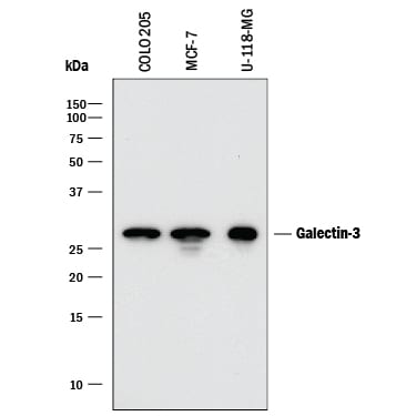
Detection of Human Galectin‑3 by Western Blot.
Western blot shows lysates of COLO 205 human colorectal adenocarcinoma cell line, MCF-7 human breast cancer cell line, and U-118-MG human glioblastoma/astrocytoma cell line. PVDF membrane was probed with 0.2 µg/mL of Mouse Anti-Human Galectin-3 Monoclonal Antibody (Catalog # MAB11541) followed by HRP-conjugated Anti-Mouse IgG Secondary Antibody (Catalog # HAF018). A specific band was detected for Galectin-3 at approximately 28 kDa (as indicated). This experiment was conducted under reducing conditions and using Immunoblot Buffer Group 1.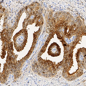
Galectin‑3 in Human Prostate Cancer Tissue.
Galectin-3 was detected in immersion fixed paraffin-embedded sections of human prostate cancer tissue using Mouse Anti-Human Galectin-3 Monoclonal Antibody (Catalog # MAB11541) at 1.7 µg/mL for 1 hour at room temperature followed by incubation with the Anti-Mouse IgG VisUCyte™ HRP Polymer Antibody (Catalog # VC001). Tissue was stained using DAB (brown) and counterstained with hematoxylin (blue). Specific staining was localized to cytoplasm and plasma membrane in epithelial cells. View our protocol for IHC Staining with VisUCyte HRP Polymer Detection Reagents.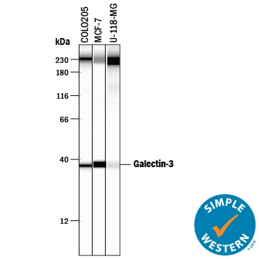
Detection of Human Galectin‑3 by Simple WesternTM.
Simple Western lane view shows lysates of COLO 205 human colorectal adenocarcinoma cell line, MCF‑7 human breast cancer cell line, and U‑118‑MG human glioblastoma/astrocytoma cell line, loaded at 0.2 mg/mL. A specific band was detected for Galectin‑3 at approximately 38 kDa (as indicated) using 10 µg/mL of Mouse Anti-Human Galectin‑3 Monoclonal Antibody (Catalog # MAB11541). This experiment was conducted under reducing conditions and using the 12-230 kDa separation system. Non-specific interaction with the 230 kDa Simple Western standard may be seen with this antibody.
Human Galectin-3 ELISA Standard Curve
Recombinant Human Galectin‑3 (Catalog # 1154-GA) was serially diluted and captured by Mouse Anti-Human Galectin‑3 Monoclonal Antibody (Catalog # MAB11541) coated on a Clear Polystyrene Microplate (Catalog # DY990). Goat Anti-Human Galectin‑3 Antigen Affinity-purified Polyclonal Antibody (Catalog # AF1154) was biotinylated and incubated with the protein captured on the plate. Detection of the standard curve was achieved by incubating Streptavidin-HRP (Catalog # DY998)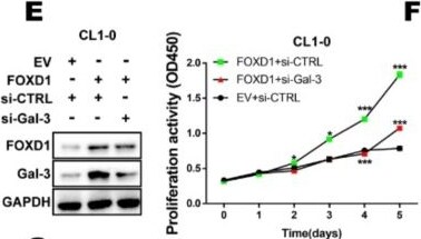
Detection of Galectin-3 by Western Blot
FOXD1 is a transcription factor of Gal-3. (A) CL1-0 cells were co-transfected with plasmids of the Gal-3 promoter reporter (pGL3-Gal-3 2000 bp) or Gal-3 promoter mutation reporter (pGL3-Gal-3 2000 bp-mut) and FOXD1 (pFOXD1). The cell lysates were collected, and the promoter activity of Gal-3 was analyzed by luciferase assay. (B) ChIP-qPCR assay using IgG as a control or FOXD1 antibody in CL1-0 and CL1-5 showed the binding of FOXD1 on the Gal-3 promoter. (C) CL1-0 cells transfected with the empty vector or plasmid of FOXD1 (pFOXD1) for 48 h and (D) CL1-5 cells transfected with scramble or siRNA targeting FOXD1 (si-FOXD1) for 72 h were collected. The mRNA and protein levels of FOXD1 and Gal-3 were analyzed by qPCR (left) and immunoblotting (right), respectively. (E) CCK-8 assay in different groups. FOXD1-stimulated CL1-0 proliferation ability was reduced by knocking down Gal-3. (F) The colony-formation ability of FOXD1-stimulated CL1-0 cells was suppressed by the depletion of Gal-3. (G) The numbers of migrating or invading cells were increased in the FOXD1 group compared with the FOXD1-stimulated CL1-0-siGal-3 group and control group. Image collected and cropped by CiteAb from the following open publication (https://pubmed.ncbi.nlm.nih.gov/31795213), licensed under a CC-BY license. Not internally tested by R&D Systems.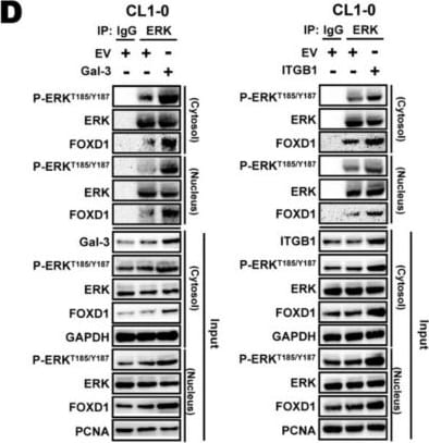
Detection of Galectin-3 by Western Blot
Gal-3 regulates FOXD1 expression through integrin-beta 1 (ITG beta 1) signaling. (A,B) CL1-0 and CL1-5 cells were transfected with the empty vector or plasmid of Gal-3 (pGal-3) for 48 h or scramble and siRNA targeting Gal-3 (si-Gal-3) for 72 h, respectively, as indicated. The mRNA and protein levels of FOXD1 and Gal-3 were analyzed by qPCR and immunoblotting. CL1-5 cells were transfected with scramble and siRNA targeting Gal-3 for 72 h or empty vector or plasmid of Gal-3 for 48 h, respectively, as indicated, and immunoprecipitation was analyzed by immunoblotting. Input, the whole cell lysates. (C) CL1-0 cells were transfected with scramble, Gal-3, ITG beta 1 or si-Gal-3, and si-ITG beta 1 for 72 h, as indicated. Cells lysates were collected and analyzed by immunoblotting. (D) CL1-0 cells were transfected with Gal-3 or ITG beta 1 expression vector for 48 h, and immunoprecipitation was analyzed by immunoblotting. Image collected and cropped by CiteAb from the following open publication (https://pubmed.ncbi.nlm.nih.gov/31795213), licensed under a CC-BY license. Not internally tested by R&D Systems.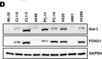
Detection of Galectin-3 by Western Blot
High FOXD1 and Gal-3 expression are associated with the poor prognosis in lung cancer. (A) The ranking of candidate molecules and the corresponding z-score and p-value using the Ingenuity Pathway Analyses (IPA) database from a microarray experiment performed with CL1-0 FOXD1 compared to CL1-0 vector and CL1-5 scramble compared to CL1-5 siRNA targeting FOXD1 (si-FOXD1) using a 5-fold change cut-off. (B) Statistical p-values and fold change of the expression of Gal-3 (LGALS3) and its relative molecules are shown. Prediction legend: upregulation (red round icon), downregulation (green round icon), predicted activation (brown round icon), predicted inhibition (blue round icon), leads to activation (brown line), leads to inhibition (blue line), findings inconsistent with stage of downstream molecule (yellow line), effect not predicted (gray line). (C) RT-qPCR analysis of Gal-3 and FOXD1 mRNA expression in various lung cancer cell lines. GAPDH was used as an internal control for mRNA loading. (D) Western blot analysis of Gal-3 and FOXD1 protein expression in various lung cancer cell lines. GAPDH was used as an internal control for protein loading. (E) Lung cancer patients with high gene level transcription of Gal-3 or FOXD1 expression level show a correlation with high risk and (F) poor disease-free survival. The data were retrieved and analyzed from the TCGA-LUAD samples (n = 475) and lung meta-base (n = 1044) of the SurvExpress database (http://bioinformatica.mty.itesm.mx:8080/Biomatec/SurvivaX.jsp). Image collected and cropped by CiteAb from the following open publication (https://pubmed.ncbi.nlm.nih.gov/31795213), licensed under a CC-BY license. Not internally tested by R&D Systems.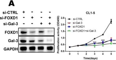
Detection of Galectin-3 by Western Blot
FOXD1 and Gal-3 form a positive regulatory loop to promote lung cancer cell growth and motility. CL1-5 cells were transfected with scramble, si-FOXD1, or si-Gal-3 or si-FOXD1 with si-Gal-3 for 72 h. Cells lysates were collected and analyzed by immunoblotting. (A) The proliferation ability was analyzed by CCK-8. (B) Representative images of the colony-forming assay. (C) Representative images of the migration and invasion assay. CL1-0 cells were transfected with the empty vector, FOXD1, Gal-3, or both FOXD1 and Gal-3 expression vector for 48 h. (D) Proliferation ability was analyzed by CCK-8 assay. (E) Representative images of colony-forming assay. (F) Representative images of migration and invasion assay. Image collected and cropped by CiteAb from the following open publication (https://pubmed.ncbi.nlm.nih.gov/31795213), licensed under a CC-BY license. Not internally tested by R&D Systems.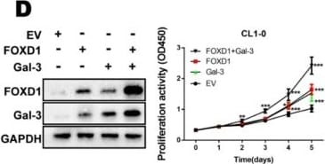
Detection of Galectin-3 by Western Blot
FOXD1 and Gal-3 form a positive regulatory loop to promote lung cancer cell growth and motility. CL1-5 cells were transfected with scramble, si-FOXD1, or si-Gal-3 or si-FOXD1 with si-Gal-3 for 72 h. Cells lysates were collected and analyzed by immunoblotting. (A) The proliferation ability was analyzed by CCK-8. (B) Representative images of the colony-forming assay. (C) Representative images of the migration and invasion assay. CL1-0 cells were transfected with the empty vector, FOXD1, Gal-3, or both FOXD1 and Gal-3 expression vector for 48 h. (D) Proliferation ability was analyzed by CCK-8 assay. (E) Representative images of colony-forming assay. (F) Representative images of migration and invasion assay. Image collected and cropped by CiteAb from the following open publication (https://pubmed.ncbi.nlm.nih.gov/31795213), licensed under a CC-BY license. Not internally tested by R&D Systems.Applications for Human Galectin-3 Antibody (194805)
Immunohistochemistry
Sample: Immersion fixed paraffin-embedded sections of human prostate cancer tissue
Simple Western
Sample: COLO 205 human colorectal adenocarcinoma cell line, MCF‑7 human breast cancer cell line, and U‑118‑MG human glioblastoma/astrocytoma cell line
Western Blot
Sample: COLO 205 human colorectal adenocarcinoma cell line, MCF‑7 human breast cancer cell line, and U‑118‑MG human glioblastoma/astrocytoma cell line
Human Galectin-3 Sandwich Immunoassay
Reviewed Applications
Read 4 reviews rated 4.5 using MAB11541 in the following applications:
Formulation, Preparation, and Storage
Purification
Reconstitution
Reconstitute at 0.5 mg/mL in sterile PBS. For liquid material, refer to CoA for concentration.
Formulation
Shipping
Stability & Storage
- 12 months from date of receipt, -20 to -70 °C as supplied.
- 1 month, 2 to 8 °C under sterile conditions after reconstitution.
- 6 months, -20 to -70 °C under sterile conditions after reconstitution.
Calculators
Background: Galectin-3
Alternate Names
Gene Symbol
UniProt
Additional Galectin-3 Products
Product Documents for Human Galectin-3 Antibody (194805)
Certificate of Analysis
To download a Certificate of Analysis, please enter a lot or batch number in the search box below.
Note: Certificate of Analysis not available for kit components.
Product Specific Notices for Human Galectin-3 Antibody (194805)
For research use only
Citations for Human Galectin-3 Antibody (194805)
 Powered by Bioz
Customer Reviews for Human Galectin-3 Antibody (194805) (4)
Have you used Human Galectin-3 Antibody (194805)?
Submit a review and receive an Amazon gift card!
$25/€18/£15/$25CAN/¥2500 for a review with an image
$10/€7/£6/$10CAN/¥1110 for a review without an image
Submit a review
Customer Images
-
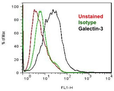
Application: Flow CytometrySample Tested: Human cell lineSpecies: HumanVerified Customer | Posted 10/26/2015Galectin-3 is detectable on the surface of the cells and is suitable at lower dilutions also. <br />Specificity: Specific<br />Sensitivity: Sensitive<br />Buffer: PBS<br />Dilution: 1:1000

-
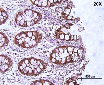
Application: ImmunohistochemistrySample Tested: Cancer TissueSpecies: HumanVerified Customer | Posted 10/26/2015The antibody is specific and detects only cells but not the background. <br />Specificity: Specific<br />Sensitivity: Sensitive<br />Buffer: PBS<br />Dilution: 1:1000
-
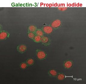
Application: Immunocytochemistry/ImmunofluorescenceSample Tested: Colon cancer cell lineSpecies: HumanVerified Customer | Posted 10/26/2015Very good binding ability. Detects galectin-3 at low dilutions also. <br />Specificity: Reasonably specific<br />Sensitivity: Sensitive<br />Buffer: PBS<br />Dilution: 1:1000
-
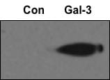
Application: Western BlotSample Tested: Cell lysate from colon cancer cell lineVerified Customer | Posted 10/26/2015Very sensitive, followed Milk block, primary BSA, Secondary Milk. As low as 1:2500 works too. <br />Specificity: Specific<br />Sensitivity: Sensitive<br />Buffer: Milk, BSA, Milk<br />Dilution: 1:1000
There are no reviews that match your criteria.
Protocols
Find general support by application which include: protocols, troubleshooting, illustrated assays, videos and webinars.
- Antigen Retrieval Protocol (PIER)
- Antigen Retrieval for Frozen Sections Protocol
- Appropriate Fixation of IHC/ICC Samples
- Cellular Response to Hypoxia Protocols
- Chromogenic IHC Staining of Formalin-Fixed Paraffin-Embedded (FFPE) Tissue Protocol
- Chromogenic Immunohistochemistry Staining of Frozen Tissue
- ClariTSA™ Fluorophore Kits
- Detection & Visualization of Antibody Binding
- Fluorescent IHC Staining of Frozen Tissue Protocol
- Graphic Protocol for Heat-induced Epitope Retrieval
- Graphic Protocol for the Preparation and Fluorescent IHC Staining of Frozen Tissue Sections
- Graphic Protocol for the Preparation and Fluorescent IHC Staining of Paraffin-embedded Tissue Sections
- Graphic Protocol for the Preparation of Gelatin-coated Slides for Histological Tissue Sections
- IHC Sample Preparation (Frozen sections vs Paraffin)
- Immunofluorescent IHC Staining of Formalin-Fixed Paraffin-Embedded (FFPE) Tissue Protocol
- Immunohistochemistry (IHC) and Immunocytochemistry (ICC) Protocols
- Immunohistochemistry Frozen Troubleshooting
- Immunohistochemistry Paraffin Troubleshooting
- Preparing Samples for IHC/ICC Experiments
- Preventing Non-Specific Staining (Non-Specific Binding)
- Primary Antibody Selection & Optimization
- Protocol for Heat-Induced Epitope Retrieval (HIER)
- Protocol for Making a 4% Formaldehyde Solution in PBS
- Protocol for VisUCyte™ HRP Polymer Detection Reagent
- Protocol for the Preparation & Fixation of Cells on Coverslips
- Protocol for the Preparation and Chromogenic IHC Staining of Frozen Tissue Sections
- Protocol for the Preparation and Chromogenic IHC Staining of Frozen Tissue Sections - Graphic
- Protocol for the Preparation and Chromogenic IHC Staining of Paraffin-embedded Tissue Sections
- Protocol for the Preparation and Chromogenic IHC Staining of Paraffin-embedded Tissue Sections - Graphic
- Protocol for the Preparation and Fluorescent IHC Staining of Frozen Tissue Sections
- Protocol for the Preparation and Fluorescent IHC Staining of Paraffin-embedded Tissue Sections
- Protocol for the Preparation of Gelatin-coated Slides for Histological Tissue Sections
- R&D Systems Quality Control Western Blot Protocol
- TUNEL and Active Caspase-3 Detection by IHC/ICC Protocol
- The Importance of IHC/ICC Controls
- Troubleshooting Guide: Immunohistochemistry
- Troubleshooting Guide: Western Blot Figures
- Western Blot Conditions
- Western Blot Protocol
- Western Blot Protocol for Cell Lysates
- Western Blot Troubleshooting
- Western Blot Troubleshooting Guide
- View all Protocols, Troubleshooting, Illustrated assays and Webinars