
Two alternatively spliced forms of HAI-2 have been found in human tissues (1). HAI-2A, the full-length molecule and also known as placental bikunin, is a major form expressed in human tissues. Encoded by the SPINT2 gene, HAI-2A consists of two Kunitz domains, and a C-terminal transmembrane domain (1-5). Both Kunitz domains can function as inhibitors independent of each other. In addition to HGF activator and trysin, HAI-2A strongly inhibits plasmin, tissue and plasma kallikreins, and factor XIa. In comparison, HAI-2A is a weaker inhibitor of factor VIIa-tissue factor, factors IXa, Xa, and XIIa. Recombinant HAI-2A prolonged the clotting time in an activated partial thromboplastin time assay.

Scientific images may be resized, cropped, or adjusted for brightness or contrast for presentation purposes. These updates are limited to presentation and do not affect the underlying scientific interpretation of the data. Where available, additional source data may be provided upon request.
Key Product Details
Species Reactivity
Validated:
Human
Cited:
Human
Applications
Validated:
Western Blot, Immunoprecipitation
Cited:
Western Blot
Label
Unconjugated
Antibody Source
Polyclonal Goat IgG
Loading...
Product Specifications
Immunogen
Mouse myeloma cell line NS0-derived recombinant human HAI-2A
Ala28-Lys197
Accession # O43291
Ala28-Lys197
Accession # O43291
Specificity
Detects human HAI-2 in direct ELISAs and Western blots. In direct ELISAs and Western blots, less than 5% cross-reactivity with recombinant human HAI‑1 and recombinant mouse HAI-2B is observed.
Clonality
Polyclonal
Host
Goat
Isotype
IgG
Scientific Data Images for Human HAI-2 Antibody
Detection of HAI-2 by Western Blot
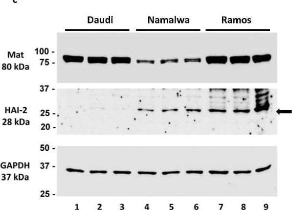
Expression analysis of matriptase, HAI-1, HAI-2, and prostasin in B cancer cells by reverse-transcription/qPCR (a,b), western blotting (c–e), and flow cytometry (f). (a) Bar graph of relative mRNA expression levels of matriptase (Mat), HAI-1, HAI-2, prostasin (Pro) in Daudi (n = 4), Namalwa (n = 5), Ramos (n = 3), Raji (n = 3), JeKo-1 (n = 3), and RS4;11 cells (n = 2) using GAPDH as the reference. The prostasin bars do not appear in the bar graph, as the actual qPCR readouts were registered as “N/A” by the instrument. (b) Bar graph of mRNA quantity ratio of HAI-2 to matriptase after normalization with the GAPDH level in each cell line in (a). (c) Western blotting images of matriptase (Ab: A300-221A), HAI-2, and GAPDH. Twenty micrograms of total protein from the cell lysate of each individual culture (including 2 repeats) were analyzed. Daudi, lanes 1–3; Namalwa, lanes 4–6; Ramos, lanes 7–9. Top panel, matriptase (Mat); middle panel, HAI-2; bottom panel, GAPDH. (d) Densitometry bar graph of relative protein quantities of matriptase and HAI-2 using GAPDH as the reference. (e) The quantitative ratio of HAI-2 to matriptase in each cell line. (f) Flow cytometry histogram of matriptase expression evaluation in Ramos cells. The Ramos cells (4 × 10⁵) were labeled with the matriptase antibody as described in the Materials and Methods section. The matriptase-positive cells are shown in the PE-A subset (blue peak). Cells without the matriptase antibody labeling (red peak) were not detected in the PE-A subset and were used as the gating control. Image collected and cropped by CiteAb from the following open publication (https://pubmed.ncbi.nlm.nih.gov/37568664), licensed under a CC-BY license. Not internally tested by R&D Systems.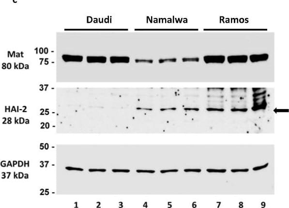
Detection of HAI-2 by Western Blot
Expression analysis of matriptase, HAI-1, HAI-2, and prostasin in B cancer cells by reverse-transcription/qPCR (a,b), western blotting (c–e), and flow cytometry (f). (a) Bar graph of relative mRNA expression levels of matriptase (Mat), HAI-1, HAI-2, prostasin (Pro) in Daudi (n = 4), Namalwa (n = 5), Ramos (n = 3), Raji (n = 3), JeKo-1 (n = 3), and RS4;11 cells (n = 2) using GAPDH as the reference. The prostasin bars do not appear in the bar graph, as the actual qPCR readouts were registered as “N/A” by the instrument. (b) Bar graph of mRNA quantity ratio of HAI-2 to matriptase after normalization with the GAPDH level in each cell line in (a). (c) Western blotting images of matriptase (Ab: A300-221A), HAI-2, and GAPDH. Twenty micrograms of total protein from the cell lysate of each individual culture (including 2 repeats) were analyzed. Daudi, lanes 1–3; Namalwa, lanes 4–6; Ramos, lanes 7–9. Top panel, matriptase (Mat); middle panel, HAI-2; bottom panel, GAPDH. (d) Densitometry bar graph of relative protein quantities of matriptase and HAI-2 using GAPDH as the reference. (e) The quantitative ratio of HAI-2 to matriptase in each cell line. (f) Flow cytometry histogram of matriptase expression evaluation in Ramos cells. The Ramos cells (4 × 10⁵) were labeled with the matriptase antibody as described in the Materials and Methods section. The matriptase-positive cells are shown in the PE-A subset (blue peak). Cells without the matriptase antibody labeling (red peak) were not detected in the PE-A subset and were used as the gating control. Image collected and cropped by CiteAb from the following open publication (https://pubmed.ncbi.nlm.nih.gov/37568664), licensed under a CC-BY license. Not internally tested by R&D Systems.
Human HAI-2 ELISA Standard Curve
Recombinant Human HAI‑2A (Catalog # 1106-PI) was serially diluted and captured by Mouse Anti-Human HAI‑2 Monoclonal Antibody (Catalog # MAB1106) coated on a Clear Polystyrene Microplate (Catalog # DY990). Goat Anti-Human HAI‑2 Antigen Affinity-purified Polyclonal Antibody (Catalog # AF1106) was biotinylated and incubated with the protein captured on the plate. Detection of the standard curve was achieved by incubating Streptavidin-HRP (Catalog # DY998)Applications for Human HAI-2 Antibody
Application
Recommended Usage
Immunoprecipitation
25 µg/mL
Sample: Conditioned cell culture medium spiked with Recombinant Human HAI‑2A (Catalog # 1106‑PI), see our available Western blot detection antibodies
Sample: Conditioned cell culture medium spiked with Recombinant Human HAI‑2A (Catalog # 1106‑PI), see our available Western blot detection antibodies
Western Blot
0.1 µg/mL
Sample: Recombinant Human HAI‑2A (Catalog # 1106-PI)
Sample: Recombinant Human HAI‑2A (Catalog # 1106-PI)
Formulation, Preparation, and Storage
Purification
Antigen Affinity-purified
Reconstitution
Reconstitute at 0.2 mg/mL in sterile PBS. For liquid material, refer to CoA for concentration.
Loading...
Formulation
Lyophilized from a 0.2 μm filtered solution in PBS with Trehalose. *Small pack size (SP) is supplied either lyophilized or as a 0.2 µm filtered solution in PBS.
Shipping
Lyophilized product is shipped at ambient temperature. Liquid small pack size (-SP) is shipped with polar packs. Upon receipt, store immediately at the temperature recommended below.
Stability & Storage
Use a manual defrost freezer and avoid repeated freeze-thaw cycles.
- 12 months from date of receipt, -20 to -70 °C as supplied.
- 1 month, 2 to 8 °C under sterile conditions after reconstitution.
- 6 months, -20 to -70 °C under sterile conditions after reconstitution.
Calculators
Background: HAI-2
References
- Kataoka, H. et al. (2002) Biochem. Biophys. Res. Comm. 290:1096.
- Kawaguchi, T. et al. (1997) J. Biol. Chem. 272:27558.
- Marlor. C.W. et al. (1997) J. Biol. Chem. 272:12202.
- Muller-Pillasch, F. et al. (1998) Biochim. Biophys. Acta 1395:88.
- Delaria, K.A. et al. (1997) J. Biol. Chem. 272:12209.
Long Name
HGF Activator Inhibitor Type 2
Alternate Names
HAI2
Gene Symbol
SPINT2
UniProt
Additional HAI-2 Products
Product Documents for Human HAI-2 Antibody
Certificate of Analysis
To download a Certificate of Analysis, please enter a lot or batch number in the search box below.
Note: Certificate of Analysis not available for kit components.
Product Specific Notices for Human HAI-2 Antibody
For research use only
Related Research Areas
Citations for Human HAI-2 Antibody
 Powered by Bioz
Customer Reviews for Human HAI-2 Antibody
There are currently no reviews for this product. Be the first to review Human HAI-2 Antibody and earn rewards!
Have you used Human HAI-2 Antibody?
Submit a review and receive an Amazon gift card!
$25/€18/£15/$25CAN/¥2500 for a review with an image
$10/€7/£6/$10CAN/¥1110 for a review without an image
Submit a review
Protocols
Find general support by application which include: protocols, troubleshooting, illustrated assays, videos and webinars.
- Cellular Response to Hypoxia Protocols
- Immunoprecipitation Protocol
- R&D Systems Quality Control Western Blot Protocol
- Troubleshooting Guide: Western Blot Figures
- Western Blot Conditions
- Western Blot Protocol
- Western Blot Protocol for Cell Lysates
- Western Blot Troubleshooting
- Western Blot Troubleshooting Guide
- View all Protocols, Troubleshooting, Illustrated assays and Webinars
Loading...