
HTRA2/Omi is the mammalian homologue of bacterial high temperature requirement protein (HtrA). HTRA2/Omi localizes to the mitochondria and is processed to expose an amino-terminal Reaper-like motif similar to SMAC/Diablo. HTRA2/Omi is released from the mitochondria in response to apoptotic insult and can interact with the BIR2 or BIR3 domains of XIAP to relieve caspase-IAP inhibition. This effect can be measured by reversing XIAP-BIR2 (R&D Systems, Catalog # 786-XB) inhibition of Caspase-7 (R&D Systems, Catalog # 823-C7) cleavage of a fluorogenic peptide (DEVD-AFC, MP Bio, Catalog # AFC-138). IC50 values for this effect are typically between 0.2 and 1.5 μM. HTRA2/Omi is trimeric and functions as a serine protease. The serine protease activity may play a more central role in apoptosis than its IAP antagonizing function. A PDZ domain regulates the serine protease activity by blocking access to the active site.
Human HTRA2/Omi Antibody (229926)
R&D Systems | Catalog # MAB1458

Scientific images may be resized, cropped, or adjusted for brightness or contrast for presentation purposes. These updates are limited to presentation and do not affect the underlying scientific interpretation of the data. Where available, additional source data may be provided upon request.
Key Product Details
Species Reactivity
Validated:
Cited:
Applications
Validated:
Cited:
Label
Antibody Source
Product Specifications
Immunogen
Ala134-Glu458
Accession # O43464
Specificity
Clonality
Host
Isotype
Scientific Data Images for Human HTRA2/Omi Antibody (229926)
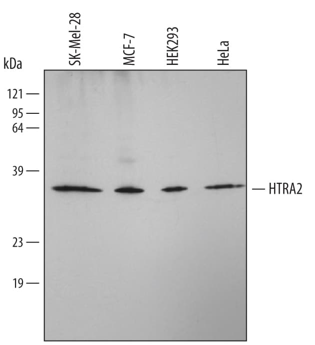
Detection of Human HTRA2/Omi by Western Blot.
Western blot shows lysates of SK-Mel-28 human malignant melanoma cell line, MCF-7 human breast cancer cell line, HEK293 human embryonic kidney cell line, and HeLa human cervical epithelial carcinoma cell line. PVDF membrane was probed with 1 µg/mL of Mouse Anti-Human HTRA2/Omi Monoclonal Antibody (Catalog # MAB1458) followed by HRP-conjugated Anti-Mouse IgG Secondary Antibody (Catalog # HAF007). A specific band was detected for HTRA2/Omi at approximately 35 kDa (as indicated). This experiment was conducted under reducing conditions and using Immunoblot Buffer Group 4.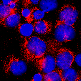
HTRA2/Omi in K562 Human Cell Line.
HTRA2/Omi was detected in immersion fixed K562 human chronic myelogenous leukemia cell line using Mouse Anti-Human HTRA2/Omi Monoclonal Antibody (Catalog # MAB1458) at 25 µg/mL for 3 hours at room temperature. Cells were stained using the NorthernLights™ 557-conjugated Anti-Mouse IgG Secondary Antibody (red; NL007) and counterstained with DAPI (blue). Specific staining was localized to cytoplasm. Staining was performed using our protocol for Fluorescent ICC Staining of Non-adherent Cells.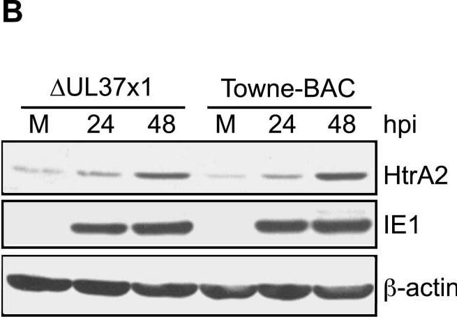
Detection of HTRA2/Omi by Western Blot
HtrA2/Omi expression increases following CMV infection.(A–B) Immunoblot analyses of HtrA2/Omi in lysates of mock-infected HF (M) or HF infected (MOI of 3) with Towne-BAC or delta UL37x1 for 24, 48, 72, 96, or 120 h and control cell lysates from HeLa cells transfected with HtrA2/Omi expression plasmid (+) or control plasmid (−). The expected migration of the 49 kDa immature, pro-HtrA2/Omi and 36 kDA mature, HtrA2/Omi was calculated based on migration of molecular weight markers (not shown). Immunoblot detection of HtrA2, beta -actin and IE1 are shown. (C–N) Immunofluorescent images of HtrA2/Omi (C, F) and cytochrome c (I, L) (red), and GFP fluorescence (green) (D, G, J, M) and merged images (E, H, K, N) at 72 h (C–H) or 96 h (I–N) postinfection (MOI of 0.001) with delta UL37x1 (C–E, I–K) or Towne-BAC (F–H, L–N). Original magnification ×1000. Image collected and cropped by CiteAb from the following open publication (https://pubmed.ncbi.nlm.nih.gov/18769594), licensed under a CC-BY license. Not internally tested by R&D Systems.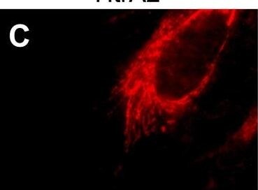
Detection of HTRA2/Omi by Immunocytochemistry/ Immunofluorescence
HtrA2/Omi expression increases following CMV infection.(A–B) Immunoblot analyses of HtrA2/Omi in lysates of mock-infected HF (M) or HF infected (MOI of 3) with Towne-BAC or delta UL37x1 for 24, 48, 72, 96, or 120 h and control cell lysates from HeLa cells transfected with HtrA2/Omi expression plasmid (+) or control plasmid (−). The expected migration of the 49 kDa immature, pro-HtrA2/Omi and 36 kDA mature, HtrA2/Omi was calculated based on migration of molecular weight markers (not shown). Immunoblot detection of HtrA2, beta -actin and IE1 are shown. (C–N) Immunofluorescent images of HtrA2/Omi (C, F) and cytochrome c (I, L) (red), and GFP fluorescence (green) (D, G, J, M) and merged images (E, H, K, N) at 72 h (C–H) or 96 h (I–N) postinfection (MOI of 0.001) with delta UL37x1 (C–E, I–K) or Towne-BAC (F–H, L–N). Original magnification ×1000. Image collected and cropped by CiteAb from the following open publication (https://pubmed.ncbi.nlm.nih.gov/18769594), licensed under a CC-BY license. Not internally tested by R&D Systems.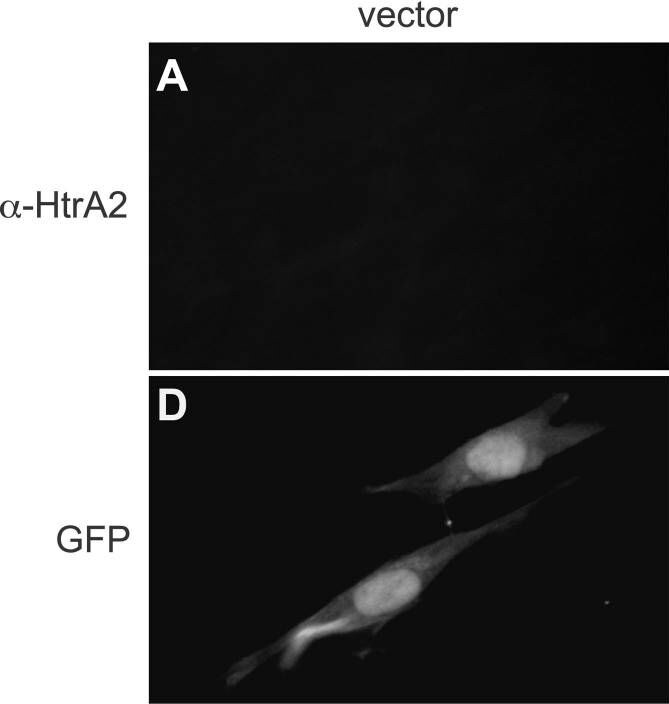
Detection of HTRA2/Omi by Immunocytochemistry/ Immunofluorescence
Transient overexpression of HtrA2/Omi induces early death in CMV-infected but not uninfected cells.(A–F) Immunofluorescent images of HtrA2/Omi (A–C) and GFP fluorescence (D–F) following cotransfection of HF with GFP expression plasmid together with empty vector, HtrA2/Omi, or HtrA2S306A expression plasmids. Original magnification ×400. (G) Immunoblot depicting 49 kDa, immature and 36 kDA, mature forms of HtrA2/Omi and HtrA2S306A as well as beta -actin control in transfected HeLa cell lysates. (H) Percentage of live HFs at 120 h post cotransfection of GFP expression plasmid with vector, HtrA2/Omi, or HtrA2S306A expression plasmids. (I) Number of viral plaques 10 days following cotransfection of delta UL37x1 or Towne-BAC DNA (500 ng) with 800 ng vector, HtrA2/Omi, or HtrA2S306A expression plasmids. (J) Number of live (intact) cells in cultures at 48 or 72 h following transfection with Towne-BAC DNA (300 ng) alone or with 333 ng of HtrA2 or HtrA2S306A expression plasmids. (K) Number of live (intact) cells (±range) at 48 or 72 h post transfection with Towne-BAC DNA (500 ng) and 300 ng of vector or HtrA2/Omi expression plasmid with or without addition of 10 µM UCF-101 from 6 h. Image collected and cropped by CiteAb from the following open publication (https://pubmed.ncbi.nlm.nih.gov/18769594), licensed under a CC-BY license. Not internally tested by R&D Systems.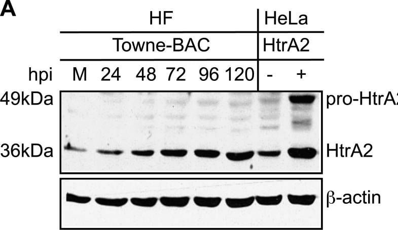
Detection of HTRA2/Omi by Western Blot
HtrA2/Omi expression increases following CMV infection.(A–B) Immunoblot analyses of HtrA2/Omi in lysates of mock-infected HF (M) or HF infected (MOI of 3) with Towne-BAC or delta UL37x1 for 24, 48, 72, 96, or 120 h and control cell lysates from HeLa cells transfected with HtrA2/Omi expression plasmid (+) or control plasmid (−). The expected migration of the 49 kDa immature, pro-HtrA2/Omi and 36 kDA mature, HtrA2/Omi was calculated based on migration of molecular weight markers (not shown). Immunoblot detection of HtrA2, beta -actin and IE1 are shown. (C–N) Immunofluorescent images of HtrA2/Omi (C, F) and cytochrome c (I, L) (red), and GFP fluorescence (green) (D, G, J, M) and merged images (E, H, K, N) at 72 h (C–H) or 96 h (I–N) postinfection (MOI of 0.001) with delta UL37x1 (C–E, I–K) or Towne-BAC (F–H, L–N). Original magnification ×1000. Image collected and cropped by CiteAb from the following open publication (https://pubmed.ncbi.nlm.nih.gov/18769594), licensed under a CC-BY license. Not internally tested by R&D Systems.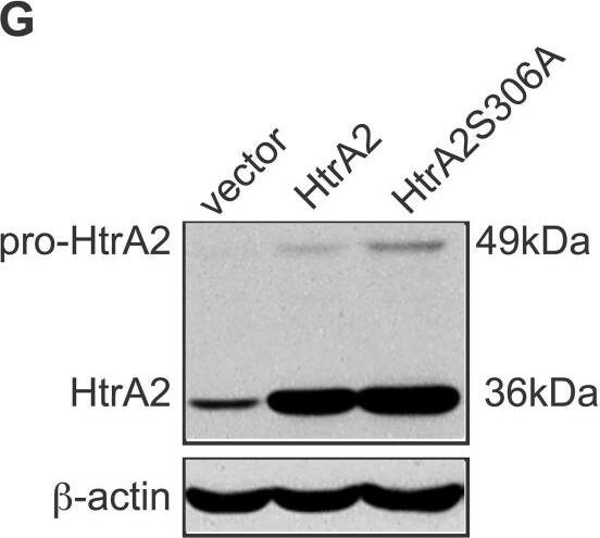
Detection of HTRA2/Omi by Western Blot
Transient overexpression of HtrA2/Omi induces early death in CMV-infected but not uninfected cells.(A–F) Immunofluorescent images of HtrA2/Omi (A–C) and GFP fluorescence (D–F) following cotransfection of HF with GFP expression plasmid together with empty vector, HtrA2/Omi, or HtrA2S306A expression plasmids. Original magnification ×400. (G) Immunoblot depicting 49 kDa, immature and 36 kDA, mature forms of HtrA2/Omi and HtrA2S306A as well as beta -actin control in transfected HeLa cell lysates. (H) Percentage of live HFs at 120 h post cotransfection of GFP expression plasmid with vector, HtrA2/Omi, or HtrA2S306A expression plasmids. (I) Number of viral plaques 10 days following cotransfection of delta UL37x1 or Towne-BAC DNA (500 ng) with 800 ng vector, HtrA2/Omi, or HtrA2S306A expression plasmids. (J) Number of live (intact) cells in cultures at 48 or 72 h following transfection with Towne-BAC DNA (300 ng) alone or with 333 ng of HtrA2 or HtrA2S306A expression plasmids. (K) Number of live (intact) cells (±range) at 48 or 72 h post transfection with Towne-BAC DNA (500 ng) and 300 ng of vector or HtrA2/Omi expression plasmid with or without addition of 10 µM UCF-101 from 6 h. Image collected and cropped by CiteAb from the following open publication (https://pubmed.ncbi.nlm.nih.gov/18769594), licensed under a CC-BY license. Not internally tested by R&D Systems.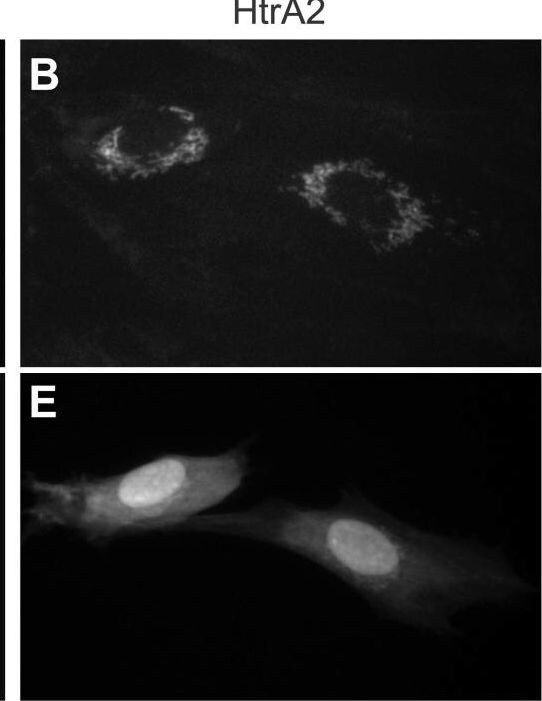
Detection of HTRA2/Omi by Immunocytochemistry/ Immunofluorescence
Transient overexpression of HtrA2/Omi induces early death in CMV-infected but not uninfected cells.(A–F) Immunofluorescent images of HtrA2/Omi (A–C) and GFP fluorescence (D–F) following cotransfection of HF with GFP expression plasmid together with empty vector, HtrA2/Omi, or HtrA2S306A expression plasmids. Original magnification ×400. (G) Immunoblot depicting 49 kDa, immature and 36 kDA, mature forms of HtrA2/Omi and HtrA2S306A as well as beta -actin control in transfected HeLa cell lysates. (H) Percentage of live HFs at 120 h post cotransfection of GFP expression plasmid with vector, HtrA2/Omi, or HtrA2S306A expression plasmids. (I) Number of viral plaques 10 days following cotransfection of delta UL37x1 or Towne-BAC DNA (500 ng) with 800 ng vector, HtrA2/Omi, or HtrA2S306A expression plasmids. (J) Number of live (intact) cells in cultures at 48 or 72 h following transfection with Towne-BAC DNA (300 ng) alone or with 333 ng of HtrA2 or HtrA2S306A expression plasmids. (K) Number of live (intact) cells (±range) at 48 or 72 h post transfection with Towne-BAC DNA (500 ng) and 300 ng of vector or HtrA2/Omi expression plasmid with or without addition of 10 µM UCF-101 from 6 h. Image collected and cropped by CiteAb from the following open publication (https://pubmed.ncbi.nlm.nih.gov/18769594), licensed under a CC-BY license. Not internally tested by R&D Systems.Applications for Human HTRA2/Omi Antibody (229926)
Immunocytochemistry
Sample: Immersion fixed K562 human chronic myelogenous leukemia cell line
Western Blot
Sample: SK-Mel-28 human malignant melanoma cell line, MCF-7 human breast cancer cell line, HEK293 human embryonic kidney cell line, and HeLa human cervical epithelial carcinoma cell line
Formulation, Preparation, and Storage
Purification
Reconstitution
Reconstitute at 0.5 mg/mL in sterile PBS. For liquid material, refer to CoA for concentration.
Formulation
Shipping
Stability & Storage
- 12 months from date of receipt, -20 to -70 °C as supplied.
- 1 month, 2 to 8 °C under sterile conditions after reconstitution.
- 6 months, -20 to -70 °C under sterile conditions after reconstitution.
Calculators
Background: HTRA2/Omi
References
- Suzuki, Y. et al. (2001) Mol. Cell. 8:613.
- van Loo, G. et al. (2002) Cell Death & Diff. 9:20.
- Hedge, R. et al. (2001) J. Biol. Chem. 277:432.
- Verhagen, A. et al. (2001) J. Biol. Chem. 277:445.
- Martins, L. et al. (2002) J. Biol. Chem. 277:439.
- Silke, J., and A. Verhagen (2002) Cell Death & Diff. 9:362.
- Savopoulos, J. et al. (2000) Protein Expression & Purification 19:227.
Long Name
Alternate Names
Gene Symbol
UniProt
Additional HTRA2/Omi Products
Product Documents for Human HTRA2/Omi Antibody (229926)
Certificate of Analysis
To download a Certificate of Analysis, please enter a lot or batch number in the search box below.
Note: Certificate of Analysis not available for kit components.
Product Specific Notices for Human HTRA2/Omi Antibody (229926)
For research use only
Related Research Areas
Citations for Human HTRA2/Omi Antibody (229926)
 Powered by Bioz
Customer Reviews for Human HTRA2/Omi Antibody (229926)
There are currently no reviews for this product. Be the first to review Human HTRA2/Omi Antibody (229926) and earn rewards!
Have you used Human HTRA2/Omi Antibody (229926)?
Submit a review and receive an Amazon gift card!
$25/€18/£15/$25CAN/¥2500 for a review with an image
$10/€7/£6/$10CAN/¥1110 for a review without an image
Submit a review
Protocols
Find general support by application which include: protocols, troubleshooting, illustrated assays, videos and webinars.
- Appropriate Fixation of IHC/ICC Samples
- Cellular Response to Hypoxia Protocols
- ClariTSA™ Fluorophore Kits
- Detection & Visualization of Antibody Binding
- ICC Cell Smear Protocol for Suspension Cells
- ICC Immunocytochemistry Protocol Videos
- ICC for Adherent Cells
- Immunocytochemistry (ICC) Protocol
- Immunocytochemistry Troubleshooting
- Immunofluorescence of Organoids Embedded in Cultrex Basement Membrane Extract
- Immunohistochemistry (IHC) and Immunocytochemistry (ICC) Protocols
- Preparing Samples for IHC/ICC Experiments
- Preventing Non-Specific Staining (Non-Specific Binding)
- Primary Antibody Selection & Optimization
- Protocol for VisUCyte™ HRP Polymer Detection Reagent
- Protocol for the Fluorescent ICC Staining of Cell Smears - Graphic
- Protocol for the Fluorescent ICC Staining of Cultured Cells on Coverslips - Graphic
- Protocol for the Preparation and Fluorescent ICC Staining of Cells on Coverslips
- Protocol for the Preparation and Fluorescent ICC Staining of Non-adherent Cells
- Protocol for the Preparation and Fluorescent ICC Staining of Stem Cells on Coverslips
- Protocol for the Preparation of a Cell Smear for Non-adherent Cell ICC - Graphic
- R&D Systems Quality Control Western Blot Protocol
- TUNEL and Active Caspase-3 Detection by IHC/ICC Protocol
- The Importance of IHC/ICC Controls
- Troubleshooting Guide: Western Blot Figures
- Western Blot Conditions
- Western Blot Protocol
- Western Blot Protocol for Cell Lysates
- Western Blot Troubleshooting
- Western Blot Troubleshooting Guide
- View all Protocols, Troubleshooting, Illustrated assays and Webinars
Associated Pathways
