
As a member of the sulfatase family, IDS is required for the lysosomal degradation of the glycosaminoglycans (GAG) heparan sulfate and dermatan sulfate (2, 3). It hydrolyzes the 2‑sulfate group of the L-iduronate 2-sulfate units of the GAG. The IDS deficiency results in mucopolysaccharidosis II (MPS II or Hunter syndrome), an X-linked inborn error leading to lysosomal accumulation of the GAG and its excretion in urine. MPS II has a wide spectrum of clinical manifestations ranging from mild to severe. The deduced amino acid sequence of human IDS consists of a signal peptide (residues 1-25), a pro peptide (residues 26-33) and a mature chain (residues 34-550) that may be further processed into the 42 kDa chain (residues 34-455) and the 14 kDa chain (residues 456-550) (1). Recombinant human IDS corresponds to the single chain and has sulfatase activity described above.
Human Iduronate 2-Sulfatase/IDS Antibody
R&D Systems | Catalog # AF2449


Scientific images may be resized, cropped, or adjusted for brightness or contrast for presentation purposes. These updates are limited to presentation and do not affect the underlying scientific interpretation of the data. Where available, additional source data may be provided upon request.
Key Product Details
Species Reactivity
Validated:
Cited:
Applications
Validated:
Cited:
Label
Antibody Source
Product Specifications
Immunogen
Ser26-Pro550
Accession # P22304
Specificity
Clonality
Host
Isotype
Scientific Data Images for Human Iduronate 2-Sulfatase/IDS Antibody
Detection of Human Iduronate 2‑Sulfatase/IDS by Western Blot.
Western blot shows lysates of human peripheral blood mononuclear cells (PBMCs). PVDF membrane was probed with 1 µg/mL of Goat Anti-Human Iduronate 2-Sulfatase/IDS Antigen Affinity-purified Polyclonal Antibody (Catalog # AF2449) followed by HRP-conjugated Anti-Goat IgG Secondary Antibody (Catalog # HAF017). A specific band was detected for Iduronate 2-Sulfatase/IDS at approximately 42 kDa (as indicated). This experiment was conducted under reducing conditions and using Immunoblot Buffer Group 1.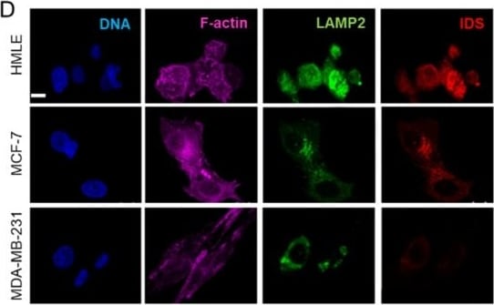
Detection of Iduronate 2-Sulfatase by Immunocytochemistry/ Immunofluorescence
Levels of iduronate-2 sulfatase (IDS) are decreased in cancer epithelia in vivo and in culture. (A) Scatter plot of dermatan sulfate (DS) levels normalized to total protein measured using ELISA in HMLE, MCF-7 and MDA-MB-231 cells cultured in 3D Type 1 collagen scaffolds. (B) Scatter plot of IDS mRNA levels in HMLE, MCF-7 and MDA-MB-231 cells cultured in 3D Type 1 collagen scaffolds, as determined by real time PCR with 18S rRNA as internal control. (C) (left) Epifluorescence micrographs of matched normal breast sections (top) and breast cancer tissues (bottom) stained for DNA (using DAPI; blue), and IDS (using antibody; red), scale = 200 μm. (middle) 3D profile plots of IDS levels in normal (top) and cancer (bottom) sections with bright colored peaks showing higher staining for IDS in nontransformed cells. (right) Bar graph showing a statistically significant decrease in IDS levels in breast cancer tissues compared with adjacent breast epithelial cells (50 cells, 5 fields, 2 sample sets). (D) Confocal micrographs of HMLE, MCF-7 and MDA-MB-231 cells cultured on top of Type 1 collagen scaffolds and stained for DNA (with DAPI; blue), F-actin (with phalloidin; pink), acidic compartment (with antibody against LAMP2; green), and for IDS (antibody; red), scale = 20 µm. Significance was measured using one-way ANOVA and student’s t test (* p < 0.05, ** p < 0.01, *** p < 0.001). Image collected and cropped by CiteAb from the following open publication (https://pubmed.ncbi.nlm.nih.gov/31574977), licensed under a CC-BY license. Not internally tested by R&D Systems.Applications for Human Iduronate 2-Sulfatase/IDS Antibody
Immunoprecipitation
Sample: Conditioned cell culture medium spiked with Recombinant Human Iduronate 2-Sulfatase/IDS (Catalog # 2449-SU), see our available Western blot detection antibodies
Western Blot
Sample: Human peripheral blood mononuclear cells (PBMCs)
Reviewed Applications
Read 1 review rated 4 using AF2449 in the following applications:
Formulation, Preparation, and Storage
Purification
Reconstitution
Reconstitute at 0.2 mg/mL in sterile PBS. For liquid material, refer to CoA for concentration.
Formulation
Shipping
Stability & Storage
- 12 months from date of receipt, -20 to -70 °C as supplied.
- 1 month, 2 to 8 °C under sterile conditions after reconstitution.
- 6 months, -20 to -70 °C under sterile conditions after reconstitution.
Calculators
Background: Iduronate 2-Sulfatase/IDS
References
- Wilson, P.J. et al. (1990) Proc. Natl. Acad. Sci. USA 87:8531.
- Parenti, G. et al. (1997) Curr. Opin. Genet. & Dev. 7:386.
- Neufeld, E.F. and J. Muenzer (2001) in The Metabolic and Molecular Basis of Inherited Disease, Scriver, C.R. et al. (eds.) pp. 3421-3452, New York, McGraw-Hill.
Alternate Names
Gene Symbol
UniProt
Additional Iduronate 2-Sulfatase/IDS Products
Product Documents for Human Iduronate 2-Sulfatase/IDS Antibody
Certificate of Analysis
To download a Certificate of Analysis, please enter a lot or batch number in the search box below.
Note: Certificate of Analysis not available for kit components.
Product Specific Notices for Human Iduronate 2-Sulfatase/IDS Antibody
For research use only
Related Research Areas
Citations for Human Iduronate 2-Sulfatase/IDS Antibody
 Powered by Bioz
Customer Reviews for Human Iduronate 2-Sulfatase/IDS Antibody (1)
Have you used Human Iduronate 2-Sulfatase/IDS Antibody?
Submit a review and receive an Amazon gift card!
$25/€18/£15/$25CAN/¥2500 for a review with an image
$10/€7/£6/$10CAN/¥1110 for a review without an image
Submit a review
-
Application: Western BlotSample Tested: See PMID 19602578Species: RatVerified Customer | Posted 01/07/2015
There are no reviews that match your criteria.
Protocols
Find general support by application which include: protocols, troubleshooting, illustrated assays, videos and webinars.
- Cellular Response to Hypoxia Protocols
- Immunoprecipitation Protocol
- R&D Systems Quality Control Western Blot Protocol
- Troubleshooting Guide: Western Blot Figures
- Western Blot Conditions
- Western Blot Protocol
- Western Blot Protocol for Cell Lysates
- Western Blot Troubleshooting
- Western Blot Troubleshooting Guide
- View all Protocols, Troubleshooting, Illustrated assays and Webinars