
The type 2 insulin-like growth factor receptor (also known as cation-independent mannose-6 phosphate receptor/CI-MPR) is a 300 kDa member of the P-type lectin family of molecules. P-type lectins generate functional eukaryotic lysosomes by binding and sorting lysosomal enzymes expressing phosphorylated mannose residues (M6P) (1-3). IGF-II R is a type I transmembrane glycoprotein that contains a 2,264 amino acid (aa) extracellular region, a 23 aa transmembrane segment and a 124 aa cytoplasmic tail (4, 5). The extracellular region consists of 15 contiguous “binding” repeats of about 150 aa each. The odd-numbered repeats interact with “ligands” while the even-numbered repeats likely generate a nondisulfide homodimer in the membrane (1). Repeat #11 binds IGF-II, while repeats 3 and 9 bind mannose-6 phosphate; repeat #13 contains a fibronectin type II motif and assists in IGF-II binding (1, 2). In the extracellular region of IGF-II R expressed by R&D Systems (600 aa’s), human IGF-II R is 85% aa identical to both mouse and bovine IGF-II R. This expressed region includes binding repeats #11, 12, and 13. In addition to IGF-II, CI-MPR/IGF-II R binds non-M6P containing ligands such as retinoic acid, urokinase-type plasminogen-activator receptor and plasminogen, plus M6P-containing molecules such as lysosomal enzymes, TGF-beta 1 precursor, proliferin, LIF, CD26, herpes simplex glycoprotein D and granzymes A and B (2, 6). IGF-II R regulates many diverse biological functions that range from intracellular trafficking to the internalization of extracellular factors and modulation of cellular responses. It delivers newly synthesized M6P-tagged lysosomal enzymes from the trans-golgi network to endosomes, and facilitates the clearance of extracellular lysosomal and matrix degrading enzymes by internalization into clathrin-coated vesicles and delivery into endosomes. With respect to IGF-II biology, it would appear that IGF-II R is principally a regulator of local IGF-II levels, targeting IGF-II for destruction in lysosomes (2). However, some evidence suggests the receptor will signal via G‑proteins, an effect that has yet to be conclusively shown (6).
Human IGF-II R/IGF2R Antibody (372604)
R&D Systems | Catalog # MAB2447

Scientific images may be resized, cropped, or adjusted for brightness or contrast for presentation purposes. These updates are limited to presentation and do not affect the underlying scientific interpretation of the data. Where available, additional source data may be provided upon request.
Key Product Details
Species Reactivity
Validated:
Cited:
Applications
Validated:
Cited:
Label
Antibody Source
Product Specifications
Immunogen
Ser1510-Phe2108
Accession # P11717.2
Specificity
Clonality
Host
Isotype
Scientific Data Images for Human IGF-II R/IGF2R Antibody (372604)
IGF-II R/IGF2R in A172 Human Cell Line.
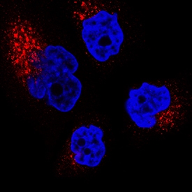
IGF-II R/IGF2R was detected in immersion fixed A172 human glioblastoma cell line using Mouse Anti-Human IGF-II R/IGF2R Monoclonal Antibody (Catalog # MAB2447) at 3 µg/mL for 3 hours at room temperature. Cells were stained using the NorthernLights™ 557-conjugated Anti-Mouse IgG Secondary Antibody (red; Catalog # NL007) and counterstained with DAPI (blue). Specific staining was localized to cytoplasm. View our protocol for Fluorescent ICC Staining of Cells on Coverslips.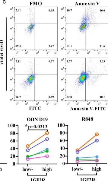
Detection of IGF-II R/IGF2R by Flow Cytometry
Analysis of the IGF2R expression and apoptosis in human circulating DCs. Purified human CD1c+ DCs were stimulated with stimuli as indicated and assessed IGF2R expression (A) and the percentages of apoptotic cells (all Annexin V+ cells and ViViD+ cells) within IGF2Rhigh and IGF2Rlow subsets (B, C). Apoptotic cells were gated against appropriate fluorescence minus one (FMO) controls. (D) Comparison of cell death percentages between IGF2Rhigh and IGF2Rlow human DCs from five volunteers. *p-value < 0.05 by Wilcoxon signed rank test (one-tailed). Image collected and cropped by CiteAb from the following open publication (https://pubmed.ncbi.nlm.nih.gov/33424850), licensed under a CC-BY license. Not internally tested by R&D Systems.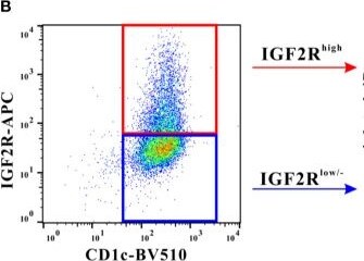
Detection of IGF-II R/IGF2R by Flow Cytometry
Analysis of the IGF2R expression and apoptosis in human circulating DCs. Purified human CD1c+ DCs were stimulated with stimuli as indicated and assessed IGF2R expression (A) and the percentages of apoptotic cells (all Annexin V+ cells and ViViD+ cells) within IGF2Rhigh and IGF2Rlow subsets (B, C). Apoptotic cells were gated against appropriate fluorescence minus one (FMO) controls. (D) Comparison of cell death percentages between IGF2Rhigh and IGF2Rlow human DCs from five volunteers. *p-value < 0.05 by Wilcoxon signed rank test (one-tailed). Image collected and cropped by CiteAb from the following open publication (https://pubmed.ncbi.nlm.nih.gov/33424850), licensed under a CC-BY license. Not internally tested by R&D Systems.Applications for Human IGF-II R/IGF2R Antibody (372604)
Immunocytochemistry
Sample: Immersion fixed A172 human glioblastoma cell line
Western Blot
Sample: Recombinant Human IGF-II R/IGF2R (Catalog # 2447-GR)
under non-reducing conditions only
Reviewed Applications
Read 1 review rated 5 using MAB2447 in the following applications:
Formulation, Preparation, and Storage
Purification
Reconstitution
Reconstitute at 0.5 mg/mL in sterile PBS. For liquid material, refer to CoA for concentration.
Formulation
Shipping
Stability & Storage
- 12 months from date of receipt, -20 to -70 °C as supplied.
- 1 month, 2 to 8 °C under sterile conditions after reconstitution.
- 6 months, -20 to -70 °C under sterile conditions after reconstitution.
Calculators
Background: IGF-II R/IGF2R
References
- Ghosh, P. et al. (2003) Nat. Rev. Mol. Cell. Biol. 4:202.
- Dahms, N.M. and M.K. Hancock (2002) Biochim. Biophys. Acta. 1572:317.
- Zaina, S. and J. Nilsson (2003) Curr. Opin. Lipidol. 14:483.
- Morgan, D.O. et al. (1987) Nature 329:301.
- Oshima, A. et al. (1988) J. Biol. Chem. 263:2553.
- Hawkes, C. and S. Kar (2004) Brain Res. Rev. 44:117.
Long Name
Alternate Names
Entrez Gene IDs
Gene Symbol
UniProt
Additional IGF-II R/IGF2R Products
Product Documents for Human IGF-II R/IGF2R Antibody (372604)
Certificate of Analysis
To download a Certificate of Analysis, please enter a lot or batch number in the search box below.
Note: Certificate of Analysis not available for kit components.
Product Specific Notices for Human IGF-II R/IGF2R Antibody (372604)
For research use only
Related Research Areas
Citations for Human IGF-II R/IGF2R Antibody (372604)
 Powered by Bioz
Customer Reviews for Human IGF-II R/IGF2R Antibody (372604) (1)
Have you used Human IGF-II R/IGF2R Antibody (372604)?
Submit a review and receive an Amazon gift card!
$25/€18/£15/$25CAN/¥2500 for a review with an image
$10/€7/£6/$10CAN/¥1110 for a review without an image
Submit a review
Customer Images
-
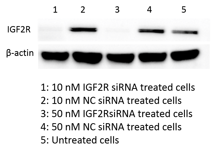
Application: Western BlotSample Tested: Cell LysatesSpecies: HumanVerified Customer | Posted 04/15/2016

There are no reviews that match your criteria.
Protocols
Find general support by application which include: protocols, troubleshooting, illustrated assays, videos and webinars.
- Appropriate Fixation of IHC/ICC Samples
- Cellular Response to Hypoxia Protocols
- ClariTSA™ Fluorophore Kits
- Detection & Visualization of Antibody Binding
- ICC Cell Smear Protocol for Suspension Cells
- ICC Immunocytochemistry Protocol Videos
- ICC for Adherent Cells
- Immunocytochemistry (ICC) Protocol
- Immunocytochemistry Troubleshooting
- Immunofluorescence of Organoids Embedded in Cultrex Basement Membrane Extract
- Immunohistochemistry (IHC) and Immunocytochemistry (ICC) Protocols
- Preparing Samples for IHC/ICC Experiments
- Preventing Non-Specific Staining (Non-Specific Binding)
- Primary Antibody Selection & Optimization
- Protocol for VisUCyte™ HRP Polymer Detection Reagent
- Protocol for the Fluorescent ICC Staining of Cell Smears - Graphic
- Protocol for the Fluorescent ICC Staining of Cultured Cells on Coverslips - Graphic
- Protocol for the Preparation and Fluorescent ICC Staining of Cells on Coverslips
- Protocol for the Preparation and Fluorescent ICC Staining of Non-adherent Cells
- Protocol for the Preparation and Fluorescent ICC Staining of Stem Cells on Coverslips
- Protocol for the Preparation of a Cell Smear for Non-adherent Cell ICC - Graphic
- R&D Systems Quality Control Western Blot Protocol
- TUNEL and Active Caspase-3 Detection by IHC/ICC Protocol
- The Importance of IHC/ICC Controls
- Troubleshooting Guide: Western Blot Figures
- Western Blot Conditions
- Western Blot Protocol
- Western Blot Protocol for Cell Lysates
- Western Blot Troubleshooting
- Western Blot Troubleshooting Guide
- View all Protocols, Troubleshooting, Illustrated assays and Webinars
Associated Pathways


