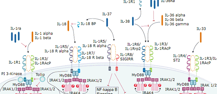
Interleukin 18 binding protein (IL-18 BP) is a secreted glycoprotein, which functions as an IL-18 antagonist by binding to IL-18 and blocking its biological activity. IL‑18 BP bears no amino acid sequence homology to the membrane-associated IL-18 and IL-1 receptor proteins. The gene for human IL-18 BP has been localized to chromosome 11q13. It encodes for at least four isoforms by alternative splicing. The IL-18 BP isoforms a and c each contain one immunoglobulin (Ig)-like C2-type domain while isoforms b and d lack a complete Ig domain. The complete Ig domain has been shown to be essential to the binding and neutralizing properties of the binding proteins. Two isoforms of mouse IL18 BP (c and d) containing the complete Ig domain have also been isolated and shown to neutralize IL-18 bioactivity. Human and mouse IL-18 BPs share approximately 61% amino acid sequence identity. Several poxviruses also encode proteins with sequence similarity to the human and mouse IL-18 BP. Viral IL-18 BPs have been shown to bind and inhibit IL-18 responses and may be involved in modulating host immune responses. The expression of IL-18 BP is markedly upregulated by IFN-gamma, suggesting that IL-18 activity is modulated by a negative feedback mechanism mediated by IL-18 BP.
Human IL-18 BPa Antibody (136033)
R&D Systems | Catalog # MAB119


Scientific images may be resized, cropped, or adjusted for brightness or contrast for presentation purposes. These updates are limited to presentation and do not affect the underlying scientific interpretation of the data. Where available, additional source data may be provided upon request.
Key Product Details
Species Reactivity
Validated:
Cited:
Applications
Validated:
Cited:
Label
Antibody Source
Product Specifications
Immunogen
Specificity
Clonality
Host
Isotype
Scientific Data Images for Human IL-18 BPa Antibody (136033)
Detection of IL-18 BPa by Western Blot
TNFR1-Fc-enhanced axon growth occurs independently of action potential generation & the requirement for PKC activation. (a) Bar charts of length & branch number & Sholl profiles of P0 SCG neurons cultured for 24 h with either 10 ng ml−1 NGF alone (control), NGF & 10 ng ml−1 TNFR1-Fc, NGF & 100 nM TTX or NGF, TNFR1-Fc & TTX. Mean ± s.e.m of neurite arbour data of 150 neurons per condition combined from three separate experiments of each type (***p < 0.001, statistical comparison with control). (b) Representative immunoblot probed for phospho-(Ser660) PKC, total PKC & beta -III tubulin of lysates of P0 SCG neurons that were cultured for 12 h in medium containing 10 ng ml−1 NGF before being treated for 15 min with TNFR1-Fc & 1 µM mibefradil. The bar chart plots the densitometry from four separate WBs of the ratio of pPKC to total PKC, normalized to 100% for the controls, mean ± s.e.m. (c) Representative immunoblot probed for phospho-ERK1/phospho-ERK2, total ERK1/ERK2 & beta -III tubulin of lysates of P0 SCG neurons that were cultured for 12 h in medium containing 10 ng ml−1 NGF before being treated for 15 min with 10 ng ml−1 TNFR1-Fc & 10 µM GF 109203X. The bar chart plots the densitometry from four separate WBs of the ratio of pERK1/2 to total ERK1/2, normalized to 100% for the controls (*p < 0.05 & **p < 0.01, statistical comparison with control). (d) Bar charts of length & branch point number & the Sholl profiles of P0 SCG neurons cultured for 24 h with either NGF alone (control), NGF & TNFR1-Fc, NGF & GF 109203X or NGF, TNFR1-Fc & GF 109203X. The data shown represent the mean ± s.e.m. of neurite arbour data of 150 neurons per condition combined from three separate experiments of each type (***p < 0.001, statistical comparison with control). Image collected & cropped by CiteAb from the following open publication (https://pubmed.ncbi.nlm.nih.gov/28100666), licensed under a CC-BY license. Not internally tested by R&D Systems.
Detection of IL-18 BPa by Western Blot
TNFR1-Fc-enhanced axon growth occurs independently of action potential generation & the requirement for PKC activation. (a) Bar charts of length & branch number & Sholl profiles of P0 SCG neurons cultured for 24 h with either 10 ng ml−1 NGF alone (control), NGF & 10 ng ml−1 TNFR1-Fc, NGF & 100 nM TTX or NGF, TNFR1-Fc & TTX. Mean ± s.e.m of neurite arbour data of 150 neurons per condition combined from three separate experiments of each type (***p < 0.001, statistical comparison with control). (b) Representative immunoblot probed for phospho-(Ser660) PKC, total PKC & beta -III tubulin of lysates of P0 SCG neurons that were cultured for 12 h in medium containing 10 ng ml−1 NGF before being treated for 15 min with TNFR1-Fc & 1 µM mibefradil. The bar chart plots the densitometry from four separate WBs of the ratio of pPKC to total PKC, normalized to 100% for the controls, mean ± s.e.m. (c) Representative immunoblot probed for phospho-ERK1/phospho-ERK2, total ERK1/ERK2 & beta -III tubulin of lysates of P0 SCG neurons that were cultured for 12 h in medium containing 10 ng ml−1 NGF before being treated for 15 min with 10 ng ml−1 TNFR1-Fc & 10 µM GF 109203X. The bar chart plots the densitometry from four separate WBs of the ratio of pERK1/2 to total ERK1/2, normalized to 100% for the controls (*p < 0.05 & **p < 0.01, statistical comparison with control). (d) Bar charts of length & branch point number & the Sholl profiles of P0 SCG neurons cultured for 24 h with either NGF alone (control), NGF & TNFR1-Fc, NGF & GF 109203X or NGF, TNFR1-Fc & GF 109203X. The data shown represent the mean ± s.e.m. of neurite arbour data of 150 neurons per condition combined from three separate experiments of each type (***p < 0.001, statistical comparison with control). Image collected & cropped by CiteAb from the following open publication (https://pubmed.ncbi.nlm.nih.gov/28100666), licensed under a CC-BY license. Not internally tested by R&D Systems.Applications for Human IL-18 BPa Antibody (136033)
Western Blot
Sample: Recombinant Human IL‑18 BPa Fc Chimera (Catalog # 119-BP)
Formulation, Preparation, and Storage
Purification
Reconstitution
Reconstitute at 0.5 mg/mL in sterile PBS. For liquid material, refer to CoA for concentration.
Formulation
Shipping
Stability & Storage
- 12 months from date of receipt, -20 to -70 °C as supplied.
- 1 month, 2 to 8 °C under sterile conditions after reconstitution.
- 6 months, -20 to -70 °C under sterile conditions after reconstitution.
Calculators
Background: IL-18 BPa
References
- Mühl, H. et al. (2000) Biochem. Biophys. Res. Commun. 267:960.
- Kim, S-H. et al. (2000) Proc. Nat. Acad. Sci. USA 97:1190.
- Calderara, S. et al. (2001) Virology 279:22.
Long Name
Alternate Names
Gene Symbol
Additional IL-18 BPa Products
Product Documents for Human IL-18 BPa Antibody (136033)
Certificate of Analysis
To download a Certificate of Analysis, please enter a lot or batch number in the search box below.
Note: Certificate of Analysis not available for kit components.
Product Specific Notices for Human IL-18 BPa Antibody (136033)
For research use only
Related Research Areas
Citations for Human IL-18 BPa Antibody (136033)
 Powered by Bioz
Customer Reviews for Human IL-18 BPa Antibody (136033)
There are currently no reviews for this product. Be the first to review Human IL-18 BPa Antibody (136033) and earn rewards!
Have you used Human IL-18 BPa Antibody (136033)?
Submit a review and receive an Amazon gift card!
$25/€18/£15/$25CAN/¥2500 for a review with an image
$10/€7/£6/$10CAN/¥1110 for a review without an image
Submit a review
Protocols
Find general support by application which include: protocols, troubleshooting, illustrated assays, videos and webinars.
- Cellular Response to Hypoxia Protocols
- R&D Systems Quality Control Western Blot Protocol
- Troubleshooting Guide: Western Blot Figures
- Western Blot Conditions
- Western Blot Protocol
- Western Blot Protocol for Cell Lysates
- Western Blot Troubleshooting
- Western Blot Troubleshooting Guide
- View all Protocols, Troubleshooting, Illustrated assays and Webinars
Associated Pathways