Human IL-1 beta/IL-1F2 Antibody [Biotin]
R&D Systems | Catalog # BAF201

Scientific images may be resized, cropped, or adjusted for brightness or contrast for presentation purposes. These updates are limited to presentation and do not affect the underlying scientific interpretation of the data. Where available, additional source data may be provided upon request.
Key Product Details
Species Reactivity
Validated:
Cited:
Applications
Validated:
Cited:
Label
Antibody Source
Product Specifications
Immunogen
Ala117-Ser269
Accession # NP_000567
Specificity
Clonality
Host
Isotype
Scientific Data Images for Human IL-1 beta/IL-1F2 Antibody [Biotin]
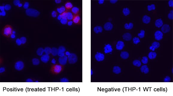
Detection of IL‑1 beta /IL‑1F2 in THP‑1 Human Cell Line.
IL‑1 beta /IL‑1F2 was detected in immersion fixed THP‑1 human acute monocytic leukemia cell line using Goat Anti-Human IL‑1 beta /IL‑1F2 Biotinylated Antigen Affinity-purified Polyclonal Antibody (Catalog # BAF201) at 5 µg/ml for 3 hours at room temperature. Cells were stained using the NorthernLights™ 557-conjugated Streptavidin (red; Catalog # NL999) and counterstained with DAPI (blue). Specific staining was localized to the cytoplasm of THP-1 cells treated with 200nM PMA for 24 hours then 10ug/mL LPS for 24 hours. View our protocol for Fluorescent ICC Staining of Cells on Coverslips.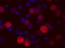
IL‑1 beta /IL‑1F2 in Human PBMCs.
IL-1 beta /IL-1F2 was detected in immersion fixed human peripheral blood mononuclear cells (PBMCs) using Human IL-1 beta /IL-1F2 Biotinylated Antigen Affinity-purified Polyclonal Antibody (Catalog # BAF201) at 10 µg/mL for 3 hours at room temperature. Cells were stained using the NorthernLights™ 557-conjugated Streptavidin (red; Catalog # NL999) and counterstained with DAPI (blue). View our protocol for Fluorescent ICC Staining of Non-adherent Cells.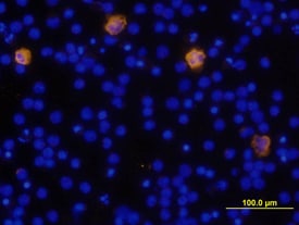
IL‑1 beta /IL‑1F2 in Human PBMCs.
IL-1 beta /IL-1F2 was detected in immersion fixed human peripheral blood mononuclear cells (PBMCs) using Human IL-1 beta /IL-1F2 Biotinylated Antigen Affinity-purified Polyclonal Antibody (Catalog # BAF201) at 10 µg/mL for 3 hours at room temperature. Cells were stained using the NorthernLights™ 557-conjugated Anti-Goat IgG Secondary Antibody (yellow; Catalog # NL001) and counterstained with DAPI (blue). View our protocol for Fluorescent ICC Staining of Non-adherent Cells.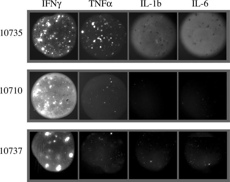
Detection of Human IL-1 beta/IL-1F2 by Functional
Differences in Functional Profiles between Three Patients with Different Clinical Outcomesgp100 209–2M spots co-spotted with anti-IFN gamma, anti-TNF alpha, anti-IL1b, and anti-IL-6 are shown for three separate patient samples. Patient 10735, who remains free of disease at this writing, displays strong IFN gamma, TNF alpha, IL-1b, and IL-6 secretion. Patients 10710 and 10737 show strong IFN gamma secretion, but weak to no TNF alpha, IL-1b, and IL-6 secretion; both patients experienced disease recurrence soon after these samples were drawn. Note the diffuse pattern of IL-1b and IL-6 capture, which differs from the focal capture of IFN gamma and TNF alpha. Image collected and cropped by CiteAb from the following publication (https://pubmed.ncbi.nlm.nih.gov/16162034), licensed under a CC-BY license. Not internally tested by R&D Systems.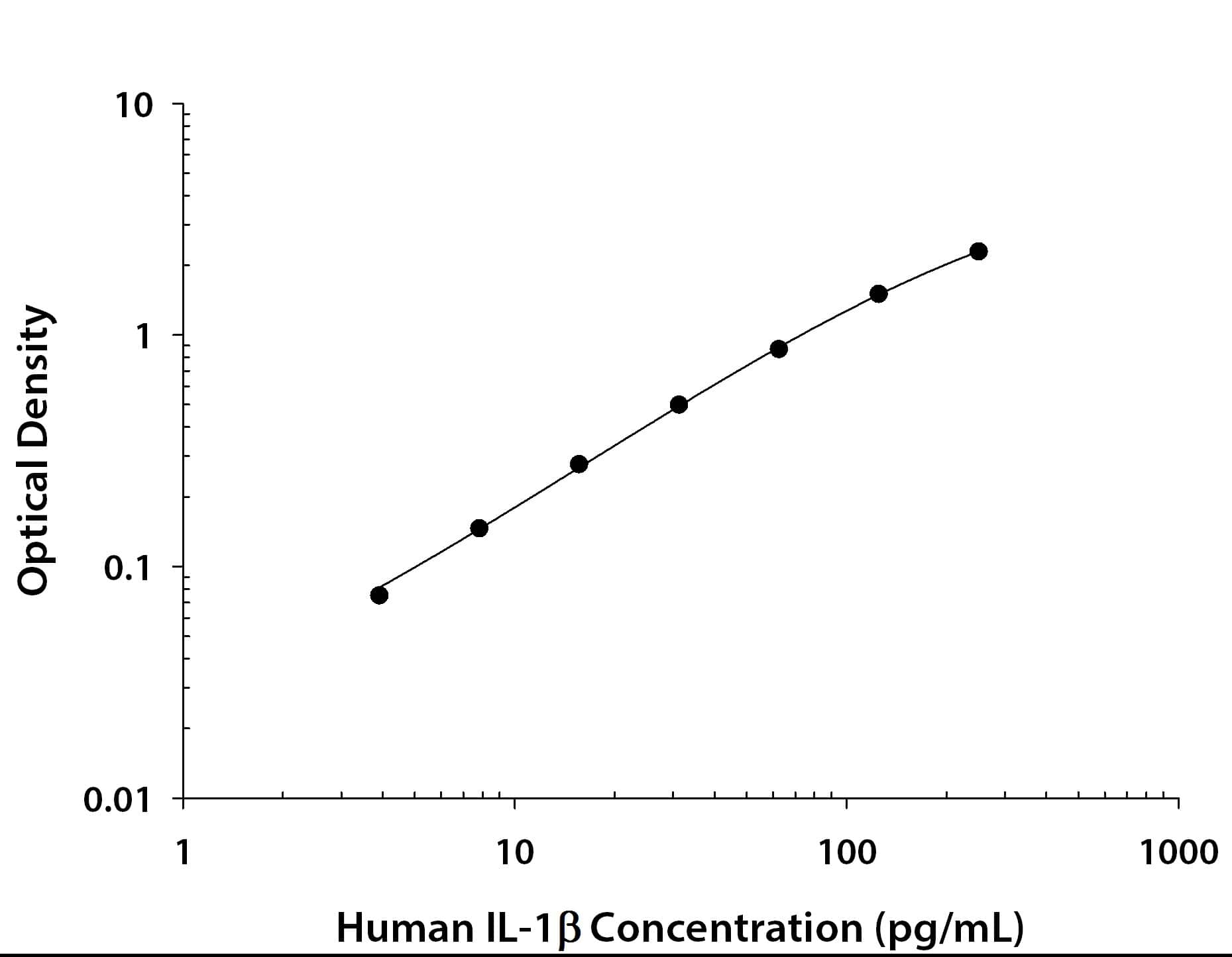
Human IL-1 beta / IL-1F2 ELISA Standard Curve
Recombinant Human IL‑1 beta /IL‑1F2 (Catalog # 201-LB) was serially diluted and captured by Mouse Anti-Human IL‑1 beta /IL‑1F2 Monoclonal Antibody (Catalog # MAB601) coated on a Clear Polystyrene Microplate (Catalog # DY990). Goat Anti-Human IL‑1 beta /IL‑1F2 Antigen Affinity-purified Polyclonal Antibody (Catalog # AF-201-NA) was biotinylated and incubated with the protein captured on the plate. Detection of the standard curve was achieved by incubating Streptavidin-HRP (Catalog # DY998)Applications for Human IL-1 beta/IL-1F2 Antibody [Biotin]
Immunocytochemistry
Sample: Immersion fixed human peripheral blood mononuclear cells (PBMCs) and THP-1 human acute monocytic leukemia cell line untreated and treated with PMA and LPS
Western Blot
Sample: Recombinant Human IL‑1 beta /IL‑1F2 (Catalog # 201-LB)
Human IL-1 beta /IL-1F2 Sandwich Immunoassay
Formulation, Preparation, and Storage
Purification
Reconstitution
Reconstitute at 0.2 mg/mL in sterile PBS.
Formulation
Shipping
Stability & Storage
- 12 months from date of receipt, -20 to -70 °C as supplied.
- 1 month, 2 to 8 °C under sterile conditions after reconstitution.
- 6 months, -20 to -70 °C under sterile conditions after reconstitution.
Calculators
Background: IL-1 beta/IL-1F2
Long Name
Alternate Names
Entrez Gene IDs
Gene Symbol
UniProt
Additional IL-1 beta/IL-1F2 Products
Product Documents for Human IL-1 beta/IL-1F2 Antibody [Biotin]
Certificate of Analysis
To download a Certificate of Analysis, please enter a lot or batch number in the search box below.
Note: Certificate of Analysis not available for kit components.
Product Specific Notices for Human IL-1 beta/IL-1F2 Antibody [Biotin]
For research use only
Related Research Areas
Citations for Human IL-1 beta/IL-1F2 Antibody [Biotin]
 Powered by Bioz
Customer Reviews for Human IL-1 beta/IL-1F2 Antibody [Biotin]
There are currently no reviews for this product. Be the first to review Human IL-1 beta/IL-1F2 Antibody [Biotin] and earn rewards!
Have you used Human IL-1 beta/IL-1F2 Antibody [Biotin]?
Submit a review and receive an Amazon gift card!
$25/€18/£15/$25CAN/¥2500 for a review with an image
$10/€7/£6/$10CAN/¥1110 for a review without an image
Submit a review
Protocols
Find general support by application which include: protocols, troubleshooting, illustrated assays, videos and webinars.
- Appropriate Fixation of IHC/ICC Samples
- Cellular Response to Hypoxia Protocols
- ClariTSA™ Fluorophore Kits
- Detection & Visualization of Antibody Binding
- ICC Cell Smear Protocol for Suspension Cells
- ICC Immunocytochemistry Protocol Videos
- ICC for Adherent Cells
- Immunocytochemistry (ICC) Protocol
- Immunocytochemistry Troubleshooting
- Immunofluorescence of Organoids Embedded in Cultrex Basement Membrane Extract
- Immunohistochemistry (IHC) and Immunocytochemistry (ICC) Protocols
- Preparing Samples for IHC/ICC Experiments
- Preventing Non-Specific Staining (Non-Specific Binding)
- Primary Antibody Selection & Optimization
- Protocol for VisUCyte™ HRP Polymer Detection Reagent
- Protocol for the Fluorescent ICC Staining of Cell Smears - Graphic
- Protocol for the Fluorescent ICC Staining of Cultured Cells on Coverslips - Graphic
- Protocol for the Preparation and Fluorescent ICC Staining of Cells on Coverslips
- Protocol for the Preparation and Fluorescent ICC Staining of Non-adherent Cells
- Protocol for the Preparation and Fluorescent ICC Staining of Stem Cells on Coverslips
- Protocol for the Preparation of a Cell Smear for Non-adherent Cell ICC - Graphic
- R&D Systems Quality Control Western Blot Protocol
- TUNEL and Active Caspase-3 Detection by IHC/ICC Protocol
- The Importance of IHC/ICC Controls
- Troubleshooting Guide: Western Blot Figures
- Western Blot Conditions
- Western Blot Protocol
- Western Blot Protocol for Cell Lysates
- Western Blot Troubleshooting
- Western Blot Troubleshooting Guide
- View all Protocols, Troubleshooting, Illustrated assays and Webinars
Associated Pathways