
Tissue kallikreins are a family of extracellular serine proteases consisting of 15 members (1, 2). The genes of this family aligned in tandem on chromosome 19q13.4 represent the largest contiguous group of proteases within the human genome. Tissue kallikreins have attracted great interest as potential biomarkers for various cancers, including prostate, ovarian, breast, testicular, and lung (2, 3). Human Kallikrein 6 (hKLK6) is a member of tissue kallikrein family observed in breast and brain tissues, colon carcinoma cells, and oligodedrocytes (1-3). Known protein substrates of hKLK6 are myelin basic protein, the precursor of the A beta amyloid peptide, and plasminogen. Its physiological functions may include the participation in demyelination processes as well as in the progression of inflammatory disease of the CNS. The level of hKLK6 is reduced in brain extracts of Alzheimer patients and increased in serum of patients with ovarian cancer.
Human Kallikrein 6/Neurosin Antibody
R&D Systems | Catalog # AF2008


Scientific images may be resized, cropped, or adjusted for brightness or contrast for presentation purposes. These updates are limited to presentation and do not affect the underlying scientific interpretation of the data. Where available, additional source data may be provided upon request.
Key Product Details
Species Reactivity
Validated:
Cited:
Applications
Validated:
Cited:
Label
Antibody Source
Product Specifications
Immunogen
Glu17-Lys244
Accession # Q92876
Specificity
Clonality
Host
Isotype
Scientific Data Images for Human Kallikrein 6/Neurosin Antibody
Kallikrein 6/Neurosin in Human Breast Cancer Tissue.
Kallikrein 6/Neurosin was detected in immersion fixed paraffin-embedded sections of human breast cancer tissue using 15 µg/mL Goat Anti-Human Kallikrein 6/Neurosin Antigen Affinity-purified Polyclonal Antibody (Catalog # AF2008) overnight at 4 °C. Tissue was stained with the Anti-Goat HRP-DAB Cell & Tissue Staining Kit (brown; Catalog # CTS008) and counterstained with hematoxylin (blue). View our protocol for Chromogenic IHC Staining of Paraffin-embedded Tissue Sections.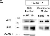
Detection of Kallikrein 6/Neurosin by Western Blot
MME and KLK6 proteases are increased in conditioned media in a stress-dependent manner. Protease profiler array membranes (R&D systems) with 35 protease capture antibodies printed in duplicate were probed with CM from (A) PC3 24 h post-stress and (B) 1532CPTX cells 16 h post-stress. Pixel density after immunoblot exposure was used to identify common proteases between the two cell lines that were significantly increased (red/purple boxes) when compared to samples without stress (dark blue/light blue boxes): Kallikrein-related peptidase 6 (KLK6) and Membrane metallo-endopeptidase (MME/CD10). (C,D) Immunoblot assays were used to detect KLK6 and MME −/+ stress in PC3 and 1532CPTX cell fraction and CM n = 3. Statistical significance was determined by unpaired Student’s t-test, * p < 0.05, ** p < 0.01. Image collected and cropped by CiteAb from the following open publication (https://www.mdpi.com/2073-4409/15/2/166), licensed under a CC-BY license. Not internally tested by R&D Systems.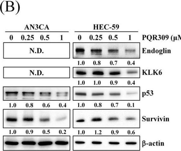
Detection of Kallikrein 6/Neurosin by Western Blot
Effect of PQR309 on the expression of c-Myc and its possible target genes. (A) After treating with PQR309 for 48 h in HEC-59 cells, Proteome Profiler Human XL Oncology Array was performed according to the manufacturer protocol. The dot intensities were quantified by ImageJ software. The ratio of cells without treatment was set at 1. (B) After treating with PQR309 in AN3CA and HEC-59 cells for 48 h, protein expressions of endoglin, KLK6, p53, and survivin were determined by Western blot. ImageJ software was used to quantify the band intensities of endoglin, KLK6, p53, and survivin. Data shown are the relative expression standardized by the beta -actin protein level. The ratio of cells without treatment was set at 1. (C) Western blot assay was performed to detect the expressions of hTERT and c-Myc in AN3CA and HEC-59 cells after PQR309 treatment for 48 h. ImageJ software was used to quantify the band intensities of hTERT and c-Myc. Data shown are the relative expression standardized by the beta -actin protein level. The ratio of cells without treatment was set at 1. (D) AN3CA (5 × 10³ cells/well of 96-well plate) and HEC-59 (6 × 10³ cells/well of 96-well plate) were treated with KJ-Pyr-9 (0, 2.5, 5, 10 and 20 μM), an inhibitor of c-Myc, for 72 h. Cell viability was analyzed by MTT assay. (E) Protein expressions of c-Myc, p53, KLK6, and survivin were determined by Western blot. ImageJ software was used to quantify the band intensities of c-Myc, p53, KLK6, and survivin. Data shown are the relative expression standardized by the beta -actin protein level. The ratio of cells without treatment was set at 1. N.D., not detected. Image collected and cropped by CiteAb from the following open publication (https://pubmed.ncbi.nlm.nih.gov/34831139), licensed under a CC-BY license. Not internally tested by R&D Systems.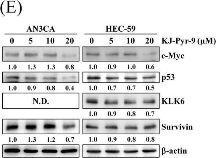
Detection of Kallikrein 6/Neurosin by Western Blot
Effect of PQR309 on the expression of c-Myc and its possible target genes. (A) After treating with PQR309 for 48 h in HEC-59 cells, Proteome Profiler Human XL Oncology Array was performed according to the manufacturer protocol. The dot intensities were quantified by ImageJ software. The ratio of cells without treatment was set at 1. (B) After treating with PQR309 in AN3CA and HEC-59 cells for 48 h, protein expressions of endoglin, KLK6, p53, and survivin were determined by Western blot. ImageJ software was used to quantify the band intensities of endoglin, KLK6, p53, and survivin. Data shown are the relative expression standardized by the beta -actin protein level. The ratio of cells without treatment was set at 1. (C) Western blot assay was performed to detect the expressions of hTERT and c-Myc in AN3CA and HEC-59 cells after PQR309 treatment for 48 h. ImageJ software was used to quantify the band intensities of hTERT and c-Myc. Data shown are the relative expression standardized by the beta -actin protein level. The ratio of cells without treatment was set at 1. (D) AN3CA (5 × 10³ cells/well of 96-well plate) and HEC-59 (6 × 10³ cells/well of 96-well plate) were treated with KJ-Pyr-9 (0, 2.5, 5, 10 and 20 μM), an inhibitor of c-Myc, for 72 h. Cell viability was analyzed by MTT assay. (E) Protein expressions of c-Myc, p53, KLK6, and survivin were determined by Western blot. ImageJ software was used to quantify the band intensities of c-Myc, p53, KLK6, and survivin. Data shown are the relative expression standardized by the beta -actin protein level. The ratio of cells without treatment was set at 1. N.D., not detected. Image collected and cropped by CiteAb from the following open publication (https://pubmed.ncbi.nlm.nih.gov/34831139), licensed under a CC-BY license. Not internally tested by R&D Systems.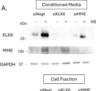
Detection of Kallikrein 6/Neurosin by Western Blot
Stress-induced KLK6 impacts MMP-2 activity and cellular invasion. (A) Immunoblot showing siRNA knockdown of KLK6 and MME in PC3 cell fraction and conditioned media 72 h post-transfection and 24 h post-stress, n = 3 (B) Gelatin DQ assay measuring MMP-2 activity in CM from siRNA samples −/+ stress. (C) Matrigel drop invasion assays with untreated PC3 cells incubated with CM from siRNA KD plates −/+ stress. Invasion was measured over 4 days. (D) Proliferation assay with untreated PC3 cells incubated with CM from untreated and stressed siRNA samples. A minimum of 2 individual biological replicates were performed for the above experiments, and the results were combined. Statistical significance was determined by Welch’s student t-test. * p < 0.05, ** p < 0.01, ns = non-significant. Image collected and cropped by CiteAb from the following open publication (https://www.mdpi.com/2073-4409/15/2/166), licensed under a CC-BY license. Not internally tested by R&D Systems.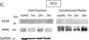
Detection of Kallikrein 6/Neurosin by Western Blot
MME and KLK6 proteases are increased in conditioned media in a stress-dependent manner. Protease profiler array membranes (R&D systems) with 35 protease capture antibodies printed in duplicate were probed with CM from (A) PC3 24 h post-stress and (B) 1532CPTX cells 16 h post-stress. Pixel density after immunoblot exposure was used to identify common proteases between the two cell lines that were significantly increased (red/purple boxes) when compared to samples without stress (dark blue/light blue boxes): Kallikrein-related peptidase 6 (KLK6) and Membrane metallo-endopeptidase (MME/CD10). (C,D) Immunoblot assays were used to detect KLK6 and MME −/+ stress in PC3 and 1532CPTX cell fraction and CM n = 3. Statistical significance was determined by unpaired Student’s t-test, * p < 0.05, ** p < 0.01. Image collected and cropped by CiteAb from the following open publication (https://www.mdpi.com/2073-4409/15/2/166), licensed under a CC-BY license. Not internally tested by R&D Systems.Applications for Human Kallikrein 6/Neurosin Antibody
Immunohistochemistry
Sample: Immersion fixed paraffin-embedded sections of human breast cancer tissue
Immunoprecipitation
Sample: Conditioned cell culture medium spiked with Recombinant Human Kallikrein 6/Neurosin (Catalog # 5164-SE), see our available Western blot detection antibodies
Western Blot
Sample: Recombinant Human Kallikrein 6/Neurosin (Catalog # 5164-SE)
Reviewed Applications
Read 2 reviews rated 4 using AF2008 in the following applications:
Formulation, Preparation, and Storage
Purification
Reconstitution
Reconstitute at 0.2 mg/mL in sterile PBS. For liquid material, refer to CoA for concentration.
Formulation
Shipping
Stability & Storage
- 12 months from date of receipt, -20 to -70 °C as supplied.
- 1 month, 2 to 8 °C under sterile conditions after reconstitution.
- 6 months, -20 to -70 °C under sterile conditions after reconstitution.
Calculators
Background: Kallikrein 6/Neurosin
References
- Emami, N. and E.P. Diamandis (2007) Clin. Chim. Acta 381:78.
- Borgoño, C.A. and E.P. Diamandis (2004) Nat. Rev. Cancer 4:876.
- Paliouras, M. et al. (2007) Cancer Lett. 249:61.
Alternate Names
Entrez Gene IDs
Gene Symbol
UniProt
Additional Kallikrein 6/Neurosin Products
Product Documents for Human Kallikrein 6/Neurosin Antibody
Certificate of Analysis
To download a Certificate of Analysis, please enter a lot or batch number in the search box below.
Note: Certificate of Analysis not available for kit components.
Product Specific Notices for Human Kallikrein 6/Neurosin Antibody
For research use only
Citations for Human Kallikrein 6/Neurosin Antibody
 Powered by Bioz
Customer Reviews for Human Kallikrein 6/Neurosin Antibody (2)
Have you used Human Kallikrein 6/Neurosin Antibody?
Submit a review and receive an Amazon gift card!
$25/€18/£15/$25CAN/¥2500 for a review with an image
$10/€7/£6/$10CAN/¥1110 for a review without an image
Submit a review
-
Application: Western BlotSample Tested: Cell lysate from colon cancer cell lineSpecies: HumanVerified Customer | Posted 04/26/2016
-
Application: Immunohistochemistry-ParaffinSample Tested: See PMID 20846516Species: HumanVerified Customer | Posted 01/06/2015
There are no reviews that match your criteria.
Protocols
Find general support by application which include: protocols, troubleshooting, illustrated assays, videos and webinars.
- Antigen Retrieval Protocol (PIER)
- Antigen Retrieval for Frozen Sections Protocol
- Appropriate Fixation of IHC/ICC Samples
- Cellular Response to Hypoxia Protocols
- Chromogenic IHC Staining of Formalin-Fixed Paraffin-Embedded (FFPE) Tissue Protocol
- Chromogenic Immunohistochemistry Staining of Frozen Tissue
- ClariTSA™ Fluorophore Kits
- Detection & Visualization of Antibody Binding
- Fluorescent IHC Staining of Frozen Tissue Protocol
- Graphic Protocol for Heat-induced Epitope Retrieval
- Graphic Protocol for the Preparation and Fluorescent IHC Staining of Frozen Tissue Sections
- Graphic Protocol for the Preparation and Fluorescent IHC Staining of Paraffin-embedded Tissue Sections
- Graphic Protocol for the Preparation of Gelatin-coated Slides for Histological Tissue Sections
- IHC Sample Preparation (Frozen sections vs Paraffin)
- Immunofluorescent IHC Staining of Formalin-Fixed Paraffin-Embedded (FFPE) Tissue Protocol
- Immunohistochemistry (IHC) and Immunocytochemistry (ICC) Protocols
- Immunohistochemistry Frozen Troubleshooting
- Immunohistochemistry Paraffin Troubleshooting
- Immunoprecipitation Protocol
- Preparing Samples for IHC/ICC Experiments
- Preventing Non-Specific Staining (Non-Specific Binding)
- Primary Antibody Selection & Optimization
- Protocol for Heat-Induced Epitope Retrieval (HIER)
- Protocol for Making a 4% Formaldehyde Solution in PBS
- Protocol for VisUCyte™ HRP Polymer Detection Reagent
- Protocol for the Preparation & Fixation of Cells on Coverslips
- Protocol for the Preparation and Chromogenic IHC Staining of Frozen Tissue Sections
- Protocol for the Preparation and Chromogenic IHC Staining of Frozen Tissue Sections - Graphic
- Protocol for the Preparation and Chromogenic IHC Staining of Paraffin-embedded Tissue Sections
- Protocol for the Preparation and Chromogenic IHC Staining of Paraffin-embedded Tissue Sections - Graphic
- Protocol for the Preparation and Fluorescent IHC Staining of Frozen Tissue Sections
- Protocol for the Preparation and Fluorescent IHC Staining of Paraffin-embedded Tissue Sections
- Protocol for the Preparation of Gelatin-coated Slides for Histological Tissue Sections
- R&D Systems Quality Control Western Blot Protocol
- TUNEL and Active Caspase-3 Detection by IHC/ICC Protocol
- The Importance of IHC/ICC Controls
- Troubleshooting Guide: Immunohistochemistry
- Troubleshooting Guide: Western Blot Figures
- Western Blot Conditions
- Western Blot Protocol
- Western Blot Protocol for Cell Lysates
- Western Blot Troubleshooting
- Western Blot Troubleshooting Guide
- View all Protocols, Troubleshooting, Illustrated assays and Webinars