

Key Product Details
Species Reactivity
Validated:
Human
Cited:
Human, Mouse
Applications
Validated:
Western Blot, Intracellular Staining by Flow Cytometry, CyTOF-ready
Cited:
Immunohistochemistry, Immunohistochemistry-Paraffin, Western Blot, Flow Cytometry, Immunocytochemistry, Bioassay, Complex Application
Label
Unconjugated
Antibody Source
Monoclonal Mouse IgG2A Clone # 278918
Loading...
Product Specifications
Immunogen
Mouse myeloma cell line NS0-derived recombinant human MFG-E8
Leu24-Cys387
Accession # Q08431
Leu24-Cys387
Accession # Q08431
Specificity
Detects human MFG-E8 in direct ELISAs and Western blots.
Clonality
Monoclonal
Host
Mouse
Isotype
IgG2A
Scientific Data Images for Human MFG-E8 Antibody (278918)
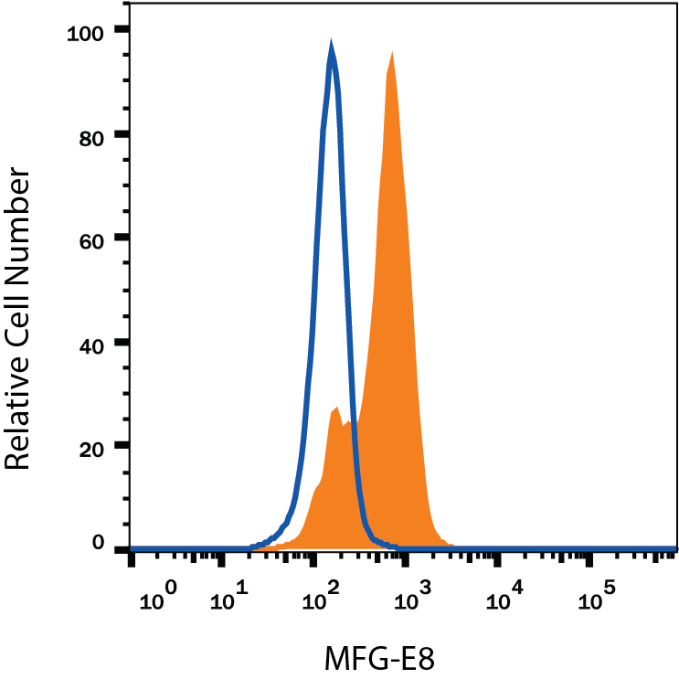
Detection of MFG‑E8 in Human Immature Dendritic Cells by Flow Cytometry.
Human immature dendritic cells were stained with Mouse Anti-Human MFG-E8 Monoclonal Antibody (Catalog # MAB27671, filled histogram) or isotype control antibody (MAB003, open histogram), followed by Phycoerythrin-conjugated Anti-Mouse IgG Secondary Antibody (F0102B). To facilitate intracellular staining, cells were fixed with Flow Cytometry Fixation Buffer (FC004) and permeabilized with Flow Cytometry Permeabilization/Wash Buffer I (FC005). View our protocol for Staining Intracellular Molecules.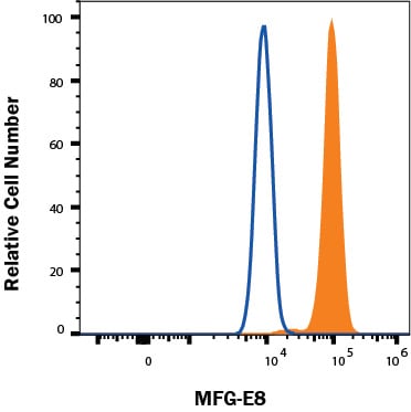
Detection of MFG‑E8 in TF-1 cells by Flow Cytometry.
TF-1 cells were stained with Mouse Anti-Human MFG‑E8 Monoclonal Antibody (Catalog # MAB27671, filled histogram) or isotype control antibody (Catalog # MAB003, open histogram), followed by Fluorescein-conjugated Anti-Mouse IgG Secondary Antibody (Catalog # F0103B). To facilitate intracellular staining, cells were fixed with FC012 and permeabilized with FoxP3 Perm. View our protocol for Staining Intracellular Molecules.Applications for Human MFG-E8 Antibody (278918)
Application
Recommended Usage
CyTOF-ready
Ready to be labeled using established conjugation methods. No BSA or other carrier proteins that could interfere with conjugation.
Intracellular Staining by Flow Cytometry
0.25 µg/106 cells
Sample: Human immature dendritic cells fixed with Flow Cytometry Fixation Buffer (Catalog # FC004) and permeabilized with Flow Cytometry Permeabilization/Wash Buffer I (Catalog # FC005) and TF-1 cells
Sample: Human immature dendritic cells fixed with Flow Cytometry Fixation Buffer (Catalog # FC004) and permeabilized with Flow Cytometry Permeabilization/Wash Buffer I (Catalog # FC005) and TF-1 cells
Western Blot
1 µg/mL
Sample: Recombinant Human MFG-E8 (Catalog # 2767-MF) under non-reducing conditions only
Sample: Recombinant Human MFG-E8 (Catalog # 2767-MF) under non-reducing conditions only
Reviewed Applications
Read 1 review rated 4 using MAB27671 in the following applications:
Flow Cytometry Panel Builder
Bio-Techne Knows Flow Cytometry
Save time and reduce costly mistakes by quickly finding compatible reagents using the Panel Builder Tool.
Advanced Features
- Spectra Viewer - Custom analysis of spectra from multiple fluorochromes
- Spillover Popups - Visualize the spectra of individual fluorochromes
- Antigen Density Selector - Match fluorochrome brightness with antigen density

Formulation, Preparation, and Storage
Purification
Protein A or G purified from hybridoma culture supernatant
Reconstitution
Reconstitute at 0.5 mg/mL in sterile PBS. For liquid material, refer to CoA for concentration.
Loading...
Formulation
Lyophilized from a 0.2 μm filtered solution in PBS with Trehalose. *Small pack size (SP) is supplied either lyophilized or as a 0.2 µm filtered solution in PBS.
Shipping
Lyophilized product is shipped at ambient temperature. Liquid small pack size (-SP) is shipped with polar packs. Upon receipt, store immediately at the temperature recommended below.
Stability & Storage
Use a manual defrost freezer and avoid repeated freeze-thaw cycles.
- 12 months from date of receipt, -20 to -70 °C as supplied.
- 1 month, 2 to 8 °C under sterile conditions after reconstitution.
- 6 months, -20 to -70 °C under sterile conditions after reconstitution.
Calculators
Background: MFG-E8
References
- Raymond, A. et al. (2009) J. Cell. Biochem. 106:957.
- Couto, J.R. et al. (1996) DNA Cell Biol. 15:281.
- Yamaguchi, H. et al. (2010) Eur. J. Immunol. 40:1778.
- Haggqvist, B. et al. (1999) Proc. Natl. Acad. Sci. USA 96:8669.
- Silvestre, J.-S. et al. (2005) Nat. Med. 11:499.
- Borges, E. et al. (2000) J. Biol. Chem. 275:39867.
- Hanayama, R. et al. (2002) Nature 417:182.
- Ait-Oufella, H. et al. (2007) Circulation 115:2168.
- Kranich, J. et al. (2010) J. Exp. Med. 207:2271.
- Hanayama, R. et al. (2004) Science 304:1147.
- Kranich, J. et al. (2010) J. Exp. Med. 205:1293.
- Atabai, K. et al. (2009) J. Clin. Invest. 119:3713.
- Bu, H.-F. et al. (2007) J. Clin. Invest. 117:3673.
- Jinushi, M. et al. (2009) J. Exp. Med. 206:1317.
- Kvistgaard, A.S. et al. (2004) J. Dairy Sci. 87:4088.
Long Name
Milk Fat Globule EGF Factor 8
Alternate Names
BA46, Breast epithelial antigen BA46, Lactahedrin, Medin, MFGE8, SED1
Gene Symbol
MFGE8
UniProt
Additional MFG-E8 Products
Product Documents for Human MFG-E8 Antibody (278918)
Certificate of Analysis
To download a Certificate of Analysis, please enter a lot or batch number in the search box below.
Note: Certificate of Analysis not available for kit components.
Product Specific Notices for Human MFG-E8 Antibody (278918)
For research use only
Citations for Human MFG-E8 Antibody (278918)
 Powered by Bioz
Customer Reviews for Human MFG-E8 Antibody (278918) (1)
4 out of 5
1 Customer Rating
Have you used Human MFG-E8 Antibody (278918)?
Submit a review and receive an Amazon gift card!
$25/€18/£15/$25CAN/¥2500 for a review with an image
$10/€7/£6/$10CAN/¥1110 for a review without an image
Submit a review
Customer Images
Showing
1
-
1 of
1 review
Showing All
Filter By:
-
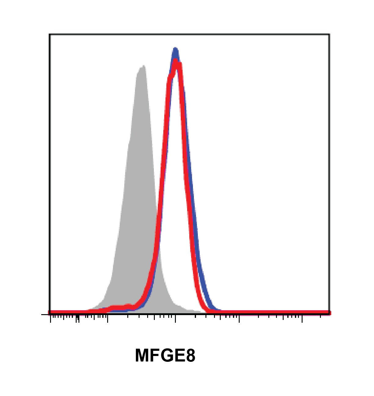
Application: Flow CytometrySample Tested: Bone marrow cellsSpecies: HumanVerified Customer | Posted 08/01/2016

There are no reviews that match your criteria.
Protocols
Find general support by application which include: protocols, troubleshooting, illustrated assays, videos and webinars.
- 7-Amino Actinomycin D (7-AAD) Cell Viability Flow Cytometry Protocol
- Cellular Response to Hypoxia Protocols
- Extracellular Membrane Flow Cytometry Protocol
- Flow Cytometry Protocol for Cell Surface Markers
- Flow Cytometry Protocol for Staining Membrane Associated Proteins
- Flow Cytometry Staining Protocols
- Flow Cytometry Troubleshooting Guide
- Intracellular Flow Cytometry Protocol Using Alcohol (Methanol)
- Intracellular Flow Cytometry Protocol Using Detergents
- Intracellular Nuclear Staining Flow Cytometry Protocol Using Detergents
- Intracellular Staining Flow Cytometry Protocol Using Alcohol Permeabilization
- Intracellular Staining Flow Cytometry Protocol Using Detergents to Permeabilize Cells
- Propidium Iodide Cell Viability Flow Cytometry Protocol
- Protocol for Liperfluo
- Protocol for the Characterization of Human Th22 Cells
- Protocol for the Characterization of Human Th9 Cells
- Protocol: Annexin V and PI Staining by Flow Cytometry
- Protocol: Annexin V and PI Staining for Apoptosis by Flow Cytometry
- R&D Systems Quality Control Western Blot Protocol
- Troubleshooting Guide: Fluorokine Flow Cytometry Kits
- Troubleshooting Guide: Western Blot Figures
- Western Blot Conditions
- Western Blot Protocol
- Western Blot Protocol for Cell Lysates
- Western Blot Troubleshooting
- Western Blot Troubleshooting Guide
- View all Protocols, Troubleshooting, Illustrated assays and Webinars
Loading...