


Scientific images may be resized, cropped, or adjusted for brightness or contrast for presentation purposes. These updates are limited to presentation and do not affect the underlying scientific interpretation of the data. Where available, additional source data may be provided upon request.
Key Product Details
Species Reactivity
Validated:
Cited:
Applications
Validated:
Cited:
Label
Antibody Source
Product Specifications
Immunogen
Ala640-Glu715
Accession # O15409
Specificity
Clonality
Host
Isotype
Scientific Data Images for FoxP2 Antibody
Detection of Human FoxP2 by Western Blot.
Western blot shows lysates of HeLa human cervical epithelial carcinoma cell line, A172 human glioblastoma cell line and U937 human histiocytic lymphoma cell line. PVDF membrane was probed with 1 µg/mL of Sheep Anti-Human/Mouse FoxP2 Antigen Affinity-purified Polyclonal Antibody (Catalog # AF5647) followed by HRP-conjugated Anti-Sheep IgG Secondary Antibody (HAF016). A specific band was detected for FoxP2 at approximately 80 kDa (as indicated). This experiment was conducted under reducing conditions and using Western Blot Buffer Group 1.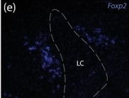
Detection of FoxP2 by Immunohistochemistry
Near the LC, Nps colocalizes with Foxp2 and is mutually exclusive with both Calca and Pdyn. (a–c) Neurons that contain Nps mRNA (red) are mutually exclusive with and medial to neurons in the LC, which contain Calca mRNA (magenta). Ubc mRNA (green) is shown for neuroanatomical background. (d–f) A subset of Foxp2‐expressing neurons (blue) also express Nps (red) medial to the LC. (g–k) This Nps‐expressing subset is mutually exclusive with Pdyn mRNA (yellow). Scale bars are 100 μm (a–c), 50 μm (d–f), 20 μm (g–k) Image collected and cropped by CiteAb from the following open publication (https://pubmed.ncbi.nlm.nih.gov/36036349), licensed under a CC-BY license. Not internally tested by R&D Systems.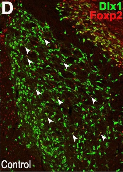
Detection of FoxP2 by Immunohistochemistry
Dlx1+ arkypallidal GABAergic projection neurons, but not prototypical neurons, are increased in the PND13 BACHD Globus Pallidus. (A/A’–D/D′) Representative immunofluorescence images of the Globus Pallidus from PND13 mice stained for the prototypical neuron marker, PV+ cells (A/A’), the arkypallidal neuron marker, Foxp2 (B/B′), EGFP driven by the Dlx1 promoter (C/C′), and combined Foxp2/Dlx1 double labeling (D/D′). (E–H) Quantification of PV+ prototypical (E), all Foxp2+ arkypallidal cells (F), Foxp2+/Dlx1+ arkypallidal cells (G), and Foxp2+/Dlx1− cells (H). Data are presented as box-and-whisker plots showing median, interquartile range, and minimum/maximum values. Individual data points represent biological replicates. Statistical comparisons were performed using unpaired Student’s t-test; p < 0.05 was considered significant. Scale bars: 200 μm (A, D). Image collected and cropped by CiteAb from the following open publication (https://pubmed.ncbi.nlm.nih.gov/41654201), licensed under a CC-BY license. Not internally tested by R&D Systems.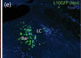
Detection of FoxP2 by Immunohistochemistry
The caudal cluster of Nps Cre‐reporter neurons is medial to the LC, with some neurons extending into Barrington's nucleus (Bar). Most L10GFP‐expressing neurons (green, b) in this region contained FoxP2 (blue, c) and not Lmx1b (red, a). FoxP2 and Lmx1b identified mutually exclusive neuronal populations, with FoxP2‐immunoreactive neurons encircling Bar and light Lmx1b immunoreactivity identifying neurons in the LC (f). (d–g) Merged images. Bar contained several L10GFP‐expressing neurons that lacked FoxP2. Scale bars in (a; also applies to b–f) and (g) are 200 μm Image collected and cropped by CiteAb from the following open publication (https://pubmed.ncbi.nlm.nih.gov/36036349), licensed under a CC-BY license. Not internally tested by R&D Systems.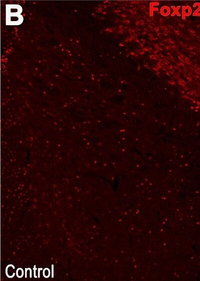
Detection of FoxP2 by Immunohistochemistry
Dlx1+ arkypallidal GABAergic projection neurons, but not prototypical neurons, are increased in the PND13 BACHD Globus Pallidus. (A/A’–D/D′) Representative immunofluorescence images of the Globus Pallidus from PND13 mice stained for the prototypical neuron marker, PV+ cells (A/A’), the arkypallidal neuron marker, Foxp2 (B/B′), EGFP driven by the Dlx1 promoter (C/C′), and combined Foxp2/Dlx1 double labeling (D/D′). (E–H) Quantification of PV+ prototypical (E), all Foxp2+ arkypallidal cells (F), Foxp2+/Dlx1+ arkypallidal cells (G), and Foxp2+/Dlx1− cells (H). Data are presented as box-and-whisker plots showing median, interquartile range, and minimum/maximum values. Individual data points represent biological replicates. Statistical comparisons were performed using unpaired Student’s t-test; p < 0.05 was considered significant. Scale bars: 200 μm (A, D). Image collected and cropped by CiteAb from the following open publication (https://pubmed.ncbi.nlm.nih.gov/41654201), licensed under a CC-BY license. Not internally tested by R&D Systems.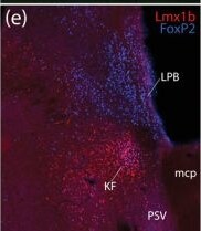
Detection of FoxP2 by Immunohistochemistry
In the rostral lateral PB, neurons expressing the L10GFP Cre‐reporter for Nps contain FoxP2 but not Lmx1b. In this region, immunoreactivity for Lmx1b (red, a) and FoxP2 (blue, c) define mutually exclusive populations of neurons except in the KF, which contains neurons coexpressing both transcription factors (e). L10GFP‐expressing neurons (green, b) are dorsal to Lmx1b‐immunolabeled neurons (d) and contain FoxP2 immunolabeling (f). These L10GFP‐expressing neurons are dorsal to the KF in the lateral PB (g). Scale bars in (f; also applies to a–e) and (g) are 500 μm. Additional mcp, middle cerebellar peduncle; DR, dorsal raphe nucleus Image collected and cropped by CiteAb from the following open publication (https://pubmed.ncbi.nlm.nih.gov/36036349), licensed under a CC-BY license. Not internally tested by R&D Systems.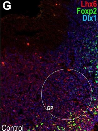
Detection of FoxP2 by Immunohistochemistry
The E12.5 BACHD subpallium displays selective alterations of GABAergic precursor cells. Representative immunofluorescence images of the lateral ganglionic eminence [LGE] and ventral pallium [VP] (A–D, A’–D′), the primordium of the globus pallidus (GP; F–G, F′–G’), and the medial ganglionic eminence (MGE) germinative zone (H/H′). Tissue sections were immunostained for Lhx6 (A/A′), Satb1 (B/B′), EGFP driven by the Dlx1 promoter (C/C′), merged staining in A to C (D/D′), Lhx6/Dlx1 (E/E′), Nkx2–1/Dlx1 (F/F′), Lhx6/Foxp2/Dlx1 (G/G′), and VZ/SVZ Nkx2–1/Dlx1 (H/H′). Quantification analyses included LGE mantle: Lhx6+ (I), Satb1+/Lhx6+ (J), and Mafb+ (K); VP: EGFP+ driven by the Dlx1 promoter (L), Lhx6+ cells (M), Satb1+/Lhx6+ (N), and Mafb+ (O); Globus Pallidus (GP): Nkx2–1+ (P) and Foxp2+/Lhx6+ (Q) cells; and MGE VZ/SVZ Nkx2–1+ progenitors (R). Data are presented as box-and-whisker plots showing the median, interquartile range, and minimum/maximum values. Individual data points represent biological replicates. Statistical comparisons were performed using Mann-Whitney U test (I,J,Q) and unpaired Student’s t-test (K, L-P, R); p < 0.05 was considered significant. Scale bars: 50 μm. Image collected and cropped by CiteAb from the following open publication (https://pubmed.ncbi.nlm.nih.gov/41654201), licensed under a CC-BY license. Not internally tested by R&D Systems.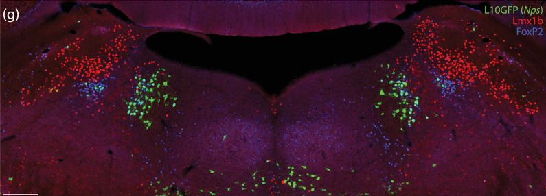
Detection of FoxP2 by Immunohistochemistry
The caudal cluster of Nps Cre‐reporter neurons is medial to the LC, with some neurons extending into Barrington's nucleus (Bar). Most L10GFP‐expressing neurons (green, b) in this region contained FoxP2 (blue, c) and not Lmx1b (red, a). FoxP2 and Lmx1b identified mutually exclusive neuronal populations, with FoxP2‐immunoreactive neurons encircling Bar and light Lmx1b immunoreactivity identifying neurons in the LC (f). (d–g) Merged images. Bar contained several L10GFP‐expressing neurons that lacked FoxP2. Scale bars in (a; also applies to b–f) and (g) are 200 μm Image collected and cropped by CiteAb from the following open publication (https://pubmed.ncbi.nlm.nih.gov/36036349), licensed under a CC-BY license. Not internally tested by R&D Systems.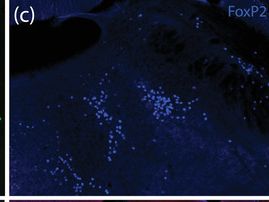
Detection of FoxP2 by Immunohistochemistry
The caudal cluster of Nps Cre‐reporter neurons is medial to the LC, with some neurons extending into Barrington's nucleus (Bar). Most L10GFP‐expressing neurons (green, b) in this region contained FoxP2 (blue, c) and not Lmx1b (red, a). FoxP2 and Lmx1b identified mutually exclusive neuronal populations, with FoxP2‐immunoreactive neurons encircling Bar and light Lmx1b immunoreactivity identifying neurons in the LC (f). (d–g) Merged images. Bar contained several L10GFP‐expressing neurons that lacked FoxP2. Scale bars in (a; also applies to b–f) and (g) are 200 μm Image collected and cropped by CiteAb from the following open publication (https://pubmed.ncbi.nlm.nih.gov/36036349), licensed under a CC-BY license. Not internally tested by R&D Systems.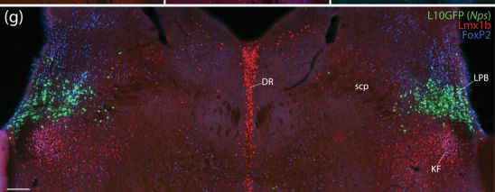
Detection of FoxP2 by Immunohistochemistry
In the rostral lateral PB, neurons expressing the L10GFP Cre‐reporter for Nps contain FoxP2 but not Lmx1b. In this region, immunoreactivity for Lmx1b (red, a) and FoxP2 (blue, c) define mutually exclusive populations of neurons except in the KF, which contains neurons coexpressing both transcription factors (e). L10GFP‐expressing neurons (green, b) are dorsal to Lmx1b‐immunolabeled neurons (d) and contain FoxP2 immunolabeling (f). These L10GFP‐expressing neurons are dorsal to the KF in the lateral PB (g). Scale bars in (f; also applies to a–e) and (g) are 500 μm. Additional mcp, middle cerebellar peduncle; DR, dorsal raphe nucleus Image collected and cropped by CiteAb from the following open publication (https://pubmed.ncbi.nlm.nih.gov/36036349), licensed under a CC-BY license. Not internally tested by R&D Systems.Applications for FoxP2 Antibody
Western Blot
Sample: HeLa human cervical epithelial carcinoma cell line, A172 human glioblastoma cell line and U937 human histiocytic lymphoma cell line
Formulation, Preparation, and Storage
Purification
Reconstitution
Reconstitute at 0.2 mg/mL in sterile PBS. For liquid material, refer to CoA for concentration.
Formulation
Shipping
Stability & Storage
- 12 months from date of receipt, -20 to -70 °C as supplied.
- 1 month, 2 to 8 °C under sterile conditions after reconstitution.
- 6 months, -20 to -70 °C under sterile conditions after reconstitution.
Calculators
Background: FoxP2
Long Name
Alternate Names
Gene Symbol
UniProt
Additional FoxP2 Products
Product Documents for FoxP2 Antibody
Certificate of Analysis
To download a Certificate of Analysis, please enter a lot or batch number in the search box below.
Note: Certificate of Analysis not available for kit components.
Product Specific Notices for FoxP2 Antibody
For research use only
Citations for FoxP2 Antibody
 Powered by Bioz
Customer Reviews for FoxP2 Antibody
There are currently no reviews for this product. Be the first to review FoxP2 Antibody and earn rewards!
Have you used FoxP2 Antibody?
Submit a review and receive an Amazon gift card!
$25/€18/£15/$25CAN/¥2500 for a review with an image
$10/€7/£6/$10CAN/¥1110 for a review without an image
Submit a review
Protocols
Find general support by application which include: protocols, troubleshooting, illustrated assays, videos and webinars.
- Cellular Response to Hypoxia Protocols
- R&D Systems Quality Control Western Blot Protocol
- Troubleshooting Guide: Western Blot Figures
- Western Blot Conditions
- Western Blot Protocol
- Western Blot Protocol for Cell Lysates
- Western Blot Troubleshooting
- Western Blot Troubleshooting Guide
- View all Protocols, Troubleshooting, Illustrated assays and Webinars