
Density Enhanced Protein Tyrosine Phosphatase (DEP-1), also known as CD148, HPTP-eta, and PTP receptor type J (PTPRJ), is an enzyme that removes phosphate groups covalently attached to tyrosine residues in proteins. A large (220 kilodalton) glycoprotein found at the cell surface, DEP-1 levels are increased with high cell density (1). DEP-1 phosphatase activity is enhanced by basement membrane proteins (2), suggesting it is involved in regulating cell adhesion and contact interactions. High levels of expression dampen PDGF (3), VEGF (4), and T-cell receptor (5) responses. DEP-1 is widely expressed in tissues, particularly ones forming epithelioid monolayers (6). In the immune system, DEP-1 is found on all cell lineages and is highest on granulocytes (7). Dep-1 is the mutated gene in the Susceptibility to Colon Cancer locus Scc1, which is altered in many human colorectal adenomas (8). Gene knockout mice lacking DEP-1 die at midgestation due to failures in cardiovascular development (9). DEP-1 dephosphorylates a variety of proteins, including the HGF (10), PDGF (11), and VEGF (4) receptors, and beta ‑catenin (12). The recombinant protein is the intracellular region of DEP-1 containing the catalytic domain. Over aa 997-337, human Dep-1 shares 95% aa sequence identity with mouse and rat Dep-1.
DEP-1/CD148 Antibody
R&D Systems | Catalog # AF1934

Scientific images may be resized, cropped, or adjusted for brightness or contrast for presentation purposes. These updates are limited to presentation and do not affect the underlying scientific interpretation of the data. Where available, additional source data may be provided upon request.
Key Product Details
Validated by
Species Reactivity
Validated:
Cited:
Applications
Validated:
Cited:
Label
Antibody Source
Product Specifications
Immunogen
Arg997-Ala1337
Accession # Q12913
Specificity
Clonality
Host
Isotype
Scientific Data Images for DEP-1/CD148 Antibody
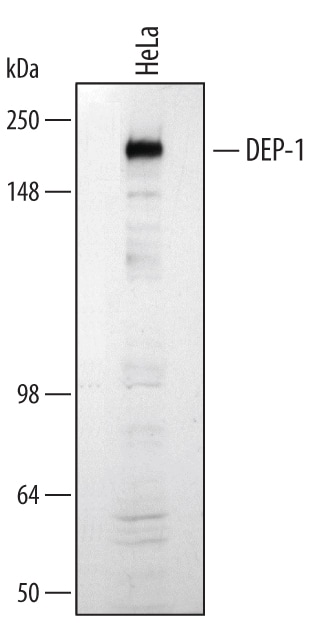
Detection of Human DEP‑1/CD148 by Western Blot.
Western blot shows lysates of HeLa human cervical epithelial carcinoma cell line. PVDF membrane was probed with 1 µg/mL of Goat Anti-Human/Mouse/Rat DEP-1/CD148 Antigen Affinity-purified Polyclonal Antibody (Catalog # AF1934) followed by HRP-conjugated Anti-Goat IgG Secondary Antibody (Catalog # HAF109). A specific band was detected for DEP-1/CD148 at approximately 220 kDa (as indicated). This experiment was conducted under reducing conditions and using Immunoblot Buffer Group 1.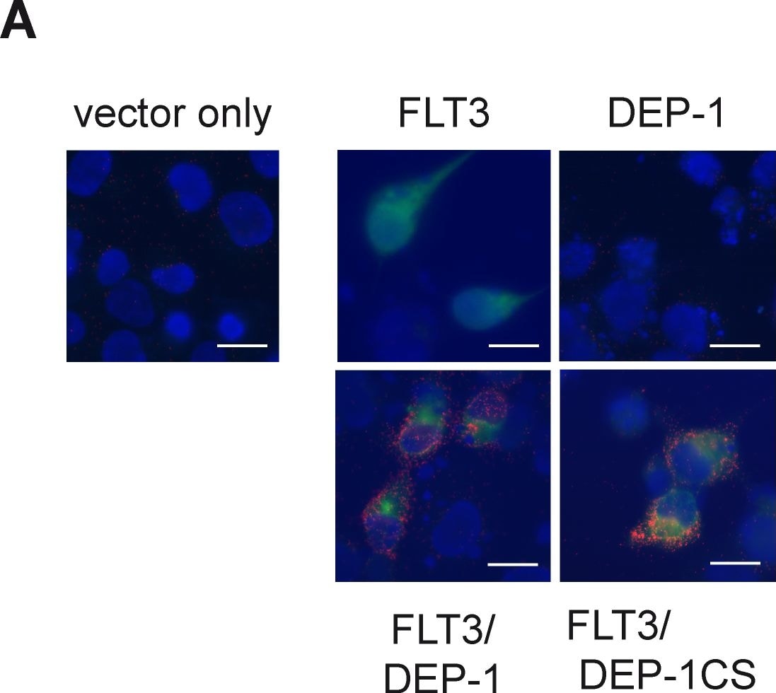
Detection of Monkey DEP-1/CD148 by Immunocytochemistry/Immunofluorescence
Proximity ligation assay reveals association of DEP-1 with its substrate FLT3.(A) COS7 cells were transiently transfected with expression constructs for FLT3, DEP-1, the catalytically inactive DEP-1 C1239S trapping mutant, or corresponding control plasmids as indicated. Complex formation was measured by in situ PLA using rabbit anti-FLT3 antibodies, goat anti-DEP-1 antibodies, and corresponding secondary reagents. In situ PLA is indicated by red signals of the rolling cycle amplification products (RCPs). FLT3 expression (green) was visualized by additional staining with FITC-labeled anti-rabbit IgG antibodies; nuclei (blue) were counterstained with Hoechst 33342. Scale bars 20 µm. (B), (C) Complex formation of endogenous DEP-1 with endogenous FLT3 in THP-1 cells. Cells were transfected with DEP-1-specific or control siRNA by Nucleofection, were then starved and either left unstimulated or were stimulated with FL (100 ng/ml) for 10 min as indicated. (B) Example images; DEP-1 knockdown efficiency was detected by immunblotting (lower panel). DEP-1-FLT3 complexes as RCPs are shown in red, nuclei are depicted in blue and scale bars represent 20 µm for the overview images and 5 µm for the insets. (C) Quantification of detected in situ PLA signals over 5 images per variant. The data are representative for 3 experiments with consistent results. Image collected and cropped by CiteAb from the following publication (https://pubmed.ncbi.nlm.nih.gov/23650535), licensed under a CC-BY license. Not internally tested by R&D Systems.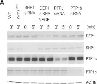
Detection of DEP-1/CD148 by Western Blot
Rescue of Defective ERK Signaling in VEGF-A-Stimulated Nrp1cyto Arterial EC by Knockdown of PTP1bPrimary arterial EC from Nrp1cyto mice transfected with siRNA specific for the indicated phosphatases were serum-starved and then stimulated with 50 ng/ml VEGF-A165.(A and B) Knockdown of the indicated phosphatases in Nrp1cyto arterial EC shown by immunoblotting (A); PTP1b knockdown was quantified in (B), dashed line indicates normal expression levels.(C and D) ERK and VEGFR2 (Y1175) phosphorylation after knockdown of the indicated phosphatases shown by immunoblotting (C). Quantification of pERK activation is shown in (D) (n = 3, mean ± SD, *p < 0.05). Image collected and cropped by CiteAb from the following open publication (https://pubmed.ncbi.nlm.nih.gov/23639442), licensed under a CC-BY license. Not internally tested by R&D Systems.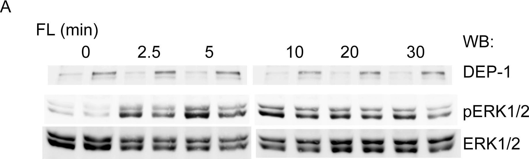
Detection of DEP-1/CD148 by Western Blot
DEP-1 regulates FL-stimulated FLT3 signaling. DEP-1 expression was stably downregulated in THP-1 cells by lentiviral transduction of shRNA.Control cells harbor a non-targeting shRNA construct. (A) FL-dependent ERK1/2 activation was assessed by immunoblotting of cell lysate samples with antibodies recognizing activated ERK1/2 (pERK1/2). Loading was analyzed by reblot with ERK1/2 antibodies. Representative experiment; the efficiency of stable DEP-1 knockdown is also shown (upper panel). (B) Quantitative data for 4 independent experiments. Numbers represent pERK1/2 signals normalized to ERK1/2 levels in the same sample. The value of control cells at 10 min was set to 1.0. Significance for the difference between responses of the two different cell pools was determined by two-way ANOVA. Image collected and cropped by CiteAb from the following open publication (https://pubmed.ncbi.nlm.nih.gov/23650535), licensed under a CC-BY license. Not internally tested by R&D Systems.Applications for DEP-1/CD148 Antibody
Immunoprecipitation
Sample: HeLa human cervical epithelial carcinoma cell line, see our available Western blot detection antibodies
Western Blot
Sample: HeLa human cervical epithelial carcinoma cell line
Formulation, Preparation, and Storage
Purification
Reconstitution
Reconstitute at 0.2 mg/mL in sterile PBS. For liquid material, refer to CoA for concentration.
Formulation
Shipping
Stability & Storage
- 12 months from date of receipt, -20 to -70 °C as supplied.
- 1 month, 2 to 8 °C under sterile conditions after reconstitution.
- 6 months, -20 to -70 °C under sterile conditions after reconstitution.
Calculators
Background: DEP-1/CD148
References
- Ostman, A. et al. (1994) Proc. Natl. Acad. Sci. USA 91:9680.
- Sorby, M. et al. (2001) Oncogene 20:5219.
- Jandt, E. et al. (2003) Oncogene 22:4175.
- Lampugnani, M.G. et al. (2003) J. Cell Biol. 161:793.
- Baker, J.E. et al. (2001) Mol. Cell. Biol. 21:2393.
- Borges, L.G. et al. (1996) Circ. Res. 79:570.
- de la Fuente-Garcia, M.A. et al. (1998) Blood 91:2800.
- Ruivenkamp, C.A. et al. (2002) Nat. Genet. 31:295.
- Takahashi, T. et al. (2003) Mol. Cell. Biol. 23:1817.
- Palka, H.L. et al. (2003) J. Biol. Chem. 278:5728.
- Kovalenko, M. et al. (2000) J. Biol. Chem. 275:16219.
- Holsinger, L.J. et al. (2002) Oncogene 21:7067.
Long Name
Alternate Names
Gene Symbol
UniProt
Additional DEP-1/CD148 Products
Product Documents for DEP-1/CD148 Antibody
Certificate of Analysis
To download a Certificate of Analysis, please enter a lot or batch number in the search box below.
Note: Certificate of Analysis not available for kit components.
Product Specific Notices for DEP-1/CD148 Antibody
For research use only
Citations for DEP-1/CD148 Antibody
 Powered by Bioz
Customer Reviews for DEP-1/CD148 Antibody
There are currently no reviews for this product. Be the first to review DEP-1/CD148 Antibody and earn rewards!
Have you used DEP-1/CD148 Antibody?
Submit a review and receive an Amazon gift card!
$25/€18/£15/$25CAN/¥2500 for a review with an image
$10/€7/£6/$10CAN/¥1110 for a review without an image
Submit a review
Protocols
Find general support by application which include: protocols, troubleshooting, illustrated assays, videos and webinars.
- Cellular Response to Hypoxia Protocols
- Immunoprecipitation Protocol
- R&D Systems Quality Control Western Blot Protocol
- Troubleshooting Guide: Western Blot Figures
- Western Blot Conditions
- Western Blot Protocol
- Western Blot Protocol for Cell Lysates
- Western Blot Troubleshooting
- Western Blot Troubleshooting Guide
- View all Protocols, Troubleshooting, Illustrated assays and Webinars