
GRP78 (Glucose-regulated protein 78 kDa; also BiP and HSPA5) is a 72 kDa member of the heat shock protein 70 family of proteins. Intracellularly, GRP78 is an endoplasmic reticulum chaperone that participates in protein folding; extracellularly, it induces IL-10 production from T cells and interacts with Cripto to block TGF-beta signaling. Human GRP78 precursor is 654 amino acids (aa) in length. It contains an 18 aa signal sequence and a 636 aa mature region that shows a hydantoinase A region (aa 145‑245) and a C-terminal KDEL motif that is present on intracellular GRP78, but absent on secreted GRP78. There is alternative splicing in the signal sequence (aa 1‑10), and multiple single aa substitituion. Over aa 1‑654, human GRP78 is more than 97% aa identical to mouse and rat GRP78.


Scientific images may be resized, cropped, or adjusted for brightness or contrast for presentation purposes. These updates are limited to presentation and do not affect the underlying scientific interpretation of the data. Where available, additional source data may be provided upon request.
Key Product Details
Species Reactivity
Validated:
Cited:
Applications
Validated:
Cited:
Label
Antibody Source
Product Specifications
Immunogen
Met1-Leu654
Accession # P11021
Specificity
Clonality
Host
Isotype
Scientific Data Images for GRP78/HSPA5 Antibody
Detection of Human and Rat GRP78/HSPA5 by Western Blot.
Western blot shows lysates of HeLa human cervical epithelial carcinoma cell line and A20 mouse B cell lymphoma cell line. PVDF membrane was probed with 0.5 µg/mL of Human/Mouse/Rat GRP78/HSPA5 Antigen Affinity-purified Polyclonal Antibody (Catalog # AF4846) followed by HRP-conjugated Anti-Goat IgG Secondary Antibody (Catalog # HAF109). A specific band was detected for GRP78/HSPA5 at approximately 80 kDa (as indicated). This experiment was conducted under reducing conditions and using Immunoblot Buffer Group 2.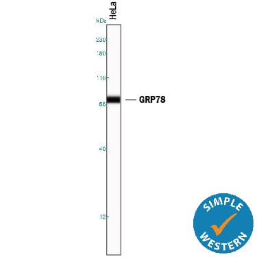
Detection of Human GRP78/HSPA5 by Simple WesternTM.
Simple Western lane view shows lysates of HeLa human cervical epithelial carcinoma cell line, loaded at 0.2 mg/mL. A specific band was detected for GRP78/HSPA5 at approximately 74 kDa (as indicated) using 5 µg/mL of Goat Anti-Human/Mouse/Rat GRP78/HSPA5 Antigen Affinity-purified Polyclonal Antibody (Catalog # AF4846) followed by 1:50 dilution of HRP-conjugated Anti-Goat IgG Secondary Antibody (Catalog # HAF109). This experiment was conducted under reducing conditions and using the 12-230 kDa separation system.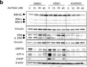
Detection of GRP78/HSPA5 by Western Blot
Increased autophagy and ER stress in A673 cells. WE-68 and A673 cells were treated with 15–45 nM of AUY922, 2 µM of VE821, 5 µM of KU55933 and their combinations. DMSO was used for control. a, b, d, e Analysis of indicated proteins was done by Western blot after 24 h; vinculin and beta -actin were used to control protein loading. Immunoblots are representative of at least two independent experiments. c, f Quantifications of selected proteins Image collected and cropped by CiteAb from the following open publication (https://pubmed.ncbi.nlm.nih.gov/33743824), licensed under a CC-BY license. Not internally tested by R&D Systems.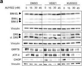
Detection of GRP78/HSPA5 by Western Blot
Increased autophagy and ER stress in A673 cells. WE-68 and A673 cells were treated with 15–45 nM of AUY922, 2 µM of VE821, 5 µM of KU55933 and their combinations. DMSO was used for control. a, b, d, e Analysis of indicated proteins was done by Western blot after 24 h; vinculin and beta -actin were used to control protein loading. Immunoblots are representative of at least two independent experiments. c, f Quantifications of selected proteins Image collected and cropped by CiteAb from the following open publication (https://pubmed.ncbi.nlm.nih.gov/33743824), licensed under a CC-BY license. Not internally tested by R&D Systems.Applications for GRP78/HSPA5 Antibody
Simple Western
Sample: HeLa human cervical epithelial carcinoma cell line
Western Blot
Sample: HeLa human cervical epithelial carcinoma cell line, A20 mouse B cell lymphoma cell line, and rat colon tissue
Reviewed Applications
Read 1 review rated 5 using AF4846 in the following applications:
Formulation, Preparation, and Storage
Purification
Reconstitution
Reconstitute at 0.2 mg/mL in sterile PBS. For liquid material, refer to CoA for concentration.
Formulation
Shipping
Stability & Storage
- 12 months from date of receipt, -20 to -70 °C as supplied.
- 1 month, 2 to 8 °C under sterile conditions after reconstitution.
- 6 months, -20 to -70 °C under sterile conditions after reconstitution.
Calculators
Background: GRP78/HSPA5
Long Name
Alternate Names
Gene Symbol
UniProt
Additional GRP78/HSPA5 Products
Product Documents for GRP78/HSPA5 Antibody
Certificate of Analysis
To download a Certificate of Analysis, please enter a lot or batch number in the search box below.
Note: Certificate of Analysis not available for kit components.
Product Specific Notices for GRP78/HSPA5 Antibody
For research use only
Related Research Areas
Citations for GRP78/HSPA5 Antibody
 Powered by Bioz
Customer Reviews for GRP78/HSPA5 Antibody (1)
Have you used GRP78/HSPA5 Antibody?
Submit a review and receive an Amazon gift card!
$25/€18/£15/$25CAN/¥2500 for a review with an image
$10/€7/£6/$10CAN/¥1110 for a review without an image
Submit a review
Customer Images

-
Application: Western BlotSample Tested: A549 human lung carcinoma cell line and H69AR human small cell lung cancer cell lineSpecies: HumanVerified Customer | Posted 10/17/2018

There are no reviews that match your criteria.
Protocols
Find general support by application which include: protocols, troubleshooting, illustrated assays, videos and webinars.
- Cellular Response to Hypoxia Protocols
- R&D Systems Quality Control Western Blot Protocol
- Troubleshooting Guide: Western Blot Figures
- Western Blot Conditions
- Western Blot Protocol
- Western Blot Protocol for Cell Lysates
- Western Blot Troubleshooting
- Western Blot Troubleshooting Guide
- View all Protocols, Troubleshooting, Illustrated assays and Webinars