


Scientific images may be resized, cropped, or adjusted for brightness or contrast for presentation purposes. These updates are limited to presentation and do not affect the underlying scientific interpretation of the data. Where available, additional source data may be provided upon request.
Key Product Details
Species Reactivity
Validated:
Cited:
Applications
Validated:
Cited:
Label
Antibody Source
Product Specifications
Immunogen
Ala31-Ser199
Accession # Q13308
Specificity
Clonality
Host
Isotype
Scientific Data Images for PTK7/CCK4 Antibody
Detection of Human, Mouse, and Rat PTK7/CCK4 by Western Blot.
Western blot shows lysates of HeLa human cervical epithelial carcinoma cell line, Rat-2 rat embryonic fibroblast cell line, and PT18 mouse mast/basophil cell line. PVDF membrane was probed with 1 µg/mL of Goat Anti-Human/Mouse/Rat PTK7/CCK4 Antigen Affinity-purified Polyclonal Antibody (Catalog # AF4499) followed by HRP-conjugated Anti-Goat IgG Secondary Antibody (Catalog # HAF109). A specific band was detected for PTK7/CCK4 at approximately 160 kDa (as indicated). This experiment was conducted using Immunoblot Buffer Group 1.
Detection of Human PTK7/CCK4 by Simple WesternTM.
Simple Western lane view shows lysates of HeLa human cervical epithelial carcinoma cell line, loaded at 0.2 mg/mL. A specific band was detected for PTK7/CCK4 at approximately 171 kDa (as indicated) using 10 µg/mL of Goat Anti-Human/Mouse/Rat PTK7/CCK4 Antigen Affinity-purified Polyclonal Antibody (Catalog # AF4499) followed by 1:50 dilution of HRP-conjugated Anti-Goat IgG Secondary Antibody (Catalog # HAF109). This experiment was conducted under reducing conditions and using the 12-230 kDa separation system.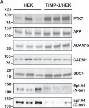
Detection of PTK7/CCK4 by Western Blot
Validation of TIMP-3/HEK cell membrane proteome analysis by immunoblotting. Immunoblots (A) and their respective quantification (B) showing levels of PTK7, APP, ADAM15, CADM1, SDC4, and EphA4 in membrane proteins isolated from TIMP-3/HEK and control HEK 293 cells, (densitometric quantifications shown as mean values ± standard deviation; * p < 0.05, ** p < 0.01, Student’s t-test; from 3 to 6 separate experiments have been performed and analyzed). Image collected and cropped by CiteAb from the following open publication (https://pubmed.ncbi.nlm.nih.gov/33673623), licensed under a CC-BY license. Not internally tested by R&D Systems.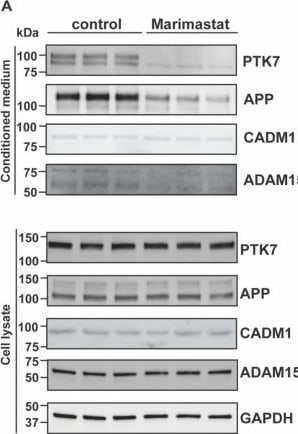
Detection of PTK7/CCK4 by Western Blot
Validation of metalloprotease-dependent shedding of TIMP-3 regulated proteins by immunoblotting. Immunoblots (A) and their respective quantification (B, conditioned media; C, cell lysates) showing levels of PTK7, APP, CADM1, and ADAM15 in conditioned media or lysates of HEK 293 cells treated with or without marimastat (control). GAPDH was used as a loading control. Densitometric quantifications shown as mean values ± standard deviation; *** p < 0.005, Student’s t-test; (n = 6). Image collected and cropped by CiteAb from the following open publication (https://pubmed.ncbi.nlm.nih.gov/33673623), licensed under a CC-BY license. Not internally tested by R&D Systems.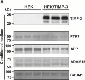
Detection of PTK7/CCK4 by Western Blot
Analysis of protein changes by immunoblotting. (A) Immunoblots showing protein abundance of TIMP-3, PTK7, APP, ADAM15, and CADM1 in the conditioned media of TIMP-3/HEK and control HEK 293 cells, as well as levels of PTK7, APP, ADAM15, CADM1 and EphA4 in the cell lysates. Calnexin and GAPDH were used as loading controls. Densitometric quantifications of specific proteins in the conditioned media (B) and cell lysates (C) are displayed as mean values ± standard deviation (** p < 0.01, *** p < 0.005, Student’s t-test; from 3 to 6 separate experiments have been performed and analyzed). Image collected and cropped by CiteAb from the following open publication (https://pubmed.ncbi.nlm.nih.gov/33673623), licensed under a CC-BY license. Not internally tested by R&D Systems.Applications for PTK7/CCK4 Antibody
Simple Western
Sample: HeLa human cervical epithelial carcinoma cell line
Western Blot
Sample: HeLa human cervical epithelial carcinoma cell line, Rat-2 rat embryonic fibroblast cell line, and PT18 mouse mast/basophil cell line
Reviewed Applications
Read 1 review rated 5 using AF4499 in the following applications:
Formulation, Preparation, and Storage
Purification
Reconstitution
Reconstitute at 0.2 mg/mL in sterile PBS. For liquid material, refer to CoA for concentration.
Formulation
Shipping
Stability & Storage
- 12 months from date of receipt, -20 to -70 °C as supplied.
- 1 month, 2 to 8 °C under sterile conditions after reconstitution.
- 6 months, -20 to -70 °C under sterile conditions after reconstitution.
Calculators
Background: PTK7/CCK4
Long Name
Alternate Names
Gene Symbol
UniProt
Additional PTK7/CCK4 Products
Product Documents for PTK7/CCK4 Antibody
Certificate of Analysis
To download a Certificate of Analysis, please enter a lot or batch number in the search box below.
Note: Certificate of Analysis not available for kit components.
Product Specific Notices for PTK7/CCK4 Antibody
For research use only
Related Research Areas
Citations for PTK7/CCK4 Antibody
 Powered by Bioz
Customer Reviews for PTK7/CCK4 Antibody (1)
Have you used PTK7/CCK4 Antibody?
Submit a review and receive an Amazon gift card!
$25/€18/£15/$25CAN/¥2500 for a review with an image
$10/€7/£6/$10CAN/¥1110 for a review without an image
Submit a review
-
Application: Western BlotSample Tested: See PMID 23095747Species: HumanVerified Customer | Posted 01/08/2015
There are no reviews that match your criteria.
Protocols
Find general support by application which include: protocols, troubleshooting, illustrated assays, videos and webinars.
- Cellular Response to Hypoxia Protocols
- R&D Systems Quality Control Western Blot Protocol
- Troubleshooting Guide: Western Blot Figures
- Western Blot Conditions
- Western Blot Protocol
- Western Blot Protocol for Cell Lysates
- Western Blot Troubleshooting
- Western Blot Troubleshooting Guide
- View all Protocols, Troubleshooting, Illustrated assays and Webinars