
RPTOR (Raptor) is a 150 kDa component of the cytosolic mammalian target of Rapamycin complex 1 (mTORC1) which also contains mTOR and GBL proteins. mTORC1 plays a dominant role in cell cycle regulation in response to metabolic conditions. The interaction of RPTOR with the kinase mTOR is stabilized under conditions of nutrient deprivation and energy stress, leading to inhibition of mTOR and cell cycle arrest. RPTOR contains multiple Ser and Thr residues whose phosphorylation regulates the activation status of mTOR. RPTOR is critical for the response of skeletal muscle and adipose tissue to insulin. It contains three RNC blocks (aa 48-511), three HEAT repeats (aa 550-667), and seven C-terminal WD40 domains (aa 1020-1335). Within aa 77-230, human RPTOR shares 100% aa sequence identity with mouse and rat RPTOR.

Scientific images may be resized, cropped, or adjusted for brightness or contrast for presentation purposes. These updates are limited to presentation and do not affect the underlying scientific interpretation of the data. Where available, additional source data may be provided upon request.
Key Product Details
Species Reactivity
Applications
Label
Antibody Source
Product Specifications
Immunogen
Lys77-Gln230
Accession # Q8N122
Specificity
Clonality
Host
Isotype
Scientific Data Images for RPTOR Antibody (514208)
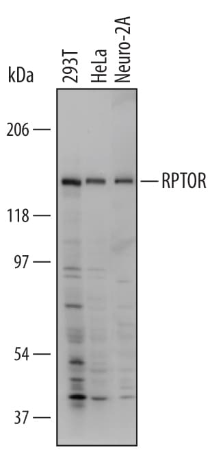
Detection of Human and Mouse RPTOR by Western Blot.
Western blot shows lysates of 293T human embryonic kidney cell line, HeLa human cervical epithelial carcinoma cell line, and Neuro-2A mouse neuroblastoma cell line. PVDF Membrane was probed with 1 µg/mL of Human/Mouse RPTOR Monoclonal Antibody (Catalog # MAB5957) followed by HRP-conjugated Anti-Mouse IgG Secondary Antibody (Catalog # HAF007). A specific band was detected for RPTOR at approximately 150 kDa (as indicated). This experiment was conducted under non-reducing conditions and using Immunoblot Buffer Group 1.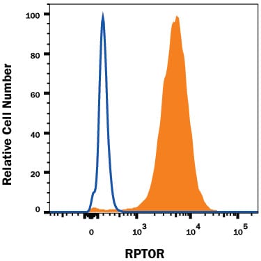
Detection of RPTOR in HeLa Human Cell Line by Flow Cytometry.
HeLa human cervical epithelial carcinoma cell line was stained with Mouse Anti-Human/Mouse RPTOR Monoclonal Antibody (Catalog # MAB5957, filled histogram) or isotype control antibody (Catalog # MAB0041, open histogram), followed by Phycoerythrin-conjugated Anti-Mouse IgG Secondary Antibody (Catalog # F0102B). To facilitate intracellular staining, cells were fixed with Flow Cytometry Fixation Buffer (Catalog # FC004) and permeabilized with Flow Cytometry Permeabilization/Wash Buffer I (Catalog # FC005). View our protocol for Staining Intracellular Molecules.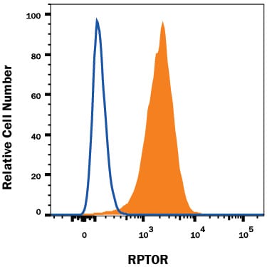
Detection of RPTOR in C2C12 Mouse Cell Line by Flow Cytometry.
C2C12 mouse myoblast cell line was stained with Mouse Anti-Human/Mouse RPTOR Monoclonal Antibody (Catalog # MAB5957, filled histogram) or isotype control antibody (Catalog # MAB0041, open histogram), followed by Phycoerythrin-conjugated Anti-Mouse IgG Secondary Antibody (Catalog # F0102B). To facilitate intracellular staining, cells were fixed with Flow Cytometry Fixation Buffer (Catalog # FC004) and permeabilized with Flow Cytometry Permeabilization/Wash Buffer I (Catalog # FC005). View our protocol for Staining Intracellular Molecules.Applications for RPTOR Antibody (514208)
CyTOF-ready
Intracellular Staining by Flow Cytometry
Sample: HeLa human cervical epithelial carcinoma cell line and C2C12 mouse myoblast cell line fixed with Flow Cytometry Fixation Buffer (Catalog # FC004) and permeabilized with Flow Cytometry Permeabilization/Wash Buffer I (Catalog # FC005)
Western Blot
Sample: 293T human embryonic kidney cell line, HeLa human cervical epithelial carcinoma cell line, and Neuro‑2A mouse neuroblastoma cell line
Reviewed Applications
Read 1 review rated 3 using MAB5957 in the following applications:
Flow Cytometry Panel Builder
Bio-Techne Knows Flow Cytometry
Save time and reduce costly mistakes by quickly finding compatible reagents using the Panel Builder Tool.
Advanced Features
- Spectra Viewer - Custom analysis of spectra from multiple fluorochromes
- Spillover Popups - Visualize the spectra of individual fluorochromes
- Antigen Density Selector - Match fluorochrome brightness with antigen density

Formulation, Preparation, and Storage
Purification
Reconstitution
Reconstitute at 0.5 mg/mL in sterile PBS. For liquid material, refer to CoA for concentration.
Formulation
Shipping
Stability & Storage
- 12 months from date of receipt, -20 to -70 °C as supplied.
- 1 month, 2 to 8 °C under sterile conditions after reconstitution.
- 6 months, -20 to -70 °C under sterile conditions after reconstitution.
Calculators
Background: RPTOR
Long Name
Alternate Names
Gene Symbol
UniProt
Additional RPTOR Products
Product Documents for RPTOR Antibody (514208)
Certificate of Analysis
To download a Certificate of Analysis, please enter a lot or batch number in the search box below.
Note: Certificate of Analysis not available for kit components.
Product Specific Notices for RPTOR Antibody (514208)
For research use only
Related Research Areas
Customer Reviews for RPTOR Antibody (514208) (1)
Have you used RPTOR Antibody (514208)?
Submit a review and receive an Amazon gift card!
$25/€18/£15/$25CAN/¥2500 for a review with an image
$10/€7/£6/$10CAN/¥1110 for a review without an image
Submit a review
Customer Images
-
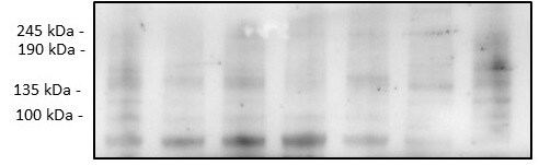
Application: Western BlotSample Tested: Adipose tissueSpecies: MouseVerified Customer | Posted 10/23/2019

There are no reviews that match your criteria.
Protocols
Find general support by application which include: protocols, troubleshooting, illustrated assays, videos and webinars.
- 7-Amino Actinomycin D (7-AAD) Cell Viability Flow Cytometry Protocol
- Cellular Response to Hypoxia Protocols
- Extracellular Membrane Flow Cytometry Protocol
- Flow Cytometry Protocol for Cell Surface Markers
- Flow Cytometry Protocol for Staining Membrane Associated Proteins
- Flow Cytometry Staining Protocols
- Flow Cytometry Troubleshooting Guide
- Intracellular Flow Cytometry Protocol Using Alcohol (Methanol)
- Intracellular Flow Cytometry Protocol Using Detergents
- Intracellular Nuclear Staining Flow Cytometry Protocol Using Detergents
- Intracellular Staining Flow Cytometry Protocol Using Alcohol Permeabilization
- Intracellular Staining Flow Cytometry Protocol Using Detergents to Permeabilize Cells
- Propidium Iodide Cell Viability Flow Cytometry Protocol
- Protocol for Liperfluo
- Protocol for the Characterization of Human Th22 Cells
- Protocol for the Characterization of Human Th9 Cells
- Protocol: Annexin V and PI Staining by Flow Cytometry
- Protocol: Annexin V and PI Staining for Apoptosis by Flow Cytometry
- R&D Systems Quality Control Western Blot Protocol
- Troubleshooting Guide: Fluorokine Flow Cytometry Kits
- Troubleshooting Guide: Western Blot Figures
- Western Blot Conditions
- Western Blot Protocol
- Western Blot Protocol for Cell Lysates
- Western Blot Troubleshooting
- Western Blot Troubleshooting Guide
- View all Protocols, Troubleshooting, Illustrated assays and Webinars
Associated Pathways


