
Semaphorin 3C (Sema3C; previously semaE) is one of six Class 3 secreted semaphorins which share 40-50% amino acid (aa) identity. Class 3 semaphorins are potent chemorepellents that function in axon and/or vascular guidance during development, and may be upregulated in tumor progression (1, 2). The 751 amino acid (aa) mouse Sema3C is highly modular. It contains a 20 aa signal sequence, an ~500 aa N-terminal Sema domain that forms a beta -propeller structure similar to that found in integrin molecules, a cysteine knot, a furin-type cleavage site, an Ig-like domain, and a C-terminal basic domain (1-3). Covalent dimerization plus cleavage at the C-terminus are required for activity of class 3 semaphorins (4). Mouse Sema3C shares at least 95% aa identity with human, rat, cow and dog Sema3C, and 89% and 75% aa identity with chick and zebrafish Sema3C, respectively. Type 3 semaphorins transduce signals through transmembrane plexins, either directly or by binding associated neuropilin receptors (1, 2). Sema3C signaling is transduced by Plexin-D1 indirectly via neuropilin-1 or neuropilin-2 receptors (5). Sema3C is expressed in all somitic motor neurons, in lung buds and in cardiac neural crest cells during development (1, 5-8). Sema3C activates integrins in certain cells so, in addition to its repulsive activities, it sometimes acts as a chemoattractant (6, 9). In the developing nervous system, this chemoattraction appears to complement Sema3A repulsion in adjacent cell layers (1, 6, 7). Sema3C also provides an attractive force opposing Sema6A and Sema6B to guide migration of neural crest endothelial cells to the cardiac outflow tract (10). Consequently, defects in aortic arch formation occur when Sema3C or Plexin-D1 genes or Sema3C-neuropilin interactions are disrupted (5, 11, 12).
Semaphorin 3C Antibody (238835)
R&D Systems | Catalog # MAB1728

Scientific images may be resized, cropped, or adjusted for brightness or contrast for presentation purposes. These updates are limited to presentation and do not affect the underlying scientific interpretation of the data. Where available, additional source data may be provided upon request.
Key Product Details
Species Reactivity
Validated:
Cited:
Applications
Validated:
Cited:
Label
Antibody Source
Product Specifications
Immunogen
Gln24-Ser751 (Arg548Ala, Arg552Ala)
Accession # Q62181
Specificity
Clonality
Host
Isotype
Scientific Data Images for Semaphorin 3C Antibody (238835)
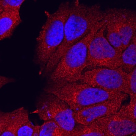
Semaphorin 3C in MCF‑7 Human Cell Line.
Semaphorin 3C was detected in immersion fixed MCF-7 human breast cancer cell line using Rat Anti-Mouse Semaphorin 3C Monoclonal Antibody (Catalog # MAB1728) at 10 µg/mL for 3 hours at room temperature. Cells were stained using the NorthernLights™ 557-conjugated Anti-Rat IgG Secondary Antibody (red; Catalog # NL013) and counterstained with DAPI (blue). Specific staining was localized to cytoplasm. View our protocol for Fluorescent ICC Staining of Cells on Coverslips.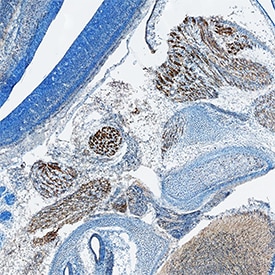
Semaphorin 3C in Mouse Embryo.
Semaphorin 3C was detected in immersion fixed frozen sections of mouse embryo (13 d.p.c.) using Rat Anti-Human/Mouse Semaphorin 3C Monoclonal Antibody (Catalog # MAB1728) at 1.7 µg/mL overnight at 4 °C. Tissue was stained using the Anti-Rat HRP-DAB Cell & Tissue Staining Kit (brown; Catalog # CTS017) and counterstained with hematoxylin (blue). Specific staining was localized to developing muscle cells. View our protocol for Chromogenic IHC Staining of Frozen Tissue Sections.Applications for Semaphorin 3C Antibody (238835)
Immunocytochemistry
Sample: Immersion fixed MCF‑7 human breast cancer cell line
Immunohistochemistry
Sample: Immersion fixed frozen sections of mouse embryo (13 d.p.c.)
Western Blot
Sample: Recombinant Mouse Semaphorin 3C Fc Chimera, Truncated (Catalog # 1728-S3)
Reviewed Applications
Read 2 reviews rated 4.5 using MAB1728 in the following applications:
Formulation, Preparation, and Storage
Purification
Reconstitution
Reconstitute at 0.5 mg/mL in sterile PBS. For liquid material, refer to CoA for concentration.
Formulation
Shipping
Stability & Storage
- 12 months from date of receipt, -20 to -70 °C as supplied.
- 1 month, 2 to 8 °C under sterile conditions after reconstitution.
- 6 months, -20 to -70 °C under sterile conditions after reconstitution.
Calculators
Background: Semaphorin 3C
References
- Hinck, L. (2004) Dev. Cell 7:783.
- Neufeld, G. et al. (2005) Front. Biosci. 10:751.
- Gherardi, E. et al. (2004) Curr. Opin. Struct. Biol. 14:669.
- Adams, R. H. et al. (1997) EMBO J. 16:6077.
- Gitler, A. D. et al. (2004) Dev. Cell 7:107.
- Bagnard, D. et al. (1998) Development 125:5043.
- Cohen, S. et al. (2005) Eur. J. Neurosci. 21:1767.
- Puschel, A. W. et al. (1995) Neuron 14:941.
- Herman, J. G. and G. G. Meadows (2007) Int. J. Oncol. 30:1231.
- Toyofuku, T. et al. (2008) Dev. Biol. 321:251.
- Feiner, L. et al. (2001) Development 128:3061.
- Gu, C. et al. (2003) Dev. Cell 5:45.
Alternate Names
Gene Symbol
UniProt
Additional Semaphorin 3C Products
Product Documents for Semaphorin 3C Antibody (238835)
Certificate of Analysis
To download a Certificate of Analysis, please enter a lot or batch number in the search box below.
Note: Certificate of Analysis not available for kit components.
Product Specific Notices for Semaphorin 3C Antibody (238835)
For research use only
Related Research Areas
Citations for Semaphorin 3C Antibody (238835)
 Powered by Bioz
Customer Reviews for Semaphorin 3C Antibody (238835) (2)
Have you used Semaphorin 3C Antibody (238835)?
Submit a review and receive an Amazon gift card!
$25/€18/£15/$25CAN/¥2500 for a review with an image
$10/€7/£6/$10CAN/¥1110 for a review without an image
Submit a review
Customer Images
-
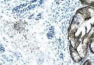
Application: ImmunohistochemistrySample Tested: Lung tissueSpecies: MouseVerified Customer | Posted 12/20/2021

-
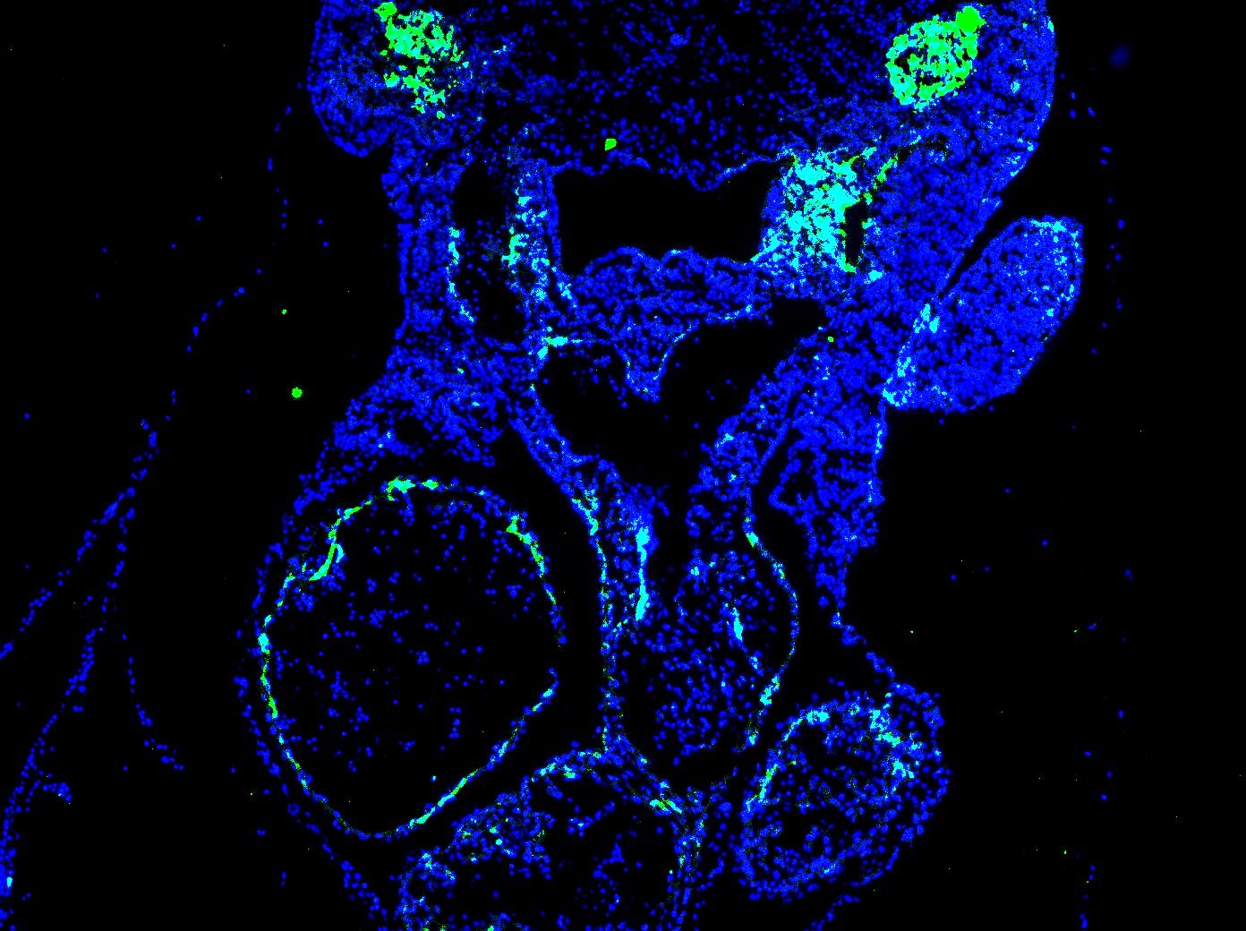
Application: Immunocytochemistry/ImmunofluorescenceSample Tested: E10.5 mouse embryo fixed in 4% PFASpecies: MouseVerified Customer | Posted 12/16/2020Dilution used - 1:200. The staining was done on an E10.5 mouse transverse section (fixed on 4% PFA overnight) and done using standard IF techniques. The staining was very good on the target region (look at the upper region of picture which shows excellent Semaphorin3C staining on supposed neural crest cells). However, there's still a little non specific staining on the arterial lining of the heart, which is a small cause of concern. Probably this could be eliminated by tweaking the fixation/blocking steps.
There are no reviews that match your criteria.
Protocols
Find general support by application which include: protocols, troubleshooting, illustrated assays, videos and webinars.
- Antigen Retrieval Protocol (PIER)
- Antigen Retrieval for Frozen Sections Protocol
- Appropriate Fixation of IHC/ICC Samples
- Cellular Response to Hypoxia Protocols
- Chromogenic IHC Staining of Formalin-Fixed Paraffin-Embedded (FFPE) Tissue Protocol
- Chromogenic Immunohistochemistry Staining of Frozen Tissue
- ClariTSA™ Fluorophore Kits
- Detection & Visualization of Antibody Binding
- Fluorescent IHC Staining of Frozen Tissue Protocol
- Graphic Protocol for Heat-induced Epitope Retrieval
- Graphic Protocol for the Preparation and Fluorescent IHC Staining of Frozen Tissue Sections
- Graphic Protocol for the Preparation and Fluorescent IHC Staining of Paraffin-embedded Tissue Sections
- Graphic Protocol for the Preparation of Gelatin-coated Slides for Histological Tissue Sections
- ICC Cell Smear Protocol for Suspension Cells
- ICC Immunocytochemistry Protocol Videos
- ICC for Adherent Cells
- IHC Sample Preparation (Frozen sections vs Paraffin)
- Immunocytochemistry (ICC) Protocol
- Immunocytochemistry Troubleshooting
- Immunofluorescence of Organoids Embedded in Cultrex Basement Membrane Extract
- Immunofluorescent IHC Staining of Formalin-Fixed Paraffin-Embedded (FFPE) Tissue Protocol
- Immunohistochemistry (IHC) and Immunocytochemistry (ICC) Protocols
- Immunohistochemistry Frozen Troubleshooting
- Immunohistochemistry Paraffin Troubleshooting
- Preparing Samples for IHC/ICC Experiments
- Preventing Non-Specific Staining (Non-Specific Binding)
- Primary Antibody Selection & Optimization
- Protocol for Heat-Induced Epitope Retrieval (HIER)
- Protocol for Making a 4% Formaldehyde Solution in PBS
- Protocol for VisUCyte™ HRP Polymer Detection Reagent
- Protocol for the Fluorescent ICC Staining of Cell Smears - Graphic
- Protocol for the Fluorescent ICC Staining of Cultured Cells on Coverslips - Graphic
- Protocol for the Preparation & Fixation of Cells on Coverslips
- Protocol for the Preparation and Chromogenic IHC Staining of Frozen Tissue Sections
- Protocol for the Preparation and Chromogenic IHC Staining of Frozen Tissue Sections - Graphic
- Protocol for the Preparation and Chromogenic IHC Staining of Paraffin-embedded Tissue Sections
- Protocol for the Preparation and Chromogenic IHC Staining of Paraffin-embedded Tissue Sections - Graphic
- Protocol for the Preparation and Fluorescent ICC Staining of Cells on Coverslips
- Protocol for the Preparation and Fluorescent ICC Staining of Non-adherent Cells
- Protocol for the Preparation and Fluorescent ICC Staining of Stem Cells on Coverslips
- Protocol for the Preparation and Fluorescent IHC Staining of Frozen Tissue Sections
- Protocol for the Preparation and Fluorescent IHC Staining of Paraffin-embedded Tissue Sections
- Protocol for the Preparation of Gelatin-coated Slides for Histological Tissue Sections
- Protocol for the Preparation of a Cell Smear for Non-adherent Cell ICC - Graphic
- R&D Systems Quality Control Western Blot Protocol
- TUNEL and Active Caspase-3 Detection by IHC/ICC Protocol
- The Importance of IHC/ICC Controls
- Troubleshooting Guide: Immunohistochemistry
- Troubleshooting Guide: Western Blot Figures
- Western Blot Conditions
- Western Blot Protocol
- Western Blot Protocol for Cell Lysates
- Western Blot Troubleshooting
- Western Blot Troubleshooting Guide
- View all Protocols, Troubleshooting, Illustrated assays and Webinars