
Semaphorin 3E (Sema3E; previously SemaH) is one of six Class 3 (secreted) semaphorins which in the human share 40-50% amino acid (aa) identity. Class 3 semaphorins are potent chemorepellents that function in axon guidance and/or vascular tip cell guidance during development (1). Sema3E is highly expressed by a subset of motor neurons in developing somites, where it acts as a repulsive cue for PlexinD1-expressing endothelial cells of adjacent intersomitic vessels (2, 3). Crystal structures of semaphorins reveal that the 500 aa N-terminal Sema domain forms a seven-blade b-propeller similar to that found in integrin molecules; 14 conserved cysteine residues and one or more N-glycosylation sites are thought critical for forming the secondary structure (4). C-terminal to the Sema domain, Sema3E has a consensus sequence for furin cleavage which, when used, creates a 61kDa form that does not dimerize and is highly expressed in tumor cell lines with metastatic potential (5, 6). Further C-terminal are a cysteine-knot plexin/semaphorin/integrin (PSI) domain, an Ig-like domain, a cysteine for dimerization and a basic domain containing another furin site. Dimerization and cleavage at the C-terminal site are required for repulsing activity of class 3 semaphorins (7). Human Sema3E shares 90%, 85% and 57% aa identity with mouse, cow and dog Sema3E, respectively. Like other semaphorins, Sema3E signaling is transduced by a transmembrane Plexin dimer, which also has a Sema domain and is coupled to kinase pathways. Unlike other Class 3 semaphorins, Sema3E binds directly to its plexin and does not require interaction with a neuropilin for activity (7). Genetic disruption of either Sema3E or PlexinD1 creates mouse mutants with excessive and disorganized vascular growth and branching, indicating the importance of this ligand-receptor pair for vascular guidance (3, 8).

Scientific images may be resized, cropped, or adjusted for brightness or contrast for presentation purposes. These updates are limited to presentation and do not affect the underlying scientific interpretation of the data. Where available, additional source data may be provided upon request.
Key Product Details
Species Reactivity
Validated:
Cited:
Applications
Validated:
Cited:
Label
Antibody Source
Product Specifications
Immunogen
Thr25-Ser775 (Arg557Ala and Arg560Ala)
Accession # O15041
Specificity
Clonality
Host
Isotype
Scientific Data Images for Semaphorin 3E Antibody
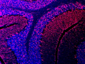
Semaphorin 3E in Mouse Brain.
Semaphorin 3E was detected in immersion fixed frozen sections of adult mouse brain using Goat Anti-Human Semaphorin 3E Antigen Affinity-purified Polyclonal Antibody (Catalog # AF3239) at 10 µg/mL overnight at 4 °C. Tissue was stained using the Northern-Lights™ 557-conjugated Anti-Goat IgG Secondary Antibody (red; Catalog # NL001) and counterstained with DAPI (blue). Specific staining was localized to cerebellum. View our protocol for Fluorescent IHC Staining of Frozen Tissue Sections.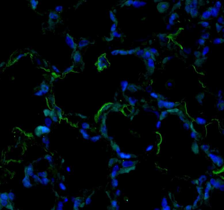
Semaphorin 3E in Mouse Lung.
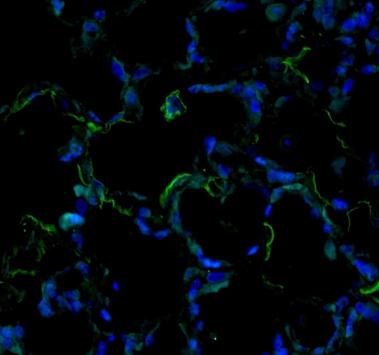
Immunohistochemistry (paraffin-embedded) on Native Adult rat lung tissue. Sema33 stained green, DAPI (blue). Image from a verified customer review.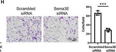
Detection of Semaphorin 3E by Immunohistochemistry
The impact of Sema3E‐Plexin D1 on the differentiation, proliferation, and migration of fibroblasts. A) Western blot analysis of Sema3E expression in primary human lung fibroblasts (PHLFs) following stimulation with different concentrations of TGF beta 1 for 48 h. B) Measurement of Sema3E concentration in culture media by ELISA. PHLFs were stimulated with TGF‐ beta 1, and Sema3E concentration was assessed after 48 h. C,D) Western blot and RT‐qPCR analysis of Fibronectin, Col1a1, and alpha ‐SMA protein and mRNA expression in Sema3E siRNA or Scrambled siRNA‐treated PHLFs following TGF‐ beta 1 induction. E,F) Western blot and RT‐qPCR analysis of Fibronectin, Col1a1, and alpha ‐SMA protein and mRNA expression in PlexinD1 siRNA or Scrambled siRNA‐treated PHLFs following TGF‐ beta 1 induction. G) Representative results for EdU staining in Sema3E siRNA or Scrambled siRNA‐treated PHLFs. H) Representative results for Transwell assay in Sema3E siRNA or Scrambled siRNA‐treated PHLFs. I) Representative results for EdU staining in Plexin D1 siRNA or Scrambled siRNA‐treated PHLFs. J) Representative results for Transwell assay in Plexin D1 siRNA or Scrambled siRNA‐treated PHLFs. Images were captured under original magnification ×200. Data are presented as the mean ± SEM of three independent experiments. Statistical analyses were performed using unpaired t‐tests and one‐way ANOVA test. *p < 0.05; **p < 0.01; ***p < 0.001. Image collected and cropped by CiteAb from the following open publication (https://pubmed.ncbi.nlm.nih.gov/40112179), licensed under a CC-BY license. Not internally tested by R&D Systems.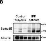
Detection of Semaphorin 3E by Western Blot
Analysis of Sema3E and Plexin D1 levels in IPF Patients. A) ELISA analysis comparing Sema3E levels in the plasma of patients with IPF (n = 63) and control subjects (n = 24). B) Representative western blot analysis depicting Sema3E expression in plasma samples from IPF patients. C) Representative western blot analysis illustrating Sema3E and Plexin D1 expression in lung tissue samples from control subjects (n = 10) and IPF patients (n = 12). D–G) Correlation analysis between Sema3E levels and clinical parameters including (D) FVC%pred, (E) DLCO%pred, (F) TLC%pred, and (G) FEV1%pred in IPF patients (n = 32). H) Co‐immunostaining of Sema3E and alpha ‐SMA in lung sections from patients with IPF(n = 3) and control subjects(n = 3). I) Co‐immunostaining of Plexin D1 and alpha ‐SMA in lung sections from patients with IPF(n = 3) and control subjects(n = 3). Nuclei were stained blue with Diamidino‐2‐phenylindole dihydrochloride (DAPI), and images were captured under original magnification ×400. Data are presented as the mean ± SEM. Statistical analyses were performed using unpaired t‐tests and Pearson's correlation. *p < 0.05, **p < 0.01, and ***p < 0.001 indicate significance levels. (FVC%pred: Forced Vital Capacity % predicted; TLC%pred: Total Lung Capacity % predicted; DLCO%pred: Diffusing Capacity of the Lung for Carbon Monoxide % predicted; FEV1%pred: Forced Expiratory Volume in 1 second % predicted.). Image collected and cropped by CiteAb from the following open publication (https://pubmed.ncbi.nlm.nih.gov/40112179), licensed under a CC-BY license. Not internally tested by R&D Systems.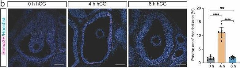
Detection of Semaphorin 3E by Immunohistochemistry
Spatial and temporal patterns of expression for Sema3e and Plxnd1 in the mouse ovary around the time of ovulation. a) RT‐qPCR shows the mRNA levels of Sema3e and Plxnd1 in mouse ovarian GCs (n = 3), OSTs (n = 3), and whole ovaries (n = 3) before and after hCG treatment from superovulated immature mice. b) Representative images of IF staining of Sema3E in mouse ovaries (n = 3 for each time point) before and post‐hCG. The areas of staining for Sema3E were quantified by normalizing the Sema3E‐positive area to the Hoechst‐positive area of preovulatory follicles. Scale bar = 100 µm. c) Representative images of FISH of Plxnd1 (red) and Vegfr2 (green) mRNA in ovaries at 8 h (n = 4) and 24 h (n = 4) post‐hCG. Red arrowheads: Plxnd1+ cells. Green arrowheads: Vegfr2+ cells. Yellow arrowheads: Plxnd1+Vegfr2+ cells. White dashed lines indicate the boundary between the GCs/CL and OST. Scale bar = 50 µm. Quantitative data are presented as mean ± SD. One‐way ANOVA with Tukey's multiple comparisons test was used for statistical analysis in a (within the same cell/tissue types), b. *p < 0.05; **p < 0.01; ***p < 0.001; ns, not significance. GC: granulosa cell; OST: ovarian stromal tissue; OV: whole ovary; F: follicle; CL: corpus lute. Image collected and cropped by CiteAb from the following open publication (https://pubmed.ncbi.nlm.nih.gov/40391781), licensed under a CC-BY license. Not internally tested by R&D Systems.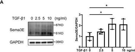
Detection of Semaphorin 3E by Western Blot
The impact of Sema3E‐Plexin D1 on the differentiation, proliferation, and migration of fibroblasts. A) Western blot analysis of Sema3E expression in primary human lung fibroblasts (PHLFs) following stimulation with different concentrations of TGF beta 1 for 48 h. B) Measurement of Sema3E concentration in culture media by ELISA. PHLFs were stimulated with TGF‐ beta 1, and Sema3E concentration was assessed after 48 h. C,D) Western blot and RT‐qPCR analysis of Fibronectin, Col1a1, and alpha ‐SMA protein and mRNA expression in Sema3E siRNA or Scrambled siRNA‐treated PHLFs following TGF‐ beta 1 induction. E,F) Western blot and RT‐qPCR analysis of Fibronectin, Col1a1, and alpha ‐SMA protein and mRNA expression in PlexinD1 siRNA or Scrambled siRNA‐treated PHLFs following TGF‐ beta 1 induction. G) Representative results for EdU staining in Sema3E siRNA or Scrambled siRNA‐treated PHLFs. H) Representative results for Transwell assay in Sema3E siRNA or Scrambled siRNA‐treated PHLFs. I) Representative results for EdU staining in Plexin D1 siRNA or Scrambled siRNA‐treated PHLFs. J) Representative results for Transwell assay in Plexin D1 siRNA or Scrambled siRNA‐treated PHLFs. Images were captured under original magnification ×200. Data are presented as the mean ± SEM of three independent experiments. Statistical analyses were performed using unpaired t‐tests and one‐way ANOVA test. *p < 0.05; **p < 0.01; ***p < 0.001. Image collected and cropped by CiteAb from the following open publication (https://pubmed.ncbi.nlm.nih.gov/40112179), licensed under a CC-BY license. Not internally tested by R&D Systems.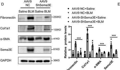
Detection of Semaphorin 3E by Western Blot
Downregulation of Sema3E attenuates BLM‐induced pulmonary fibrosis. A,B) Histological analysis of lung fibrosis severity in mice following BLM induction. (A) Representative images of lung sections stained with H&E, Masson's trichrome, and Sirius red to assess fibrosis severity. (B) Bar graph showing the quantitative mean score of fibrosis severity. Samples include AAV9‐NC saline mice (n = 5), AAV9‐ShSema3E saline mice (n = 5), AAV9‐NC BLM mice (n = 5), and AAV9‐ShSema3E BLM mice (n = 5). C) Quantification of hydroxyproline contents in AAV9‐NC mice and AAV9‐ShSema3E mice after BLM challenge. D,E) Western blot and RT‐qPCR analysis of Fibronectin, Col1a1, and alpha ‐SMA protein and mRNA expression in lung homogenates from the mentioned mouse groups. F) Western blot analysis of ErbB2, P‐ErbB2, AKT, P‐AKT, ERK1/2, and P‐ERK1/2 expression in lung homogenates from the mentioned mouse groups. Samples include AAV9‐NC saline mice (n = 3), AAV9‐ShSema3E saline mice (n = 3), AAV9‐NC BLM mice (n = 3), and AAV9‐ShSema3E BLM mice (n = 3). Data are represented as the mean ± SEM. Statistical analyses were performed using one‐way ANOVA tests. *p < 0.05; **p < 0.01; ***p < 0.001. Image collected and cropped by CiteAb from the following open publication (https://pubmed.ncbi.nlm.nih.gov/40112179), licensed under a CC-BY license. Not internally tested by R&D Systems.Applications for Semaphorin 3E Antibody
Immunocytochemistry
Sample: Immersion fixed MDA-MB-453 human breast cancer cell line
Immunohistochemistry
Sample: Immersion fixed frozen sections of adult mouse brain
Western Blot
Sample: Recombinant Human Semaphorin 3E (Catalog # 3239-S3)
Reviewed Applications
Read 1 review rated 4 using AF3239 in the following applications:
Formulation, Preparation, and Storage
Purification
Reconstitution
Reconstitute at 0.2 mg/mL in sterile PBS. For liquid material, refer to CoA for concentration.
Formulation
Shipping
Stability & Storage
- 12 months from date of receipt, -20 to -70 °C as supplied.
- 1 month, 2 to 8 °C under sterile conditions after reconstitution.
- 6 months, -20 to -70 °C under sterile conditions after reconstitution.
Calculators
Background: Semaphorin 3E
References
- Eichmann, A. et al. (2005) Genes Dev. 19:1013.
- Cohen, S. et al. (2005) Eur. J. Neurosci. 21:1767.
- Gu, C. et al. (2005) Science 307:265.
- Gherardi, E. et al. (2004) Curr. Opin. Struct. Biol. 14:669.
- Christensen, C. et al. (1998) Cancer Res. 58:1238.
- Christensen, C. et al. (2005) Cancer Res. 65:6167.
- Adams, R. H. et al. (1997) EMBO J. 16:6077.
- Gitler, A. D. et al. (2004) Developmental Cell 7:107.
Alternate Names
Gene Symbol
UniProt
Additional Semaphorin 3E Products
Product Documents for Semaphorin 3E Antibody
Certificate of Analysis
To download a Certificate of Analysis, please enter a lot or batch number in the search box below.
Note: Certificate of Analysis not available for kit components.
Product Specific Notices for Semaphorin 3E Antibody
For research use only
Related Research Areas
Citations for Semaphorin 3E Antibody
 Powered by Bioz
Customer Reviews for Semaphorin 3E Antibody (1)
Have you used Semaphorin 3E Antibody?
Submit a review and receive an Amazon gift card!
$25/€18/£15/$25CAN/¥2500 for a review with an image
$10/€7/£6/$10CAN/¥1110 for a review without an image
Submit a review
Customer Images
-
Application: Immunohistochemistry (paraffin-embedded) on Native Adult rat lung tissue. Sema33 stained green, DAPI (blue).Sample Tested: Lung tissueSpecies: RatVerified Customer | Posted 07/19/2025
 Bio-Techne ResponseThis review reflects a new species or application tested on a primary antibody.
There are no reviews that match your criteria.
Protocols
Find general support by application which include: protocols, troubleshooting, illustrated assays, videos and webinars.
- Antigen Retrieval Protocol (PIER)
- Antigen Retrieval for Frozen Sections Protocol
- Appropriate Fixation of IHC/ICC Samples
- Cellular Response to Hypoxia Protocols
- Chromogenic IHC Staining of Formalin-Fixed Paraffin-Embedded (FFPE) Tissue Protocol
- Chromogenic Immunohistochemistry Staining of Frozen Tissue
- ClariTSA™ Fluorophore Kits
- Detection & Visualization of Antibody Binding
- Fluorescent IHC Staining of Frozen Tissue Protocol
- Graphic Protocol for Heat-induced Epitope Retrieval
- Graphic Protocol for the Preparation and Fluorescent IHC Staining of Frozen Tissue Sections
- Graphic Protocol for the Preparation and Fluorescent IHC Staining of Paraffin-embedded Tissue Sections
- Graphic Protocol for the Preparation of Gelatin-coated Slides for Histological Tissue Sections
- ICC Cell Smear Protocol for Suspension Cells
- ICC Immunocytochemistry Protocol Videos
- ICC for Adherent Cells
- IHC Sample Preparation (Frozen sections vs Paraffin)
- Immunocytochemistry (ICC) Protocol
- Immunocytochemistry Troubleshooting
- Immunofluorescence of Organoids Embedded in Cultrex Basement Membrane Extract
- Immunofluorescent IHC Staining of Formalin-Fixed Paraffin-Embedded (FFPE) Tissue Protocol
- Immunohistochemistry (IHC) and Immunocytochemistry (ICC) Protocols
- Immunohistochemistry Frozen Troubleshooting
- Immunohistochemistry Paraffin Troubleshooting
- Preparing Samples for IHC/ICC Experiments
- Preventing Non-Specific Staining (Non-Specific Binding)
- Primary Antibody Selection & Optimization
- Protocol for Heat-Induced Epitope Retrieval (HIER)
- Protocol for Making a 4% Formaldehyde Solution in PBS
- Protocol for VisUCyte™ HRP Polymer Detection Reagent
- Protocol for the Fluorescent ICC Staining of Cell Smears - Graphic
- Protocol for the Fluorescent ICC Staining of Cultured Cells on Coverslips - Graphic
- Protocol for the Preparation & Fixation of Cells on Coverslips
- Protocol for the Preparation and Chromogenic IHC Staining of Frozen Tissue Sections
- Protocol for the Preparation and Chromogenic IHC Staining of Frozen Tissue Sections - Graphic
- Protocol for the Preparation and Chromogenic IHC Staining of Paraffin-embedded Tissue Sections
- Protocol for the Preparation and Chromogenic IHC Staining of Paraffin-embedded Tissue Sections - Graphic
- Protocol for the Preparation and Fluorescent ICC Staining of Cells on Coverslips
- Protocol for the Preparation and Fluorescent ICC Staining of Non-adherent Cells
- Protocol for the Preparation and Fluorescent ICC Staining of Stem Cells on Coverslips
- Protocol for the Preparation and Fluorescent IHC Staining of Frozen Tissue Sections
- Protocol for the Preparation and Fluorescent IHC Staining of Paraffin-embedded Tissue Sections
- Protocol for the Preparation of Gelatin-coated Slides for Histological Tissue Sections
- Protocol for the Preparation of a Cell Smear for Non-adherent Cell ICC - Graphic
- R&D Systems Quality Control Western Blot Protocol
- TUNEL and Active Caspase-3 Detection by IHC/ICC Protocol
- The Importance of IHC/ICC Controls
- Troubleshooting Guide: Immunohistochemistry
- Troubleshooting Guide: Western Blot Figures
- Western Blot Conditions
- Western Blot Protocol
- Western Blot Protocol for Cell Lysates
- Western Blot Troubleshooting
- Western Blot Troubleshooting Guide
- View all Protocols, Troubleshooting, Illustrated assays and Webinars