

Scientific images may be resized, cropped, or adjusted for brightness or contrast for presentation purposes. These updates are limited to presentation and do not affect the underlying scientific interpretation of the data. Where available, additional source data may be provided upon request.
Key Product Details
Species Reactivity
Applications
Label
Antibody Source
Product Specifications
Immunogen
Accession # P52701
Specificity
Clonality
Host
Isotype
Scientific Data Images for Human MSH6 Antibody (1128063)
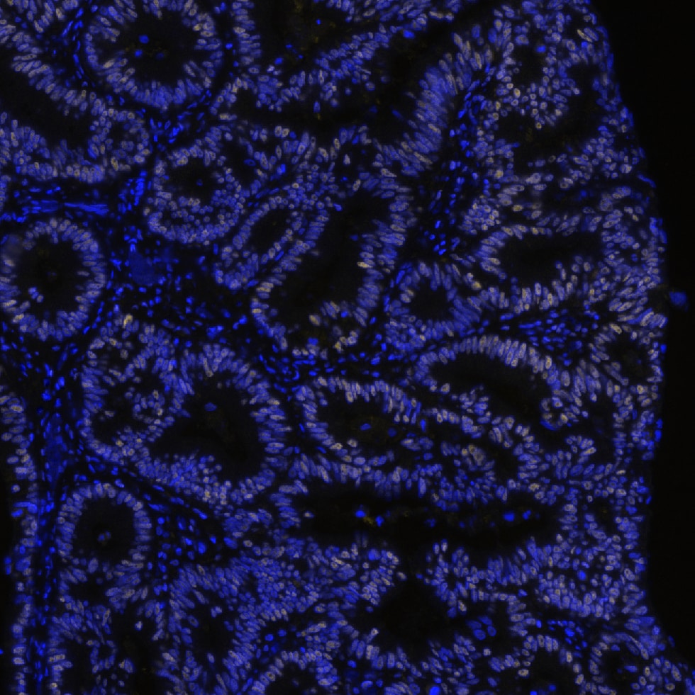
MSH6 in Human Colon Tumor via seqIF™ staining on COMET™
MSH6 was detected in immersion fixed paraffin-embedded sections of human colon tumor using Mouse Anti-Human MSH6, Monoclonal Antibody (Catalog # MAB11832) at 25ug/mL at 37 ° Celsius for 4 minutes. Before incubation with the primary antibody, tissue underwent an all-in-one dewaxing and antigen retrieval preprocessing using PreTreatment Module (PT Module) and Dewax and HIER Buffer H (pH 9; Epredia Catalog # TA-999-DHBH). Tissue was stained using the Alexa Fluor™ 647 Goat anti-Mouse IgG Secondary Antibody at 1:200 at 37 ° Celsius for 2 minutes. (Yellow; Lunaphore Catalog # DR647MS) and counterstained with DAPI (blue; Lunaphore Catalog # DR100). Specific staining was localized to the nucleus. Protocol available in COMET™ Panel Builder.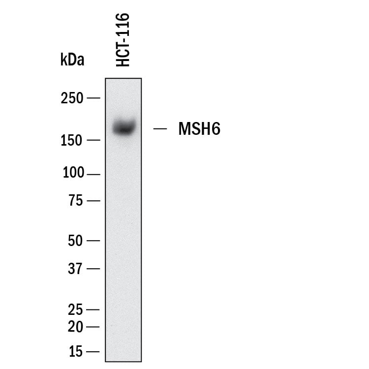
Detection of Human MSH6 by Western Blot.
Western Blot shows lysates of HCT‑116 human colorectal carcinoma cell line. PVDF membrane was probed with 1 µg/ml of Mouse Anti-Human MSH6 Monoclonal Antibody (Catalog # MAB11832) followed by HRP-conjugated Anti-Mouse IgG Secondary Antibody (HAF018). A specific band was detected for MSH6 at approximately 160 kDa (as indicated). This experiment was conducted under reducing conditions and using Western Blot Buffer Group 1.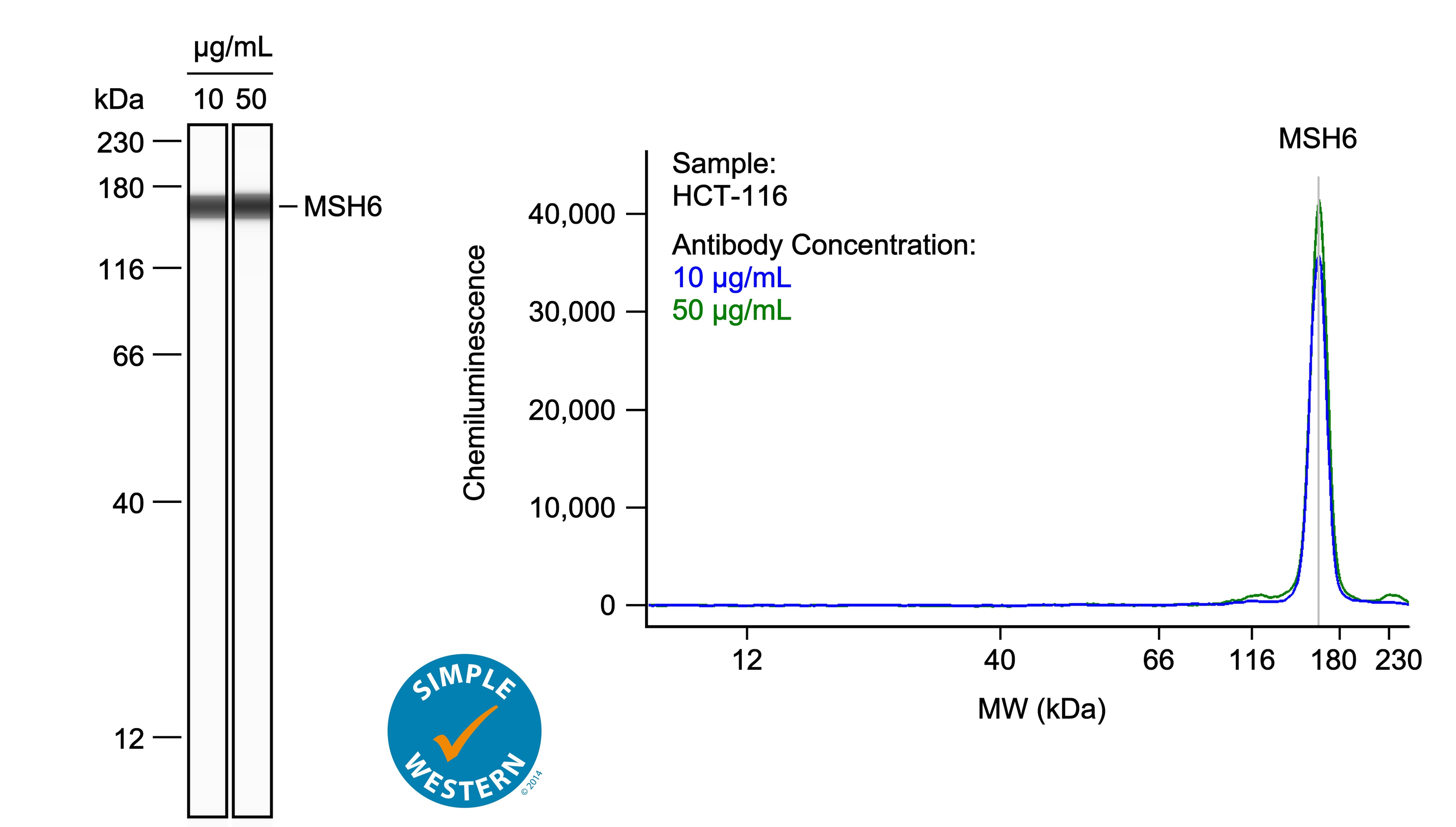
Detection of Human MSH6 by Simple WesternTM.
Left: Simple Western lane view shows lysates of HCT‑116 human colorectal carcinoma cell line, loaded at 0.1 mg/ml. A specific band was detected for MSH6 at approximately 165 kDa (as indicated) using both 10 µg/ml and 50 µg/ml of Mouse Anti-Human MSH6 Monoclonal Antibody (Catalog # MAB11832) followed by HRP-conjugated Goat Anti-Mouse Secondary Antibody (042-205). This experiment was conducted under reducing conditions and using the 12-230kDa separation system. Right: Simple Western electropherogram showing the same Mouse Anti-Human MSH6 Monoclonal Antibody (Catalog # MAB11832) tested at 10 µg/ml (blue line) and 50 µg/ml (green line) in the HCT‑116 human colorectal carcinoma cell line.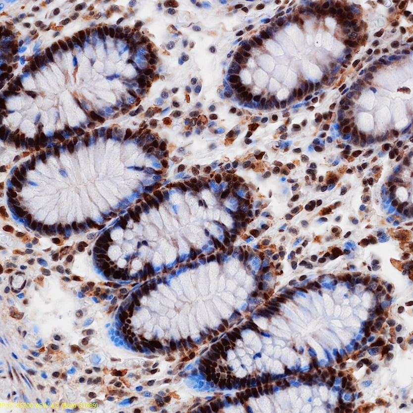
Immersion fixed paraffin-embedded sections of colon
MSH6 was detected in immersion fixed paraffin-embedded sections of colon using Mouse Anti-Human MSH6 Monoclonal Antibody (Catalog # MAB11832) at 5 µg/ml overnight at 4 °C. Before incubation with the primary antibody, tissue was subjected to heat-induced epitope retrieval using VisUCyte Antigen Retrieval Reagent-Basic (VCTS021). Tissue was stained using the HRP-conjugated Anti-Mouse IgG Secondary Antibody (HAF007) and counterstained with hematoxylin (blue). Specific staining was localized to the nucleus. View our protocol for Chromogenic IHC Staining of Paraffin-embedded Tissue Sections.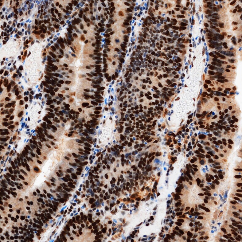
Immersion fixed paraffin-embedded sections of colon tumor
MSH6 was detected in immersion fixed paraffin-embedded sections of colon tumor using Mouse Anti-Human MSH6 Monoclonal Antibody (Catalog # MAB11832) at 5 µg/ml overnight at 4 °C. Before incubation with the primary antibody, tissue was subjected to heat-induced epitope retrieval using VisUCyte Antigen Retrieval Reagent-Basic (VCTS021). Tissue was stained using the HRP-conjugated Anti-Mouse IgG Secondary Antibody (HAF007) and counterstained with hematoxylin (blue). Specific staining was localized to the nucleus. View our protocol for Chromogenic IHC Staining of Paraffin-embedded Tissue Sections.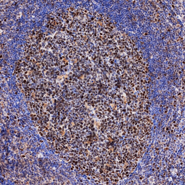
Immersion fixed paraffin-embedded sections of tonsil
MSH6 was detected in immersion fixed paraffin-embedded sections of tonsil using Mouse Anti-Human MSH6 Monoclonal Antibody (Catalog # MAB11832) at 5 µg/ml overnight at 4 °C. Before incubation with the primary antibody, tissue was subjected to heat-induced epitope retrieval using VisUCyte Antigen Retrieval Reagent-Basic (VCTS021). Tissue was stained using the HRP-conjugated Anti-Mouse IgG Secondary Antibody (HAF007) and counterstained with hematoxylin (blue). Specific staining was localized to the nucleus. View our protocol for Chromogenic IHC Staining of Paraffin-embedded Tissue Sections.Applications for Human MSH6 Antibody (1128063)
COMET
Sample: Immersion fixed paraffin-embedded sections of human colon tumor
Immunohistochemistry
Sample: Immersion fixed paraffin-embedded sections of normal colon, colon tumor, and tonsil
Simple Western
Sample: HCT-116 human colorectal carcinoma cell line
Western Blot
Sample: HCT-116 human colorectal carcinoma cell line
Formulation, Preparation, and Storage
Purification
Reconstitution
Reconstitute lyophilized material at 0.2 mg/ml in sterile PBS. For liquid material, refer to CoA for concentration.
Formulation
Shipping
Stability & Storage
- 12 months from date of receipt, -20 to -70 °C as supplied.
- 1 month, 2 to 8 °C under sterile conditions after reconstitution.
- 6 months, -20 to -70 °C under sterile conditions after reconstitution.
Calculators
Background: MSH6
References
- Modrich P, Lahue R. Mismatch repair in replication fidelity, genetic recombination, and cancer biology. Annu Rev Biochem. 1996;65:101-33. doi: 10.1146/annurev.bi.65.070196.000533. PMID: 8811176.
- Bonadona V, Bonaïti B, Olschwang S, Grandjouan S, Huiart L, Longy M, Guimbaud R, Buecher B, Bignon YJ, Caron O, Colas C, Noguès C, Lejeune-Dumoulin S, Olivier-Faivre L, Polycarpe-Osaer F, Nguyen TD, Desseigne F, Saurin JC, Berthet P, Leroux D, Duffour J, Manouvrier S, Frébourg T, Sobol H, Lasset C, Bonaïti-Pellié C; French Cancer Genetics Network. Cancer risks associated with germline mutations in MLH1, MSH2, and MSH6 genes in Lynch syndrome. JAMA. 2011 Jun 8;305(22):2304-10. doi: 10.1001/jama.2011.743. PMID: 21642682.
- Jiricny J. The multifaceted mismatch-repair system. Nat Rev Mol Cell Biol. 2006 May;7(5):335-46. doi: 10.1038/nrm1907. PMID: 16612326.
Long Name
Alternate Names
Gene Symbol
UniProt
Additional MSH6 Products
Product Documents for Human MSH6 Antibody (1128063)
Certificate of Analysis
To download a Certificate of Analysis, please enter a lot or batch number in the search box below.
Note: Certificate of Analysis not available for kit components.
Product Specific Notices for Human MSH6 Antibody (1128063)
For research use only
Customer Reviews for Human MSH6 Antibody (1128063)
There are currently no reviews for this product. Be the first to review Human MSH6 Antibody (1128063) and earn rewards!
Have you used Human MSH6 Antibody (1128063)?
Submit a review and receive an Amazon gift card!
$25/€18/£15/$25CAN/¥2500 for a review with an image
$10/€7/£6/$10CAN/¥1110 for a review without an image
Submit a review
Protocols
Find general support by application which include: protocols, troubleshooting, illustrated assays, videos and webinars.
- Antigen Retrieval Protocol (PIER)
- Antigen Retrieval for Frozen Sections Protocol
- Appropriate Fixation of IHC/ICC Samples
- Cellular Response to Hypoxia Protocols
- Chromogenic IHC Staining of Formalin-Fixed Paraffin-Embedded (FFPE) Tissue Protocol
- Chromogenic Immunohistochemistry Staining of Frozen Tissue
- ClariTSA™ Fluorophore Kits
- Detection & Visualization of Antibody Binding
- Fluorescent IHC Staining of Frozen Tissue Protocol
- Graphic Protocol for Heat-induced Epitope Retrieval
- Graphic Protocol for the Preparation and Fluorescent IHC Staining of Frozen Tissue Sections
- Graphic Protocol for the Preparation and Fluorescent IHC Staining of Paraffin-embedded Tissue Sections
- Graphic Protocol for the Preparation of Gelatin-coated Slides for Histological Tissue Sections
- IHC Sample Preparation (Frozen sections vs Paraffin)
- Immunofluorescent IHC Staining of Formalin-Fixed Paraffin-Embedded (FFPE) Tissue Protocol
- Immunohistochemistry (IHC) and Immunocytochemistry (ICC) Protocols
- Immunohistochemistry Frozen Troubleshooting
- Immunohistochemistry Paraffin Troubleshooting
- Preparing Samples for IHC/ICC Experiments
- Preventing Non-Specific Staining (Non-Specific Binding)
- Primary Antibody Selection & Optimization
- Protocol for Heat-Induced Epitope Retrieval (HIER)
- Protocol for Making a 4% Formaldehyde Solution in PBS
- Protocol for VisUCyte™ HRP Polymer Detection Reagent
- Protocol for the Preparation & Fixation of Cells on Coverslips
- Protocol for the Preparation and Chromogenic IHC Staining of Frozen Tissue Sections
- Protocol for the Preparation and Chromogenic IHC Staining of Frozen Tissue Sections - Graphic
- Protocol for the Preparation and Chromogenic IHC Staining of Paraffin-embedded Tissue Sections
- Protocol for the Preparation and Chromogenic IHC Staining of Paraffin-embedded Tissue Sections - Graphic
- Protocol for the Preparation and Fluorescent IHC Staining of Frozen Tissue Sections
- Protocol for the Preparation and Fluorescent IHC Staining of Paraffin-embedded Tissue Sections
- Protocol for the Preparation of Gelatin-coated Slides for Histological Tissue Sections
- R&D Systems Quality Control Western Blot Protocol
- TUNEL and Active Caspase-3 Detection by IHC/ICC Protocol
- The Importance of IHC/ICC Controls
- Troubleshooting Guide: Immunohistochemistry
- Troubleshooting Guide: Western Blot Figures
- Western Blot Conditions
- Western Blot Protocol
- Western Blot Protocol for Cell Lysates
- Western Blot Troubleshooting
- Western Blot Troubleshooting Guide
- View all Protocols, Troubleshooting, Illustrated assays and Webinars