
Nidogen-1 (also entactin) is a 150 kDa, secreted, monomeric glycoprotein that serves as a major linking component of basement membranes (1-4). It is synthesized as a 1247 amino acid (aa) precursor with a 28 aa signal sequence and a 1219 aa mature protein. The molecule is modular in structure with five distinct regions. There are three globular domains (G1-3) separated by a mucin region and an extended rod-shaped segment (5-7). The N-terminal globular domain (G1) is 200 aa in length and seemingly unrelated to any known motif (8). The mucin region is nearly 160 aa in length and presumably O-glycosylated (2, 8). G2 and G3 are both approximately 300 aa in length. G2 is described as a Nidogen ( beta -barrel) domain, while C-terminal G3 assumes a beta -propeller configuration (1). The 250 aa rod-shaped segment has multiple EGF-like motifs and two thyroglobulin type 1 domains. Functionally, G1 is reported to bind type IV collagen (2, 7). The mucin region contains a short peptide that ligates alpha 3 beta 1 integrins (9, 10). G2 interacts with perlecan, and an RGD motif in the rod-shaped segment serves as a binding site for alpha v beta 3 integrins (9, 10). Finally, G3 is associated with laminin binding (2, 7). As a full-length molecule, the multiple extracellular matrix-binding sites of Nidogen-1 are well positioned to serve as anchor sites for basement membrane molecules. Nidogen-1 also undergoes proteolytic processing by at least two MMPs, MMP-7 and MMP-19 (10, 11). While this destroys the integrity of Nidogen-associated matrices, it also generates peptide fragments that are capable of inducing neutrophil chemotaxis and phagocytosis (10). Nidogen-2 is related to Nidogen-1 (≈ 50% aa identity) and shares many of the same adhesive properties as Nidogen-1 (12). Both bind perlecan plus collagens I and IV. Nidogen‑2, however, does not bind fibulin-1 or 2, and shows only modest interaction with laminin. Thus, although coexpressed, Nidogen-2 serves as only a partial substitute for Nidogen-1 (2, 12). Human Nidogen-1 shares 85% aa sequence identity with both mouse and rat Nidogen-1, and 88% aa sequence identity with canine Nidogen-1.
Human Nidogen-1/Entactin Antibody (302117)
R&D Systems | Catalog # MAB2570


Scientific images may be resized, cropped, or adjusted for brightness or contrast for presentation purposes. These updates are limited to presentation and do not affect the underlying scientific interpretation of the data. Where available, additional source data may be provided upon request.
Key Product Details
Species Reactivity
Validated:
Cited:
Applications
Validated:
Cited:
Label
Antibody Source
Product Specifications
Immunogen
Leu29-Lys1114 (Gln1113Arg)
Accession # AAH45606.1
Specificity
Clonality
Host
Isotype
Scientific Data Images for Human Nidogen-1/Entactin Antibody (302117)
Detection of Nidogen‑1/Entactin in Human Heart.
Nidogen‑1/Entactin was detected in immersion fixed paraffin-embedded sections of human heart using Mouse Anti-Human Nidogen‑1/Entactin Monoclonal Antibody (Catalog # MAB2570) at 5 µg/ml for 1 hour at room temperature followed by incubation with the HRP-conjugated Anti-Mouse IgG Secondary Antibody (Catalog # HAF007) or the Anti-Mouse IgG VisUCyte™ HRP Polymer Antibody (Catalog # VC001). Before incubation with the primary antibody, tissue was subjected to heat-induced epitope retrieval using VisUCyte Antigen Retrieval Reagent-Basic (Catalog # VCTS021). Tissue was stained using DAB (brown) and counterstained with hematoxylin (blue). Specific staining was localized to the membrane. View our protocol for Chromogenic IHC Staining of Paraffin-embedded Tissue Sections.
Nidogen‑1/Entactin in Human Chondrocytes.
Nidogen-1/Entactin was detected in immersion fixed human mesenchymal stem cells differentiated into chondrocytes using Mouse Anti-Human Nidogen-1/Entactin Monoclonal Antibody (Catalog # MAB2570) at 10 µg/mL for 3 hours at room temperature. Cells were stained using the NorthernLights™ 557-conjugated Anti-Mouse IgG Secondary Antibody (yellow; NL007) and counterstained with DAPI (blue). View our protocol for Fluorescent ICC Staining of Cells on Coverslips.
Human Nidogen-1 / Entactin ELISA Standard Curve
Recombinant Human Nidogen‑1/Entactin (Catalog # 2570-ND) was serially diluted and captured by Mouse Anti-Human Nidogen‑1/Entactin Monoclonal Antibody (Catalog # MAB2570) coated on a Clear Polystyrene Microplate (Catalog # DY990). Goat Anti-Human Nidogen‑1/Entactin Antigen Affinity-purified Polyclonal Antibody (Catalog # AF2570) was biotinylated and incubated with the protein captured on the plate. Detection of the standard curve was achieved by incubating Streptavidin-HRP (Catalog # DY998)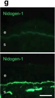
Detection of Nidogen-1/Entactin by Immunohistochemistry
Zebularine improves wound healing and limbal stem cell phenotype in diabetic organ-cultured corneas via Wnt-5a. (a) Wound healing time was significantly decreased in heptanol-wounded diabetic corneas treated with zebularine (20 µmol/l) vs DMSO (control) (n=4). (b–g) Immunostaining (n=3) showed that zebularine decreased DNMT1 (b) but increased Wnt-5a (c) levels in healed corneas. Zebularine also increased limbal levels of putative stem cell markers, K15 (d) and K17 (e), as well as diabetic markers integrin alpha 3 beta 1 (f) and nidogen-1 (g). Values are mean ± SEM. **p<0.01 vs control (paired t test). Scale bar, 20 µm. e, epithelium; s, stroma Image collected and cropped by CiteAb from the following open publication (https://link.springer.com/10.1007/s00125-023-05960-1), licensed under a CC-BY license. Not internally tested by R&D Systems.Applications for Human Nidogen-1/Entactin Antibody (302117)
Immunocytochemistry
Immunohistochemistry
Sample: Immersion fixed paraffin-embedded sections of human heart
Western Blot
Sample: Recombinant Human Nidogen‑1/Entactin (Catalog # 2570-ND)
Human Nidogen-1/Entactin Sandwich Immunoassay
Reviewed Applications
Read 7 reviews rated 4 using MAB2570 in the following applications:
Formulation, Preparation, and Storage
Purification
Reconstitution
Reconstitute at 0.5 mg/mL in sterile PBS. For liquid material, refer to CoA for concentration.
Formulation
Shipping
Stability & Storage
- 12 months from date of receipt, -20 to -70 °C as supplied.
- 1 month, 2 to 8 °C under sterile conditions after reconstitution.
- 6 months, -20 to -70 °C under sterile conditions after reconstitution.
Calculators
Background: Nidogen-1/Entactin
References
- Hohenester, E. and J. Engel (2002) Matrix Biol. 21:115.
- Miosge, N. et al. (2001) Histochem. J. 33:523.
- Charonis, A. et al. (2005) Curr. Med. Chem. 12:1495.
- Timpl, R. and J.C. Brown (1996) BioEssays 18:123.
- Nagayoshi, T. et al. (1989) DNA 8:581.
- Zimmerman, K. et al. (1995) Genomics 27:245.
- Fox, J.W. et al. (1991) EMBO J. 10:3137.
- Mayer, U. et al. (1995) Eur. J. Biochem. 227:681.
- Gresham, H.D. et al. (1996) J. Biol. Chem. 271:30587.
- Dong, L-J. et al. (1995) J. Biol. Chem. 270:15383.
- Titz, B. et al. (2004) Cell. Mol. Life Sci. 61:1826.
- Kohfeldt, K. et al. (1998) J. Mol. Biol. 282:99.
Alternate Names
Entrez Gene IDs
Gene Symbol
UniProt
Additional Nidogen-1/Entactin Products
Product Documents for Human Nidogen-1/Entactin Antibody (302117)
Certificate of Analysis
To download a Certificate of Analysis, please enter a lot or batch number in the search box below.
Note: Certificate of Analysis not available for kit components.
Product Specific Notices for Human Nidogen-1/Entactin Antibody (302117)
For research use only
Citations for Human Nidogen-1/Entactin Antibody (302117)
 Powered by Bioz
Customer Reviews for Human Nidogen-1/Entactin Antibody (302117) (7)
Have you used Human Nidogen-1/Entactin Antibody (302117)?
Submit a review and receive an Amazon gift card!
$25/€18/£15/$25CAN/¥2500 for a review with an image
$10/€7/£6/$10CAN/¥1110 for a review without an image
Submit a review
Customer Images

-
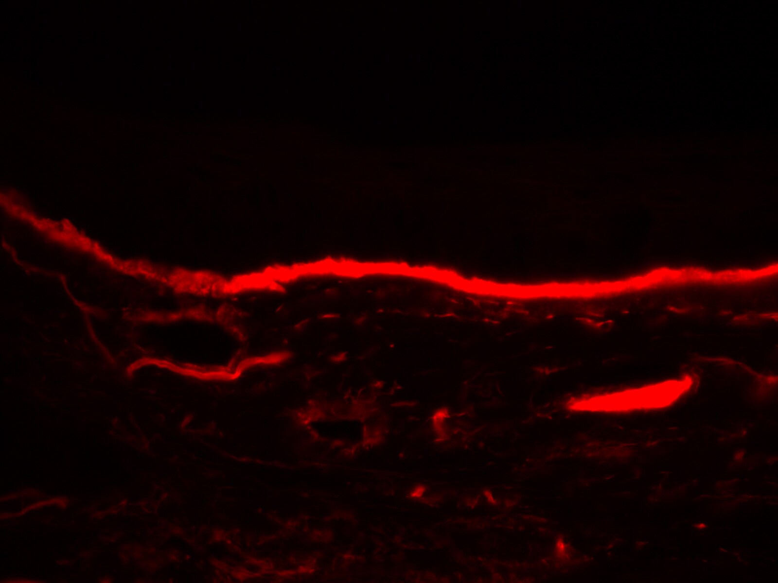
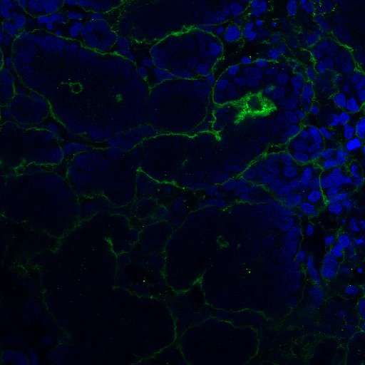
Application: ImmunohistochemistrySample Tested: corneal tissueSpecies: HumanVerified Customer | Posted 03/21/20231% formalin fixed

-
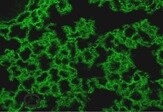
Application: Immunocytochemistry/ImmunofluorescenceSample Tested: Lung tissueSpecies: HumanVerified Customer | Posted 11/01/2021
-
Application: MicroarraysSample Tested: EDTA PlasmaSpecies: HumanVerified Customer | Posted 01/14/2021
-
Application: Immunocytochemistry/ImmunofluorescenceSample Tested: Differentiated embryonic stem cellsSpecies: HumanVerified Customer | Posted 12/02/2020Staining for basement membrane
-
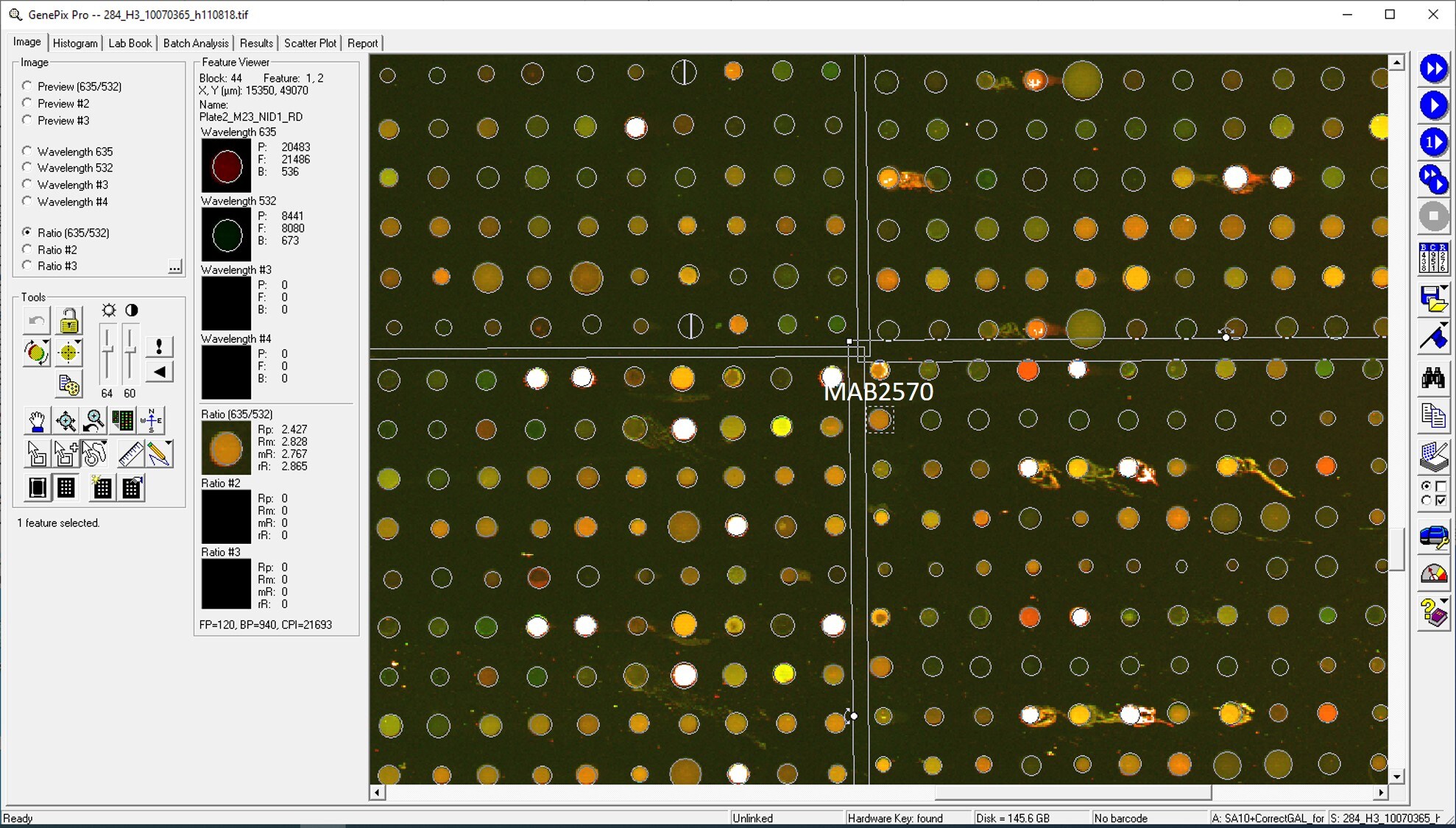
Application: MicroarraySample Tested: EDTA PlasmaSpecies: HumanVerified Customer | Posted 02/07/2020Antibody was printed on custom arrays and incubated with fluorescently labeled human EDTA plasma
-
Application: MicroarraysSample Tested: EDTA PlasmaSpecies: HumanVerified Customer | Posted 11/14/2018
-
Application: ELISASample Tested: PlasmaSpecies: HumanVerified Customer | Posted 11/10/2018
There are no reviews that match your criteria.
Protocols
Find general support by application which include: protocols, troubleshooting, illustrated assays, videos and webinars.
- Antigen Retrieval Protocol (PIER)
- Antigen Retrieval for Frozen Sections Protocol
- Appropriate Fixation of IHC/ICC Samples
- Cellular Response to Hypoxia Protocols
- Chromogenic IHC Staining of Formalin-Fixed Paraffin-Embedded (FFPE) Tissue Protocol
- Chromogenic Immunohistochemistry Staining of Frozen Tissue
- ClariTSA™ Fluorophore Kits
- Detection & Visualization of Antibody Binding
- Fluorescent IHC Staining of Frozen Tissue Protocol
- Graphic Protocol for Heat-induced Epitope Retrieval
- Graphic Protocol for the Preparation and Fluorescent IHC Staining of Frozen Tissue Sections
- Graphic Protocol for the Preparation and Fluorescent IHC Staining of Paraffin-embedded Tissue Sections
- Graphic Protocol for the Preparation of Gelatin-coated Slides for Histological Tissue Sections
- ICC Cell Smear Protocol for Suspension Cells
- ICC Immunocytochemistry Protocol Videos
- ICC for Adherent Cells
- IHC Sample Preparation (Frozen sections vs Paraffin)
- Immunocytochemistry (ICC) Protocol
- Immunocytochemistry Troubleshooting
- Immunofluorescence of Organoids Embedded in Cultrex Basement Membrane Extract
- Immunofluorescent IHC Staining of Formalin-Fixed Paraffin-Embedded (FFPE) Tissue Protocol
- Immunohistochemistry (IHC) and Immunocytochemistry (ICC) Protocols
- Immunohistochemistry Frozen Troubleshooting
- Immunohistochemistry Paraffin Troubleshooting
- Preparing Samples for IHC/ICC Experiments
- Preventing Non-Specific Staining (Non-Specific Binding)
- Primary Antibody Selection & Optimization
- Protocol for Heat-Induced Epitope Retrieval (HIER)
- Protocol for Making a 4% Formaldehyde Solution in PBS
- Protocol for VisUCyte™ HRP Polymer Detection Reagent
- Protocol for the Fluorescent ICC Staining of Cell Smears - Graphic
- Protocol for the Fluorescent ICC Staining of Cultured Cells on Coverslips - Graphic
- Protocol for the Preparation & Fixation of Cells on Coverslips
- Protocol for the Preparation and Chromogenic IHC Staining of Frozen Tissue Sections
- Protocol for the Preparation and Chromogenic IHC Staining of Frozen Tissue Sections - Graphic
- Protocol for the Preparation and Chromogenic IHC Staining of Paraffin-embedded Tissue Sections
- Protocol for the Preparation and Chromogenic IHC Staining of Paraffin-embedded Tissue Sections - Graphic
- Protocol for the Preparation and Fluorescent ICC Staining of Cells on Coverslips
- Protocol for the Preparation and Fluorescent ICC Staining of Non-adherent Cells
- Protocol for the Preparation and Fluorescent ICC Staining of Stem Cells on Coverslips
- Protocol for the Preparation and Fluorescent IHC Staining of Frozen Tissue Sections
- Protocol for the Preparation and Fluorescent IHC Staining of Paraffin-embedded Tissue Sections
- Protocol for the Preparation of Gelatin-coated Slides for Histological Tissue Sections
- Protocol for the Preparation of a Cell Smear for Non-adherent Cell ICC - Graphic
- R&D Systems Quality Control Western Blot Protocol
- TUNEL and Active Caspase-3 Detection by IHC/ICC Protocol
- The Importance of IHC/ICC Controls
- Troubleshooting Guide: Immunohistochemistry
- Troubleshooting Guide: Western Blot Figures
- Western Blot Conditions
- Western Blot Protocol
- Western Blot Protocol for Cell Lysates
- Western Blot Troubleshooting
- Western Blot Troubleshooting Guide
- View all Protocols, Troubleshooting, Illustrated assays and Webinars