
NKp44, along with NKp30 and NKp46, constitute a group of receptors termed "Natural Cytotoxicity Receptors" (NCR) (1). These receptors are expressed almost exclusively by NK cells and play a major role in triggering NK-mediated killing of most tumor cell lines. No rodent ortholog to NKp44 has been identified. Human NKp44, also known as NCR2, is a 44 kDa type I transmembrane glycoprotein that is characterized by the presence of one extracellular V-like immunoglobulin domain (2). It is synthesized as a 276 amino acid (aa) precursor that contains a 21 aa signal sequence, a 171 aa extracellular region, a 21 aa transmembrane segment and a 63 aa cytoplasmic tail. Alternate splicing in both the cytoplasmic tail and extracellular region generates multiple isoforms of unknown significance. The Ig-like region is unaffected. A physical association with the ITAM-bearing accessory protein, DAP12, occurs via a charged residue in the NKp44 transmembrane domain. Ligation of NKp44 with a specific antibody results in phosphorylation of DAP12 (3) and activation of target cell lysis in a redirected killing assay (4). NKp44 is absent from resting NK cells but is upregulated upon activation with IL-2. Activation-induced expression occurs in the CD56dim CD16+ NK subset that accounts for more than 85% of NK cells found in peripheral blood and spleen, as well as the CD56bright CD16- NK subset that constitutes the majority of NK cells in lymph node and tonsil (5). Studies with neutralizing antibodies reveal that NKp44 is partially responsible for triggering lytic activity against several tumor cell types (2, 6). Blocking any of the individual NCRs results in partial inhibition of tumor cell lysis, but nearly complete inhibition of lysis is observed if all three receptors are blocked simultaneously (6). NKp44 has also been implicated in recognition of virus-infected cells through its capacity to bind to viral hemagglutinins (7).
Human NKp44/NCR2 Antibody (253424)
R&D Systems | Catalog # MAB22492

Scientific images may be resized, cropped, or adjusted for brightness or contrast for presentation purposes. These updates are limited to presentation and do not affect the underlying scientific interpretation of the data. Where available, additional source data may be provided upon request.
Key Product Details
Species Reactivity
Applications
Label
Antibody Source
Product Specifications
Immunogen
Glu22-Pro190
Accession # CAB52289
Specificity
Clonality
Host
Isotype
Scientific Data Images for Human NKp44/NCR2 Antibody (253424)
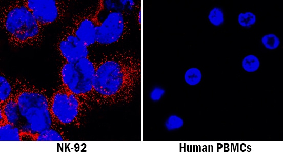
NKp44/NCR2 in NK‑92 Human Cell Line.
NKp44/NCR2 was detected in immersion fixed NK-92 human natural killer lymphoma cell line (positive staining) and human peripheral blood mononuclear cells (PBMCs; negative staining) using Mouse Anti-Human NKp44/NCR2 Monoclonal Antibody (Catalog # MAB22492) at 8 µg/mL for 3 hours at room temperature. Cells were stained using the NorthernLights™ 557-conjugated Anti-Mouse IgG Secondary Antibody (red; Catalog # NL007) and counterstained with DAPI (blue). Specific staining was localized to cytoplasm. View our protocol for Fluorescent ICC Staining of Non-adherent Cells.Applications for Human NKp44/NCR2 Antibody (253424)
Immunocytochemistry
Sample: Immersion fixed NK-92 human natural killer lymphoma cell line
Formulation, Preparation, and Storage
Purification
Reconstitution
Reconstitute at 0.5 mg/mL in sterile PBS. For liquid material, refer to CoA for concentration.
Formulation
Shipping
Stability & Storage
- 12 months from date of receipt, -20 to -70 °C as supplied.
- 1 month, 2 to 8 °C under sterile conditions after reconstitution.
- 6 months, -20 to -70 °C under sterile conditions after reconstitution.
Calculators
Background: NKp44/NCR2
References
- Moretta, L. and A. Moretta (2004) EMBO J. 23:255.
- Cantoni, C. et al. (1999) J. Exp. Med. 189:787.
- Augugliaro, R. et al. (2003) Eur. J. Immunol. 33:1235.
- Vitale, M. et al. (1998) J. Exp. Med. 187:2065.
- Ferlazzo, G. et al. (2004) J. Immunol. 172:1455.
- Pende, D. et al. (1999) J. Exp. Med. 190:1505.
- Arnon, T. et al. (2001) Eur. J. Immunol. 31:2680.
Long Name
Alternate Names
Gene Symbol
UniProt
Additional NKp44/NCR2 Products
Product Documents for Human NKp44/NCR2 Antibody (253424)
Certificate of Analysis
To download a Certificate of Analysis, please enter a lot or batch number in the search box below.
Note: Certificate of Analysis not available for kit components.
Product Specific Notices for Human NKp44/NCR2 Antibody (253424)
For research use only
Customer Reviews for Human NKp44/NCR2 Antibody (253424)
There are currently no reviews for this product. Be the first to review Human NKp44/NCR2 Antibody (253424) and earn rewards!
Have you used Human NKp44/NCR2 Antibody (253424)?
Submit a review and receive an Amazon gift card!
$25/€18/£15/$25CAN/¥2500 for a review with an image
$10/€7/£6/$10CAN/¥1110 for a review without an image
Submit a review
Protocols
Find general support by application which include: protocols, troubleshooting, illustrated assays, videos and webinars.
- Appropriate Fixation of IHC/ICC Samples
- Cellular Response to Hypoxia Protocols
- ClariTSA™ Fluorophore Kits
- Detection & Visualization of Antibody Binding
- ICC Cell Smear Protocol for Suspension Cells
- ICC Immunocytochemistry Protocol Videos
- ICC for Adherent Cells
- Immunocytochemistry (ICC) Protocol
- Immunocytochemistry Troubleshooting
- Immunofluorescence of Organoids Embedded in Cultrex Basement Membrane Extract
- Immunohistochemistry (IHC) and Immunocytochemistry (ICC) Protocols
- Preparing Samples for IHC/ICC Experiments
- Preventing Non-Specific Staining (Non-Specific Binding)
- Primary Antibody Selection & Optimization
- Protocol for VisUCyte™ HRP Polymer Detection Reagent
- Protocol for the Fluorescent ICC Staining of Cell Smears - Graphic
- Protocol for the Fluorescent ICC Staining of Cultured Cells on Coverslips - Graphic
- Protocol for the Preparation and Fluorescent ICC Staining of Cells on Coverslips
- Protocol for the Preparation and Fluorescent ICC Staining of Non-adherent Cells
- Protocol for the Preparation and Fluorescent ICC Staining of Stem Cells on Coverslips
- Protocol for the Preparation of a Cell Smear for Non-adherent Cell ICC - Graphic
- TUNEL and Active Caspase-3 Detection by IHC/ICC Protocol
- The Importance of IHC/ICC Controls
- View all Protocols, Troubleshooting, Illustrated assays and Webinars
Associated Pathways

